Abstract
BACKGROUND AND PURPOSE:
Provocative testing before AVM embolization has been shown to be a predictor of a successful endovascular treatment without neurologic deficits. Propofol has been used previously as an alternative agent in Wada testing with adequate results. The purpose of this study was to show our experience with the use of propofol as a safe and effective alternative to barbiturate provocative testing in AVM embolization procedures.
MATERIALS AND METHODS:
A series of 20 patients, undergoing 38 embolization sessions, was treated for cerebral AVMs between November 2007 and February 2009 by endovascular methods. All patients were treated under conscious sedation. Pre-embolization neurologic assessment was performed with provocative testing by using propofol at 7-mg doses by an intra-arterial route after microcathether placement in or near the AVM nidus.
RESULTS:
Among these 20 patients, 3 developed transient neurologic deficits after provocative testing, precluding initial or further embolization. One of the patients passing the provocative test developed slight paresis as a result of embolization with n-BCA, resulting in a PPV of 97%.
CONCLUSIONS:
Propofol use during provocative testing in AVM embolization procedures represents an effective alternative to barbiturate testing and can have a positive impact in improving safety under sedation.
The treatment of cerebral AVMs by endovascular techniques has been shown to be an invaluable tool in the preoperative and the preradiosurgical management of AVMs. In certain cases, it can be the definitive treatment, with cure rates varying between 14% and 49% in some series.1–5 Yet, complication rates between 10% and 50% have been reported.4 The use of provocative or superselective Wada testing may be of significant value in certain circumstances, improving the safety of arterial embolization.6 The usual trend is to use short-acting barbiturates for intracranial lesions and lidocaine for extracranial ones. Amobarbital has been the traditional drug of choice for pre-embolization testing, yet its availability has been affected by several shortages, including the present one. This has led many centers to consider the use of other drugs, such as methohexital,7 pentobarbital,8 etomidate,9 and propofol,10,11 reported primarily in Wada testing. Similar reports on the use of these or similar anesthetics in cerebral AVM pre-embolization provocative testing are absent or scarce. One study reported sequential use of amobarbital and lidocaine in pre-embolization studies, increasing the sensitivity of testing,12 yet the role and safety profile of lidocaine as a sole medication for intracranial testing has yet to be determined. Propofol is one of the readily available anesthetics that has been used intra-arterially for intracranial testing. Its safety margin, low incidence of side effects, and effectiveness in inducing controlled transitory losses of neurologic function in the perfused areas has been reported.10,11,13 In the present study, we report our experience using propofol during provocative testing in a series of patients who underwent embolization for cerebral AVMs. The purpose of this study was to establish the use of propofol as a safe and effective alternative to barbiturate provocative testing in AVM embolization procedures. Institutional review board approval was granted before submission of this study.
Materials and Methods
Patients and Techniques
The population under study included all patients treated for cerebral AVMs by endovascular methods between November 2007 and February 2009. Pediatric patients requiring endotracheal anesthesia were excluded because we were unable to perform a thorough neurologic examination during testing. A total of 20 patients were treated in 38 embolization sessions. One provocative test was performed for each embolization session. The time interval between each session was between 1.5 and 6 months. Demographics and other important clinical data, such as associated diagnoses, medications, initial presentation, and any deficits before endovascular treatments, were collected (Table 1). AVM grades and flow characteristics (transit times), by counting the angiographic frames until venous drainage, as well as any deficits and adverse reactions to propofol injection, were also recorded (Table 2).
Table 1:
Clinical and demographic characteristics of the patient population
| Demographic and Clinical Parameters | Value |
|---|---|
| Age | |
| Median (yrs) | 52 |
| Range (yrs) | 11–76 |
| Sex | |
| Male | 12 |
| Female | 8 |
| Associated conditions | |
| HBP | 7 |
| DM | 3 |
| Hypercholesterolemia | 4 |
| CAD | 1 |
| Hypothyroidism | 1 |
| BA | 1 |
| BPH | 1 |
| Medications | |
| Calcium channel blockers | 3 |
| ACE | 4 |
| β-blockers | 2 |
| α-Agonists | 1 |
| AEDs | |
| Dilantin (phenytoin) | 3 |
| Tegretol (carbamazepine) | 1 |
| Levetiracetam | 3 |
| Valproic acid | 1 |
| Lamotrigine | 1 |
| Synthroid (levothyroxine) | 1 |
| Statins | 4 |
| Other psychiatric/neurologic | 4 |
| Clinical presentation | |
| Headaches | 9 |
| Seizures | 4 |
| TIAs | 1 |
| Paresthesias | 2 |
| ICH, SAH, IVH | 4 |
| Dizziness | 4 |
| Hemianopsia | 1 |
| Incidental | 3 |
| Pre-embolization neurologic deficits | |
| Blindness | 1 |
| Dysarthria | 1 |
| Nystagmus | 1 |
| Paresis | 1 |
| Dysesthesias | 1 |
| Hemianopsia | 1 |
Table 2:
AVM angiographic characteristics of the population
| Angiographic Parameter | Value |
|---|---|
| AVM locations | |
| Frontal | 3 |
| Parietal | 6 |
| Parieto-occipital | 4 |
| Temporoparietal | 1 |
| Occipital | 3 |
| Temporal | 2 |
| Cerebellar | 1 |
| AVM S-M grades | |
| 1 | 5 |
| 2 | 9 |
| 3 | 5 |
| 4 | 1 |
| Size (cms) | |
| ≤1 | 1 |
| ≤2 and >1 | 10 |
| ≤3 and >2 | 13 |
| ≤4 and >3 | 11 |
| ≤5 and >4 | 1 |
| ≤6 and >5 | 0 |
| ≤7 and >6 | 2 |
| Transit times (secs) | |
| 0.5 | 8 |
| 0.75 | 21 |
| 0.83 | 1 |
| 1.0 | 5 |
Before treatment, patients and relatives were informed about provocative testing with propofol, an off-label use, to improve the safety of subsequent embolization. It was mentioned that its use was documented in the medical literature with an adequate safety profile. Informed consent was granted for the embolization procedure and provocative testing. All information was recorded by our neuroendovascular team on preoperative and postoperative evaluations at the time of the intervention.
All patients were treated under conscious sedation, implying an alert or verbally arousable patient, with oxygen supplementation and cardiac and respiratory monitoring. Sedative and analgesic agents included midazolam, 0.07–0.08 mg/kg IV per dose in adults (0.05–0.1 mg/kg/dose in pediatric patients) and fentanyl, 1–2 mcg/kg intravenously per dose.14 Procedures were performed after thorough case evaluations and by using standardized institutional and the Occupational Safety and Health Administration−approved safety measures and aseptic materials and techniques. Pre-embolization neurologic assessment was performed before and after anesthetic injection with propofol at 7-mg doses intra-arterially after microcatheter placement in or near the AVM nidus. The 7-mg dose was reached by trial and error, titrating from higher doses so as not to cause dosage-related somnolence in the examined patients. Doses as low as 10 mg have been used for intracarotid Wada testing11,13; therefore, a 7-mg dose would be sufficient to induce a deficit in a smaller volume of distribution. The duration of the assessment was <5 minutes because the effect of propofol is immediate after a 20-second injection and has a short duration of action. The neurologic examination was tailored to the functional areas at risk, and if necessary, provocative testing was repeated for confirmation. Muscle strength in the upper and lower extremities was tested, including facial grimacing, shoulder abduction, elbow and wrist flexion/extension, finger flexion/extension/abduction, hip flexion, knee flexion/extension, and ankle dorsiflexion/plantar flexion. Strength was graded by using the Royal Medical Research Council of Great Britain scale.15 Sensory modalities tested included light touch, pinprick, and proprioception. Any tingling or numbness was recorded. Cortical sensory functions were tested, if indicated, by the location of the lesion. Memory function, if indicated, was tested by the recollection of 5 words mentioned at the beginning of the assessment and after 3–5 minutes of testing. Language testing included number counting, word fluency, and comprehension by following commands. Visual function was evaluated by picture and color naming, as well as reading short sentences.
Digital subtraction angiography was performed with biplane units with 3D reconstruction (LCN+, GE Medical Systems, Chalfont St. Giles, United Kingdom; Allura, Philips Medical Systems, Amsterdam, the Netherlands). Catheterization and embolization materials included standard microcatheters (Excelsior SL-10 and Excel 14; Boston Scientific, Natick, Massachusetts) in case aneurysm coil placement was anticipated, flow-guided microcatheters (Spinnaker Elite; Boston Scientific), steerable microwires (0.20–0.30 mm) (Traxcess 0.014 inch, MicroVention, Tustin, California; Transend EX, Boston Scientific; Mirage, ev3, Irvine, California), guide catheters (5F and 6F) (Envoy; Cordis, Bridgewater, New Jersey), sheath introducers (5F and 6F) (Cordis), coils (GDC and Berenstein liquid coils; Boston Scientific), n-BCA (Cordis Neurovascular), and ethiodized oil (Ethiodol; Cordis Neurovascular). Conray and Optiray contrast media (Mallinckrodt, Hazelwood, Missouri) were used, the latter in the case of allergy history.
Results
A total of 20 patients underwent 38 embolization sessions. Tables 1 and 2 show the clinical, demographic, and angiographic characteristics of the patient population.
Negative Provocative Test.
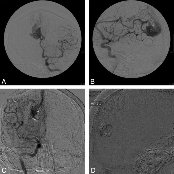
Among the 38 provocative tests, 3 resulted in transient neurologic deficits precluding embolization. One patient (Fig 1) had a right parieto-occipital AVM with 2 prior embolization procedures. On provocative testing of a PCA feeder, the patient developed transient left-legged proximal paresis. Further embolization was withheld, and the patient was referred for radiosurgery after refusing surgery. A second patient had a right temporal lesion and developed transient left-handed weakness and decreased number recall after propofol injection. The third patient had a left temporoparietal AVM and developed reversible dysarthria after propofol infusion. The latter 2 patients had not undergone any prior embolization procedures, and after we discussed management alternatives with them, they decided on further treatment with radiosurgery. All the patients recovered to baseline after 3–5 minutes.
Fig 1.

A 47-year-old man with a right parieto-occipital grade 2 AVM discovered incidentally during a work-up for head trauma after a fall. A and B, AP and lateral views of the first diagnostic angiogram. C, Note arterials feeders coming from the MCA, ACA, and PCA, as well as n-BCA from 2 prior embolizations. D, After superselective catheterization of the AVM through a PCA feeder, propofol was injected, causing proximal left-legged weakness. Further embolization was withheld, and the patient was referred for radiosurgery after refusing surgery.
Positive Provocative Test.
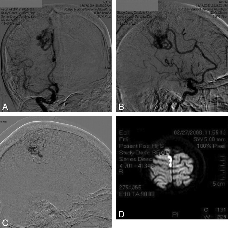
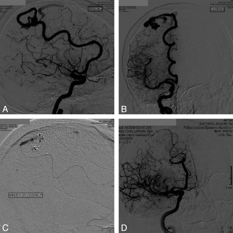
A total of 35 provocative tests (17 patients) yielded positive results (embolizable lesions). One of the patients with a positive provocative test developed slight distal contralateral leg paresis postprocedure (Fig 2). This patient had undergone prior embolization with n-BCA requiring the assistance of aneurysm coils due to the high-flow nature of the lesion. Transit time was less than half a second, suggesting a fistulous behavior. Despite this, aneurysm coils were not used to slow flow, and the concentration of n-BCA chosen may have been less than ideal for the situation (30% n-BCA/70% iodized oil [Ethiodol]). Another explanation for the postoperative deficit might have been a technical problem during embolic-agent injection with possible reflux into opening collaterals. Figure 3 shows a patient with a grade 1 parietal AVM. This patient had a positive provocative test after 7-mg propofol injection. Embolization with n-BCA and aneurysm coils then proceeded without any complications. Angiographic follow-up at 2 months showed no recanalization. Overall, a total of 34 embolization procedures proceeded uneventfully.
Fig 2.

A 54-year-old woman with a right medial parietal grade 2 AVM who presented with chronic headaches and dizziness. A and B, AP and lateral views of the first diagnostic angiogram. C, Note prior embolization, which required assistance of aneurysm coils due to the fistulous nature of the lesion. Also note the microcatheterization of the AVM nidus through the ACA feeder. After a superselective Wada test with propofol, the patient showed no neurologic changes, yet after embolization, she developed left-leg distal paresis. D, Postoperative diffusion-weighted MR image shows an ischemic insult close to the embolization area. The patient improved to baseline after physical therapy.
Fig 3.

A 63-year-old woman with a grade 1 right parietal AVM who presented with left-handed numbness. After discussion of management alternatives, she decided on an endovascular intervention. A and B, AP and lateral views of the diagnostic angiogram. A superselective Wada test with propofol was performed with positive results (embolizable ACA feeder). C, Embolization with n-BCA after placement of the aneurysm coil to slow transit time. D, Note that the follow-up angiogram after 2 months shows no recanalization.
The calculated PPV for the provocative test with propofol would be 97%. If we took into account only lesions located in or adjacent to eloquent areas, the calculated PPV would be 94.7%. Estimation of negative predictive value, sensitivity, and specificity was not possible because patients testing negative could not be embolized, and there is no criterion standard provocative test with which to compare. Therefore, the number of embolizable and nonembolizable patients testing negative is not known. There were no adverse reactions to propofol injection at the dose used.
Discussion
Provocative or superselective Wada testing has become an important part of endovascular management of numerous extracranial and intracranial vascular conditions. It can be used with neurophysiologic monitoring in cases in which patient cooperation and steadiness are important.16 Nevertheless, the value of awake neurologic assessment (wake-up test) cannot be underestimated when neurophysiologic monitoring responses are equivocal. Amobarbital, a short-acting barbiturate, has been the traditional drug of choice for pre-embolization testing, yet its availability has been affected by several shortages, including the present one. Anesthetic agents with similar performance and safety profiles need to be available in these cases in order to maintain the quality of patient care. Drugs that have been used intra-arterially and intracranially, have a rapid onset of action, are short-acting, and have a low incidence of side effects need be considered. Lidocaine is one of these medications. One report has indicated that the use of lidocaine subsequent to or in combination with amobarbital improved the sensitivity of the provocative test.12
Other barbiturates also used in Wada testing include pentobarbital8 and methohexital.7 Experience with these anesthetics in this setting has been promising but scarce. Etomidate9,17 has also been suggested, yet association with increased risk of adrenal insufficiency has been reported.18 Propofol has been cited more frequently, and more experience has been documented with its use in Wada testing.10,11,13 Propofol is an anesthetic that acts in the CNS and is delivered as an emulsified solution of soybean oil and glycerol microdroplets. Its effect may be mediated by inhibition of the N-methy D-aspartate receptor modulating calcium influx presynaptically and by direct activation and potentiation of the gamma-aminobutyric acid-A and glycine CNS receptors postsynaptically through chloride channels.19
Adverse Effects
Adverse effects of intravenous propofol include cardiopulmonary dysfunction, related to dose and infusion time; pain in the injection site; and allergic reactions. Other adverse events have been reported in the literature and are related to intracarotid propofol administration.10,11,13 Among symptoms that may be peripherally mediated, reports have listed eye pain, warm sensation in the head and face, face contortion, and lacrimation. Laughing and apathy have also been reported, possibly related to frontal lobe disinhibition. Involuntary movements, head and eye version, increased muscle tone, twitching, rhythmic movements, myoclonus, and tonic posturing have also been documented, yet may also be observed with other anesthetic agents. Correlation of these effects with age older than 55 years and total injection dose >20 mg has been documented.13
In our series, patients reported no adverse reactions during neurologic assessment or after. These outcomes may be explained by the superselective nature of the injection, avoiding peripherally mediated reactions. Other general adverse events might have not been observed due to the small sample size and, favorably, due to the small propofol doses used (<10 mg). Nevertheless, care should still be taken in the event of an adverse neurologic reaction because this may represent a failed test, depending on the area being tested (prefrontal, temporal, thalamic, basal ganglia, or brainstem area, among others). The possibility of arterial endothelial injury should also be contemplated, yet this has not been reported in intracarotid testing, and in our study the use of superselective injections in AVM feeders essentially shunts most propofol concentration to the venous system, reducing this probability. In addition, to our knowledge, no significant interactions were found in the literature regarding the anesthetic medications used for induction and provocative testing.
Flow-Related Issues
Table 3 shows the relationship between the flow characteristics of the AVM and the concentration of n-BCA used with or without coils. Note that as time to venous drainage or transit time decreases (higher flows), higher n-BCA concentrations with or without aneurysm coils tend to be used. The observation that only false-positive results were observed in a high-flow (fistulous) AVM feeder suggests that propofol might not be suitable for these cases. There is a difference in hemodynamic behavior of the AVM during provocative testing and during embolic-agent injection. Proximal collaterals not visualized before injection may appear due to a reduction in the steal phenomenon. Also, there are some inherent differences in the properties of the substances, such as the density and viscosity, as well as variations in the embolic-agent concentrations and injection techniques. Nevertheless, increments in false-negative and false-positive results have also been reported for other anesthetic agents used in provocative testing.12 The fact that a dilute n-BCA concentration was used (without coil assistance) for a high-flow feeder may explain the subsequent deficit noted in the patient shown in Fig 2. This also stresses the importance of a positive test being no substitute for a proper technique, including embolization as close as possible to the nidus and careful evaluation of the angiographic anatomy for en passage vessels and for proximal collaterals, which may be visible only after certain reductions in arteriovenous shunting, increasing the probability of a false-positive result.
Table 3:
AVM flow characteristics (in terms of transit times) and the concentration of n-BCAa used with or without aneurysm coils
| n-BCAa | 0.5 Secs | 0.75 Secs | 0.83 Secs | 1 Sec |
|---|---|---|---|---|
| 50/50 with coils | 0 | 0 | 0 | 0 |
| 50/50 | 0 | 2 | 0 | 0 |
| 60/40 with coils | 4 | 6 | 0 | 1 |
| 60/40 | 0 | 4 | 0 | 1 |
| 65/35 with coils | 1 | 1 | 0 | 1 |
| 65/35 | 1 | 3 | 0 | 0 |
| 70/30 with coils | 1 | 1 | 0 | 0 |
| 70/30 | 1 | 1 | 1 | 2 |
| Coils only | 0 | 1 | 0 | 0 |
Percentage of iodized oil (Lipiodol) over the percentage of n-BCA with or without coils.
Screening Value
In our series, the use of propofol for provocative testing demonstrated no incidence of significant side effects. Not only were risks to the patient low but also only minimal costs were incurred. The latter becomes more significant when one considers the incidence of complications4 and the potential morbidity. These criteria validate the use of provocative testing in AVM embolization procedures as well as for other lesions with potential for collateral flow to eloquent areas. The high PPV observed for provocative testing with propofol, even when considering only lesions located in or adjacent to eloquent areas, suggests that it can be a useful and reliable tool in AVM embolization procedures. Nevertheless, care should be taken when interpreting the PPV, due to the limitation of the small sample size in our study.
Conclusions
Propofol demonstrated a low incidence of side effects when administered intra-arterially for provocative or superselective Wada testing. This has also been reported in the literature for Wada testing in patients undergoing epilepsy surgery. Our results suggest that propofol can be an effective and reliable anesthetic agent during provocative testing under conscious sedation. Therefore, it represents an alternative to barbiturate testing when these agents are not available or not tolerated, and it can have a positive impact on improving safety. As is the case with other agents, care should be taken when the test is applied to fistulous AVMs because results may be equivocal and a positive test is no substitute for a proper and careful technique. Further studies should be directed to extending results to patients requiring general endotracheal anesthesia by using neurophysiologic monitoring techniques.
Acknowledgments
We appreciate the assistance of Mariely Nieves-Plaza, biostatistics core manager of the Clinical Research Center of the Puerto Rico Medical Center, and Myrna Morales-Franqui, MD, DABA, neuroanesthesia and critical care medicine specialist.
Abbreviations
- ACA
anterior cerebral artery
- ACE
acetylcholinesterase inhibitor
- AEDs
antiepileptic drugs
- AP
anteroposterior
- AVM
arteriovenous malformation
- BA
bronchial asthma
- BPH
benign prostatic hyperplasia
- CAD
coronary artery disease
- CNS
central nervous system
- DM
diabetes mellitus
- HBP
high blood pressure
- ICH
intracerebral hematoma
- IVH
intraventricular hematoma
- MCA
middle cerebral artery
- n-BCA
n-butyl cyanoacrylate
- PCA
posterior cerebral artery
- PPV
positive predictive value
- SAH
subarachnoid hemorrhage
- secs
seconds
- S-M
Spetzler-Martin
- TIAs
transient ischemic attacks
Footnotes
This work was supported by grant 5P20RR011126 from the National Center for Research Resources, a component of the National Institutes of Health. The views herein are solely the responsibility of the authors and do not necessarily represent the official view of National Center for Research Resources or National Institutes of Health.
References
- 1. Fournier D, TerBrugge KG, Willinsky R, et al. Endovascular treatment of intracerebral arteriovenous malformations: experience in 49 cases. J Neurosurg 1991;75:228–33 [DOI] [PubMed] [Google Scholar]
- 2. Hurst RW, Berenstein A, Kupersmith MJ, et al. Deep central arteriovenous malformations of the brain: the role of endovascular treatment. J Neurosurg 1995;82:190–95 [DOI] [PubMed] [Google Scholar]
- 3. Mounayer C, Hammami N, Piotin M, et al. Nidal embolization of brain arteriovenous malformations using Onyx in 94 patients. AJNR Am J Neuroradiol 2007;28:518–23 [PMC free article] [PubMed] [Google Scholar]
- 4. Valavanis A, Yaçsargil MG. The endovascular treatment of brain arteriovenous malformations. Adv Tech Stand Neurosurg 1998;24:131–214 [DOI] [PubMed] [Google Scholar]
- 5. Yu SC, Chan MS, Lam JM, et al. Complete obliteration of intracranial arteriovenous malformation with endovascular cyanoacrylate embolization: initial success and rate of permanent cure. AJNR Am J Neuroradiol 2004;25:1139–43 [PMC free article] [PubMed] [Google Scholar]
- 6. Barr JD, Mathis JM, Horton JA. Provocative pharmacologic testing during arterial embolization. Neurosurg Clin N Am 1994;5:403–11 [PubMed] [Google Scholar]
- 7. Buchtel HA, Passaro EA, Selwa LM, et al. Sodium methohexital (Brevital) as an anesthetic in the Wada test. Epilepsia 2002;43:1056–61 [DOI] [PubMed] [Google Scholar]
- 8. Kim JH, Joo EY, Han SJ, et al. Can pentobarbital replace amobarbital in the Wada test? Epilepsy Behav 2007;11:378–83 [DOI] [PubMed] [Google Scholar]
- 9. Jones-Gotman M, Sziklas V, Djordjevic J, et al. Etomidate speech and memory test (eSAM): a new drug and improved intracarotid procedure. Neurology 2005;65:1723–29 [DOI] [PubMed] [Google Scholar]
- 10. Silva TM, Hernández-Fustes OJ, Bueno ML, et al. The Wada test with propofol in a patient with epilepsy. Arq Neuropsiquiatr 2000;58:348–50 [DOI] [PubMed] [Google Scholar]
- 11. Takayama M, Miyamoto S, Ikeda A, et al. Intracarotid propofol test for speech and memory dominance in man. Neurology 2004;63:510–15 [DOI] [PubMed] [Google Scholar]
- 12. Fitzsimmons BF, Marshall RS, Pile-Spellman J, et al. Neurobehavioral differences in superselective Wada testing with amobarbital versus lidocaine. AJNR Am J Neuroradiol 2003;24:1456–60 [PMC free article] [PubMed] [Google Scholar]
- 13. Mikuni N, Takayama M, Satow T, et al. Evaluation of adverse effects in intracarotid propofol injection for Wada test. Neurology 2005;65:1813–16 [DOI] [PubMed] [Google Scholar]
- 14. Barash PG, Cullen BF, Stoelting RK, et al. Handbook of Clinical Anesthesia. 6th ed. Philadelphia: Wolters Kluwer Health/Lippincott Williams & Wilkins; 2009:1093, 1110 [Google Scholar]
- 15. Aids to the Examination of the Peripheral Nervous System. Medical Research Council. London: Her Majesty's Stationary Office, 1976. [Google Scholar]
- 16. Niimi Y, Sala F, Deletis V, et al. Neurophysiologic monitoring and pharmacologic provocative testing for embolization of spinal cord arteriovenous malformations. AJNR Am J Neuroradiol 2004;25:1131–38 [PMC free article] [PubMed] [Google Scholar]
- 17. Grote CL, Meador K. Has amobarbital expired? Considering the future of the Wada. Neurology 2005;65:1692–93 [DOI] [PubMed] [Google Scholar]
- 18. Malerba G, Romano-Girard F, Cravoisy A, et al. Risk factors of relative adrenocortical deficiency in intensive care patients needing mechanical ventilation. Intensive Care Med 2005;31:388–92 [DOI] [PubMed] [Google Scholar]
- 19. Kotani Y, Shimazawa M, Yoshimura S, et al. The experimental and clinical pharmacology of propofol, an anesthetic agent with neuroprotective properties. CNS Neurosci Ther 2008:14:95–106 [DOI] [PMC free article] [PubMed] [Google Scholar]


