Abstract
BACKGROUND AND PURPOSE:
Vertebroplasty is known for its high leakage rate compared with kyphoplasty. In recent preclinical studies, high-viscosity cements were shown to significantly enhance the uniformity of cement filling and decrease the incidence of leakage in cancellous bonelike substrates compared with low-viscosity cements. In this study, the incidence and pattern of cement leakage by using a new high-viscosity cement (Confidence spinal cement system) was compared with that of standard kyphoplasty.
MATERIALS AND METHODS:
Postoperative radiographs of patients treated with either kyphoplasty or Confidence were analyzed for cement leakage by using a stringent and thorough 4-point scale (none, minimal, moderate, or severe). When leakage was observed, the location of the cement leakage was also recorded and described as diskal, venous, paravertebral, or epidural. Sixty-two consecutive patients with 112 treated levels were included in this retrospective review. There were 46 kyphoplasty- versus 66 Confidence-treated levels, which ranged from T3 to L5.
RESULTS:
The average vertebral collapse reached 27.9 ± 20.7% in the Confidence group versus 25.0 ± 19.1% in the kyphoplasty group. There was no or mild leakage in 92% of Confidence and 91% of the kyphoplasty cases (mild, 39% Confidence versus 50% kyphoplasty). Severe leakage was only reported in 1 (2%) Confidence and 1 (2%) kyphoplasty case. In both cases, the severe leakage was found in the disk space. No significant leakage that required any surgical intervention was noticed.
CONCLUSIONS:
This finding confirms prior observations that highly viscous cements may increase the safety of vertebral augmentation techniques compared with less viscous cements. The high-viscosity Confidence cement results in a leakage rate comparable with that of kyphoplasty.
Osteoporosis, often referred to as the epidemic of Western and Asian societies, is characterized by low bone mass and structural deterioration of bone tissue. Patients with osteoporosis are at significant risk of VCF, a painful and debilitating condition that can also result from primary or metastatic spine neoplasms and benign bone tumors such as vertebral hemangiomas.
Vertebral body augmentation as a treatment option for VCFs has recently been discussed in the “Position Statement on Percutaneous Vertebral Augmentation.”1 In this document, kyphoplasty and vertebroplasty were described as safer and more effective therapies for the treatment of VCF than conservative approaches and bed rest. This conclusion was in part driven by the significant and immediate pain relief resulting from vertebral augmentations as well as the low incidence of adverse events from these procedures. In fact, a recent review reported 53% improvement in pain from the preoperative to the immediate postoperative period.2 Within 1-year follow-up, pain relief from the procedure was found to be further maintained for 72.49% of all patients.3
The major complications arising from vertebroplasty or kyphoplasty are related to leakage of cement beyond the confines of the collapsed vertebral body. The Society of Interventional Radiology defines complications as minor or major: Minor complications do not require therapy and have no short- or long-term consequence, while major complications require an unplanned increase in the level of care needed or result in ongoing permanent sequelae.4 In their meta-analysis, Hulme et al2 reported leakage rates in 41% of cases during vertebroplasty and in 9% of cases during kyphoplasty. While most leaks were clinically asymptomatic, clinical complications (ie, major complications) occurred in 3.9% and 2.2% of the vertebroplasty and kyphoplasty cases, of which pulmonary emboli accounted for 0.6% and 0.01%, respectively.
The presumed differences in leakage rates between kyphoplasty and vertebroplasty were long attributed to the kyphoplasty cavity-creation approach, a strategy believed to help contain the cement within the vertebral body.5 Others, however, hypothesized that cement viscosity and injection volume were the critical factors in controlling cement leakage.6 This hypothesis was further verified in vitro by Baroud et al,7 who demonstrated that cement leakage in a fractured vertebral body model was reduced from 50% to <10% of the total injected cement when the viscosity of the material was increased from low to medium. Leakage ceased completely when the cement reached high viscosity, defined as being comparable to that of dough. Recently, a new cement with a high viscosity was developed for vertebral body augmentation. The purpose of this study was to test the hypothesis that a high-viscosity cement with standard vertebroplasty would result in a leakage rate comparable with that of balloon kyphoplasty in the treatment of benign and malignant compression fractures.
Materials and Methods
Study Design
A single-center retrospective chart review was performed on 62 consecutive patients treated with either KyphX Expander IBT and KyphX HV-R Bone Cement (Kyphon, Sunnyvale, California) or the Confidence spinal cement system (DePuy Spine, Raynham, Massachusetts). All patients had initially presented with painful collapsed vertebrae not responding to conventional therapy. Radiographic work-up included MR imaging and plain films for all patients. All procedures were performed by the author, as described below. Institutional review board permission was obtained for this retrospective radiology and chart review.
Procedural Technique
Confidence.
The surgical approach for all patients treated with Confidence was as follows: Patients were placed in a prone position and were given conscious sedation, consisting of multiple doses of midazolam (Versed) and fentanyl. The patient's heart rate, blood pressure, PO2, and level of consciousness were measured with electronic monitors throughout the procedure. The procedure was performed under strict antiseptic conditions and intravenous coverage of antibiotics. The levels requiring treatment were identified under fluoroscopy. The skin was then prepped and draped by using standard antiseptic techniques. The periosteum was anesthetized by using 1% lidocaine. A bipedicular approach was used in all but 7 of 66 levels. The needle placement was checked by fluoroscopy, after which cement was injected. The volume of injected cement was approximately 2–4 mL per level. Patients were monitored postoperatively to ensure full recovery and were discharged the same day.
Kyphoplasty.
Kyphoplasty was performed in a manner similar to vertebroplasty. A bipedicular approach was used in all except 3 of 46 levels. The needle (a 13-gauge beveled bone marrow harvesting needle) placement was checked by fluoroscopy. The needle was exchanged over a K-wire to a standard 9-gauge working cannula (KyphX Expander, Kyphon). Through the working cannula, a drill was used to create a tract or the balloon into the center of the vertebral body. A 10- or 15-mm balloon was used. We used a mixture of bone cement (Dough-Type; Zimmer, Warsaw, Indiana) and Biotrace sterile barium sulfate (Bryan, Woburn, Massachusetts) for augmentation by using the standard bone-filler devices; an average of 2–4 mL was injected in each level.
Radiographic Analysis
Postoperative radiographs for all patients were reviewed in a blind manner by a single reviewer. All radiographs were characterized for percentage compression, as a result of the VCF. These data were collected to ensure that the nature and severity of compression fractures were comparable for both groups. The degree of compression was calculated from plain films, obtained anterior and/or central to the vertebral body. The amount of leakage was then characterized postoperative as minimal, mild, moderate, or severe. Figure 1 presents representative images of each grade of leakage. The Table provides additional description characterizing each grade of leakage. In addition, the location of the leakage was also recorded. The following locations were subject to leakage: 1) the disk space, 2) the epidural space, 3) the paravertebral areas, and 4) the peripheral veins.
Fig 1.
Radiographic examples of a mild venous leakage (A), a moderate disk leakage (B), and a severe disk leakage (C).
Description of each grade of leakage
| Leakage Characterization | Description |
|---|---|
| No | No leakage observed |
| Mild | Clear visible or possible cement extravasation was observed within ∼1–2 mm of the vertebral bodies. The minimum cement extravasation may have occurred in only 1 location. No additional medical care or observation was required. |
| Moderate | Clearly visible cement extravasation was observed, possibly beyond 3 mm but in a location that did not suggest any risk or harm to the patient. No additional medical care or observation was required. |
| Severe | Clearly visible cement extravasation was observed, possibly requiring additional patient observation and/or care. |
Cement Injection End Point
The end point of cement injection for both Confidence and kyphoplasty patients was the presence of radiologically adequate filling, the start of extravasation, and increased pressure during injection.
Statistical Analyses
Continuous values (patient age per group and compression rates) were analyzed between groups by using a Student t test. Ordinal categoric data (eg, leakage severity rates) were compared between groups by cumulative logistic regression analysis.
Results
Demographics
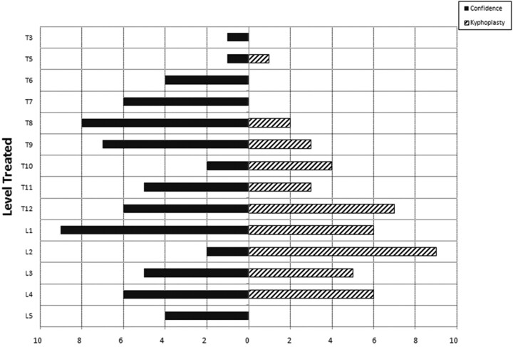
A total of 62 consecutive patients with painful collapsed vertebral bodies that did not respond to conventional therapy underwent vertebral body augmentation. The charts of all patients were reviewed. In 13 cases, the patient had undergone multiple treatment sessions, KyphX in 1 setting and Confidence in a separate setting. In total, 66 levels were treated with Confidence and 46, with KyphX. The etiology in all cases was osteoporosis, except in 3 Confidence cases, in which the vertebral body fractures were due to malignancies. In the Confidence group, there were 31 women with 50 levels (average age, 82.7 ± 9.6 years) and 9 men with 16 levels (average age, 77.6 ± 14.3 years). In the KyphX group, there were 28 women with 38 levels (average age, 82.4 ± 9.5 years) and 7 men with 8 levels (average age, 75.1 ± 19.0 years). The distribution of levels and average age of patients per level for both, Confidence and KyphX, are shown in Fig 2.
Fig 2.
Distribution of levels treated for Confidence versus kyphoplasty.
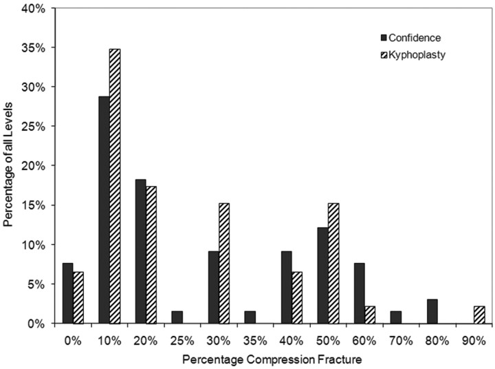
The average compression reached 26.8 ± 20.1% across all levels and treatment groups (Confidence group, average compression of 27.9 ± 20.7%; KyphX group, average compression of 25.0 ± 19.1%). A histogram showing the distribution of compression rates per treatment group is included in Fig 3. The distribution of compression rates and the overall mean and median compression rates were similar for both the Confidence and KyphX groups.
Fig 3.
Histogram of percentage compression for all levels by treatment group.
Leakage Rates and Locations
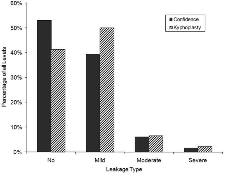
Leakage rates are presented in Fig 4. In the Confidence group, 92% of patients had no-to-mild leakage (patients with no leakage, 53%; patients with mild leakage, 39%). Similarly, in the KyphX group, 91% of cases had no-to-mild leakage (patients with no leakage, 41%; patients with mild leakage, 50%). The results between the 2 groups were not statistically different (P = .6745).
Fig 4.
Chart of leakage rates per leakage type (none, minimal, mild, moderate, and severe) and treatment group (kyphoplasty versus Confidence). No difference in leakage type or rate was observed between the groups.
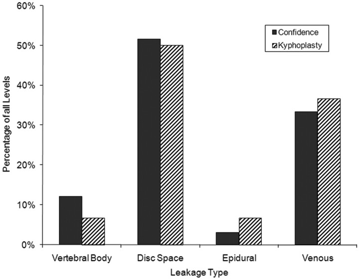
Leakage locations are presented in Fig 5. The location of the leakages was overall comparable for both cements. Most cases of leakage occurred in the disk space, followed by venous leakages. Rare cases of epidural or paravertebral space leakage were observed.
Fig 5.
Chart of leakage area per treatment group. No statistical different was observed on the basis of leakage area for each group.
Discussion
This study evaluated the safety profile of 2 different approaches to treating vertebral body fracture: vertebral body augmentation with Confidence, a high viscosity cement, or kyphoplasty with KyphX, which involved creating space inside the fracture site via inflation of a bone tamp (balloon). This retrospective study included similar sample sizes between both groups and comparable patient types (ie, mostly female patients with osteoporosis). The compression rates and distribution of compression rates between groups were also similar. The patient population studied herein was also unresponsive to conservative therapy and thus represents the patient group for whom percutaneous vertebral body augmentation is the most beneficial, as per previous reports.8
In our study, approximately 90% of both Confidence and KyphX cases had no-to-mild leakage. This finding is consistent with previously published reports for both cements9 and indicates that both procedures may have low rates of cement extravasation compared with historically reported leakage rates obtained with lower viscosity cements.2
Cement extravasation is 1 of the key safety risks of kyphoplasty or vertebroplasty. For reducing leakage rates, kyphoplasty uses a bone tamp that expels bone marrow out of the fractured vertebrae and compacts cancellous bone, thus creating a cavity—a path of least resistance—for the injected cement.10 This technology initially met significant enthusiasm because it was shown to reduce leakage rates to ∼9%2 and potentially allowed vertebral height restoration in properly positioned patients with mobile fractures.9 This technique, however, was shown to exacerbate risks of adjacent-level fractures.11 Thus, while addressing the leakage issue, kyphoplasty introduced new potential risks and limitations.
Other suggested techniques to limit polymethylmethacrylate extravasation included the use of smaller volumes of bone cement, slow injection times under low pressures, and limitation of usage above T7.12 However, none of these recommendations were found to consistently improve leakage rates.
The use of cements that would be too viscous to flow freely outside of the injection site has recently raised significant interest in the scientific and medical community, especially because of the commercial availability of devices that can safely deliver these cements to a fracture site. In fact, the impact of cement viscosity on leakage rate was recently evaluated in vitro by Baroud et al.7 This study demonstrated the impact of increased viscosity on cement leakage through a laboratory model of vertebral fracture. In this model, higher viscosity cements were shown to result in significantly lower leakage rates. The clinical relevance of this finding is significant, considering the rare yet potentially serious risks of cement leakage for patients undergoing percutaneous vertebroplasty. Higher viscosity cements may thus reduce the incidence of leakage to that observed with kyphoplasty, as reported in this study. Thus, high-viscosity cements may alleviate the need for bone tamps and cavity creation within the vertebral body, thus significantly reducing the number of steps and procedure time.
Limitations of this study include the fact that it was conducted retrospectively, and as a result, no clear methodology was followed to ensure unbiased randomization of patients in either the kyphoplasty or the high-viscosity cement groups. In addition, this study was conducted at 1 site only and may, therefore, not reflect possible interoperator variability, typical for surgical devices. Further studies with larger sample sizes would also address the issue of the relatively small sample size included in this evaluation. Despite all the limitations, observational studies are the mainstay of the literature for initial reports and for complications in general. Considering that there have not been any studies in this area, this retrospective evaluation should provide appropriate information for future controlled prospective studies
In this study, no difference in extravasation rate was observed between the high-viscosity cement and the kyphoplasty procedure. In another recently published study, the same high-viscosity cement was found to have significantly lower extravasation rates compared with a normal low-viscosity cement.13 Our study and the data of Anselmetti et al13 indicate that by simply increasing the viscosity of the cement, the safety profile of the procedure was increased to the point of being significantly better than vertebroplasty and comparable with that of kyphoplasty. Further long-term studies will be required to demonstrate whether this technique alleviates other kyphoplasty-related risks such as increased rates of adjacent-level fracture.
Conclusions
In this study, radiographic evidence of leakage was evaluated following either kyphoplasty or vertebral body augmentation with a novel high-viscosity cement (Confidence). The demographic characteristics of both cohorts were similar in terms of age, sex split, and rate of compression. Leakage rates and locations were found to be equivalent between both groups.
Augmentation by using high-viscosity cement may have a role in decreasing the risk of cement leakage during vertebroplasty and may result in leakage rates comparable with those of balloon kyphoplasty. Highly viscous cement may eliminate the need for cavity creation before vertebral cement augmentation.
Acknowledgments
I acknowledge Chantal E. Holy, MD, and Daniel H. Stoutenburgh, for editorial support.
Abbreviations
- PO2
partial pressure of oxygen
- VCF
vertebral compression fracture
Footnotes
This work was supported by DePuy Spine, Inc.
References
- 1. Jensen ME, McGraw JK, Cardella JF, et al. Position statement on percutaneous vertebral augmentation: a consensus statement developed by the American Society of Interventional and Therapeutic Neuroradiology, Society of Interventional Radiology, American Association of Neurological Surgeons/Congress of Neurological Surgeons, and American Society of Spine Radiology. J Vasc Interv Radiol 2007;18:325–30 [DOI] [PubMed] [Google Scholar]
- 2. Hulme PA, Krebs J, Ferguson SJ, et al. Vertebroplasty and kyphoplasty: a systematic review of 69 clinical studies. Spine 2006;31:1983–2001 [DOI] [PubMed] [Google Scholar]
- 3. Georgy BA. Interventional techniques in managing persistent pain after vertebral augmentation procedures: a retrospective evaluation. Pain Physician 2007;10:673–76 [PubMed] [Google Scholar]
- 4. McGraw JK, Cardella J, Barr JD, et al. Society of Interventional Radiology quality improvement guidelines for percutaneous vertebroplasty. J Vasc Interv Radiol 2003;14(9 pt 2):S311–15 [DOI] [PubMed] [Google Scholar]
- 5. Pateder DB, Khanna AJ, Lieberman IH. Vertebroplasty and kyphoplasty for the management of osteoporotic vertebral compression fractures. Orthop Clin North Am 2007;38:409–18; abstract vii [DOI] [PubMed] [Google Scholar]
- 6. Burton AW, Rhines LD, Mendel E. Vertebroplasty and kyphoplasty: a comprehensive review. Neurosurg 2005;18:e1 [DOI] [PubMed] [Google Scholar]
- 7. Baroud G, Crookshank M, Bohner M. High-viscosity cement significantly enhances uniformity of cement filling in vertebroplasty: an experimental model and study on cement leakage. Spine 2006;31:2562–68 [DOI] [PubMed] [Google Scholar]
- 8. Gangi A, Guth S, Imbert JP, et al. Percutaneous vertebroplasty: indications, technique, and results. Radiographics 2003;23:e10 [DOI] [PubMed] [Google Scholar]
- 9. Taylor RS, Fritzell P, Taylor RJ. Balloon kyphoplasty in the management of vertebral compression fractures: an updated systematic review and meta-analysis. Eur Spine J 2007;16:1085–100 [DOI] [PMC free article] [PubMed] [Google Scholar]
- 10. Phillips FM, Pfeifer BA, Lieberman IH, et al. Minimally invasive treatments of osteoporotic vertebral compression fractures: vertebroplasty and kyphoplasty. Instr Course Lect 2003;52:559–67 [PubMed] [Google Scholar]
- 11. Frankel BM, Monroe T, Wang C. Percutaneous vertebral augmentation: an elevation in adjacent-level fracture risk in kyphoplasty as compared with vertebroplasty. Spine J 2007;7:575–82 [DOI] [PubMed] [Google Scholar]
- 12. Ryu K-S, Park C-K, Kim M-K, et al. Single balloon kyphoplasty using far-lateral extrapedicular approach: technical note and preliminary results. J Spinal Disord Tech. 2007;20:392–98 [DOI] [PubMed] [Google Scholar]
- 13. Anselmetti GC, Zoarski G, Manca A, et al. Percutaneous vertebroplasty and bone cement leakage: clinical experience with a new high-viscosity bone cement and delivery system for vertebral augmentation in benign and malignant compression fractures. Cardiovasc Intervent Radiol 2008;31:937–47 [DOI] [PubMed] [Google Scholar]


