Abstract
BACKGROUND AND PURPOSE:
Endovascular brain cooling as a method for rapid and selective induction of hypothermic neuroprotection has not been systematically studied in humans. In this clinical pilot study we investigated the feasibility, safety, and physiologic responses of short-term brain cooling with IC-CSI.
MATERIALS AND METHODS:
We studied 18 patients (50 ± 10 years old, 9 women) undergoing follow-up cerebral angiography after previous treatment of vascular malformations. Isotonic saline (4–17°C) was infused into 1 internal carotid artery at 33 mL/min for 10 minutes. Brain (JVB) and bladder/esophageal temperature measurements (n = 9) were performed. Both MCAs were monitored with transcranial Doppler sonography (n = 13). Arterial and JV blood were sampled to estimate hemodilution and brain oxygen extraction.
RESULTS:
JVB temperature dropped ∼0.84 ± 0.13°C and systemic temperature by 0.15 ± 0.08°C from baseline (JVB versus systemic temperature: P = .0006). Systolic MCA-flow velocities decreased from 101 ± 27 to 73 ± 18 cm/s on the infused side and from 83 ± 24 to 78 ± 21 cm/s on the contralateral side (relative changes, −26 ± 8% versus −4 ± 27%; P = .009). Changes in hematocrit (−1.2 ± 1.1%) and cerebral arteriovenous oxygen difference (0.2 ± 1.0 mL O2/100 mL) were not significant. Doppler data showed no signs of vascular spasm or microemboli. No focal neurologic deficits occurred. Pain was not reported.
CONCLUSIONS:
The results of this pilot study suggest that brain cooling can be achieved safely, rapidly, and selectively by means of IC-CSI, opening a new potential avenue for acute neuroprotection. Clinical investigations with control of infusion parameters and measurements of CBF, oxygen consumption, and brain temperature are warranted.
The neuroprotective effect of induced hypothermia (pre-, intra-, and postischemia) has been well described in animal models of focal and global ischemia, with even slight reductions of the body temperature (mild to moderate hypothermia, 33–35°C) effective in reducing ischemic brain damage.1–4
In humans, the benefits of hypothermia have been demonstrated after cardiac arrest.5,6 Furthermore, high volumes of cold fluids are routinely used in cardiac surgery.7,8 For acute ischemic stroke systemic hypothermia has been shown to be feasible with surface cooling and intravascular systems, and clinical trials on safety and efficacy of hypothermia are ongoing.9–11
A significant subset of patients with acute ischemic stroke is eligible for local recanalization therapy, such as intra-arterial thrombolysis or mechanical clot extraction.12–16 This subset of patients is at high risk for brain injury due to their larger strokes and the potential for reperfusion injury after successful recanalization.17 Local endovascular cooling with intracarotid infusion of cold saline therefore presents a unique opportunity to provide short-term neuroprotection in such patients by rapidly and selectively cooling brain tissue at ischemic risk before, during, and after endovascular procedures. Several animal experiments (on-line Table)18–24 and theoretic models25–27 support the safety and feasibility of local endovascular brain cooling; however, experience in humans is limited,28,29 and it is yet undetermined whether selective brain cooling is feasible and safe in humans by using this endovascular method.
The goals of this translational project were 1) to assess the feasibility and safety of rapid and selective endovascular brain cooling in patients undergoing cerebral angiographic procedures, and 2) to investigate the physiologic responses induced by the local cooling. Procedures were performed with materials routinely used in the interventional angiography suite.
Materials and Methods
This report includes combined results from a 2-part prospective, nonrandomized clinical pilot study and a phantom model experiment. Because the safety of local endovascular infusion of cold saline on the human brain has not been established, a stepwise decrease in saline infusion temperature (starting with temperatures slightly lower than room temperature) was performed. Infusion rate and duration were the same in all patients. Due to the small volume of cold saline, infusion volumes were not adjusted for body weight. For part 1, patients were recruited between June 2006 and February 2007. The main goal, in addition to safety assessment, was to assess intracranial flow changes during local infusion of cold saline. Part 2, a modification of part 1, started in May 2007 and ended in January 2009. In this part, saline infusion temperature was lowered further after analysis of safety data from part 1. The main additional objective in this part was to measure the actual temperature achieved in the brain versus the body. The experimental protocols were approved by the institutional review board at Columbia University Medical Center where the study was performed.
Patient Selection
Subjects recruited for this pilot study were elective patients with partially or completely treated intracranial vascular diseases undergoing diagnostic cerebral angiograms, who were between 21 and 80 years of age, not disabled at baseline (Rankin Scale score <2), and were able to give written consent.
Exclusion criteria included significant stenosis or occlusion of the ICA, intracranial stenosis of brain-supplying vessels, major health problems such as disabling stroke at baseline, tumor or carcinoma, cardiac or renal failure, or pulmonary dysfunction. Patients with known coagulopathy, a platelet count of <75,000, a hematocrit of <30%, an imbalance of electrolytes (enough to require clinical attention), or hypersensitivity to hypothermia (eg, cryoglobulinemia, Raynaud syndrome) were also excluded.
Clinical Study Protocol
Following the diagnostic cerebral angiogram, patients remained in the supine position. The catheter (5F Angiographic Catheter, Cordis, Miami Lakes, Florida) was attached to a standard heparinized room temperature saline drip. The catheter tip remained in the extracranial part of the ICA. A 2-MHz pulse-wave TCD device (Pioneer 4040, Nicolet Vascular, Madison, Wisconsin; Marc 600, Spencer Technologies, Seattle, Washington; at 54–56 mm depths, sampling rate 100 Hz) was used for continuous bilateral MCA FV monitoring (part 1; n = 6 patients). TCD was used in both parts for on-line detection of potential microemboli and vascular spasm (n = 13 patients).30,31 MES were defined as unidirectional (direction of flow) signals, of 10–100 ms duration, and visualized inside the normal Doppler spectrum. A significant sign for vascular spasm was a MCA FV of >160 cm/s. As surrogate measures of brain and body (systemic) temperatures, JVB (pentalumen thermodilution catheter in the “dominant” JV) and bladder or esophageal temperatures were monitored for patients enrolled in part 2. The dominant draining JV was visually assessed from the venous washout phases of the initial angiograms. If both JVs showed equal drainage patterns, the ipsilateral JV was selected for the placement of the JVB catheter. From a femoral vein access the thermodilution catheter was navigated to the JVB (superior bulb of the internal jugular vein) under fluoroscopic guidance to ensure its correct placement in the JVB and avoid mixing from facial blood. Systemic temperature was assessed with either a Foley catheter or an esophageal catheter with integrated temperature probe. All temperatures were continuously monitored (displayed on patient's monitor) and recorded on a central server during the procedure. Warming blankets (Bair Hugger) were used in the last 12 patients to avoid shivering and potential systemic cooling.
Infusion Protocol
Isotonic saline bags (1000 mL, 0.9%) were degassed and cooled to ∼15°C (range, 12–17°C) for part 1 and to ∼7°C (range, 4–10°C) for part 2. After baseline TCD recording for ∼1 minute, the heparinized cold saline was infused into the ICA by using 2 continuous infusion pumps (Colleague, Baxter Healthcare, Irvine, California) at the maximum flow rate of 33 mL/min (2 × 1000 mL/h). Infusion lines were attached to 2 small-bore air filters (PFE-1207, B. Braun Medical, Bethlehem, Pennsylvania). Study duration ranged from 10 to 13 minutes.
Collected Data
Vital signs (blood pressure, heart rate, capillary oxygen saturation) were recorded every minute from the patient's monitor. Consciousness and verbal response were assessed during the study in awake patients. Neurologic examination was performed by a study neurologist before and after the study in all patients. In part 2 local brain and systemic temperatures measurements were taken every minute throughout the procedure; blood samples (blood gases, hematocrit, blood count, and electrolytes) were obtained from an arterial access and the JVB before and at the end of the cold infusion; Doppler spectra and angiographic images were studied for signs of vascular spasm or microemboli.
Data Analysis
Descriptive statistics were calculated (mean and standard deviation). FV (cm/s) and PI as well as pulse rate were recorded with the Doppler device, each at a sampling rate of 100 Hz. Thus, 60,000 data points were recorded for a study procedure of 10 minutes. For statistical purposes, 1000 data points were averaged every minute resulting in 10 values for each Doppler variable and patient during a 10-minute procedure. The Doppler variables were plotted against time for each variable. The largest deviation from the baseline value was determined (maximum change) and the relative change from baseline (percentage) was calculated. Absolute changes in JVB and bladder temperature were averaged and plotted against time. Absolute changes in hematocrit were calculated from arterial and venous samples. Arterial and venous oxygen content was calculated by using the following equation:
where CaO2 is the arterial oxygen content (CvO2 for venous), Hgb is hemoglobin, SaO2 is the arterial oxygen saturation (SvO2 for venous), and PaO2 is the arterial partial pressure of oxygen (PvO2 for venous). The difference between CaO2 and CvO2 is AvO2 (arteriovenous difference in oxygen content) and was used as a surrogate measure of oxygen extraction. Nonparametric tests were used for comparison of averaged values from variables and percentage values. All statistical analyses were performed by using SPSS (Chicago, Illinois), version 11.5.
Phantom Experiment
Because there is considerable heat transfer between cold saline and the counter-current aortic blood along the endovascular catheter from the femoral artery to the ICA, we estimated the actual temperature of the saline-blood mixture (perfusate) in the ICA by using a life-sized plastic phantom model of the aorta (Flow-Tek, Houston, Texas). Warm water (37–40°C) was pumped through the aortic arch portion of the model to simulate blood flow. Using the methods of the clinical study (standard 5F endovascular catheter, saline infusion rate of 33 mL/min) cold saline was infused for 20 minutes. Temperatures were monitored at the femoral entry (input) and carotid outflow (inside the catheter tip) with fluoroptic temperature probes (Luxtron, Santa Clara, California). The resulting temperature of the mixture of cold saline with blood (perfusate) beyond the tip of the catheter was estimated as follows:
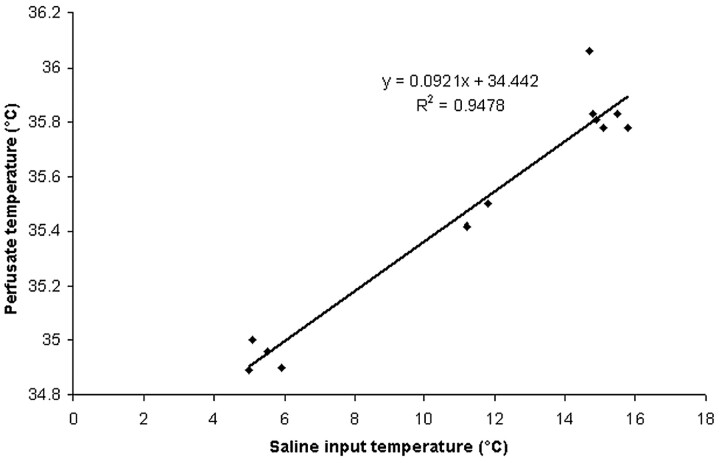
where Tcp is the temperature of the cold perfusate and T0 is the outflow temperature. ICA blood flow was assumed to be 250 mL/min. The initial femoral inflow temperature was 5.4 ± 0.41°C and the fluid in the phantom was 38 ± 0.76°C. The initial mean outflow temperature at the catheter tip was 25.21 ± 0.49°C. The calculated temperatures of the perfusate relative to the saline infusion temperature are presented in Fig 1 (r2 = 0.95).
Fig 1.
Phantom model experiment: temperature of the saline-ICA “blood” mixture.
Results
The study was performed in 18 patients (9 women, mean age, 50 ± 10 years; Table 1).
Table 1:
Patient characteristics
| Gender | Age (years) | Diagnosis | Treatment Status Before Study | Main Measurement during Study | Consciousness during Procedure | Complications | |
|---|---|---|---|---|---|---|---|
| 1 | F | 32 | AComA aneurysm | Complete | TCD | Awake | Transient facial shivering |
| 2 | F | 43 | PICA aneurysm | Complete | TCD | Awake | None |
| 3 | M | 45 | Dural AVM | Partial | TCD | Awake | Transient systemic shivering |
| 4 | F | 49 | AcomA aneurysm | Complete | TCD | Awake | None |
| 5 | M | 45 | Dural AVM | Partial | TCD | Awake | None |
| 6 | F | 56 | Facial AVM | Partial | TCD | General anesthesia | None |
| 7 | M | 45 | Dural AVM | Partial | No temporal window | Awake | None |
| 8 | M | 63 | AcomA aneurysm | Full | No temporal window | Awake | None |
| 9 | F | 49 | Facial AVM | Full | No temporal window | Awake | Transient systemic shivering |
| 10 | F | 59 | Intracranial aneurysms | Partial | JVB/systemic temperature | Awake | None |
| 11 | M | 38 | Paraclinoid aneurysm | Partial | JVB/systemic temperature | General anesthesia | None |
| 12 | F | 45 | Dural fistula | Partial | JVB/systemic temperature | Awake | None |
| 13 | F | 52 | ICA aneurysms | Partial | JVB/systemic temperature | General anesthesia | None |
| 14 | M | 45 | Dural AVM | Full | JVB/systemic temperature | Awake | None |
| 15 | M | 54 | MCA aneurysm | Full | JVB/systemic temperature | General anesthesia | None |
| 16 | F | 55 | BA aneurysm | Partial | JVB/systemic temperature | General anesthesia | None |
| 17 | M | 76 | Epistaxis | Full | JVB/systemic temperature | General anesthesia | None |
| 18 | M | 55 | ICA aneurysms | Partial | JVB/systemic temperature | General anesthesia | None |
Note:—There were no stenoses of brain-supplying vessels. AVMs and fistulas, fed by their respective external carotid artery, showed drainage via superficial veins.
Part 1
Nine patients were enrolled into part 1 of the study. Two patients had poor temporal window for TCD insonation and 1 had incomplete TCD data. Thus, TCD data were analyzed from 6 patients (Table 1). However, clinical data (assessment of neurologic deficits, consciousness, verbal response, and pain during procedure) were available from all patients.
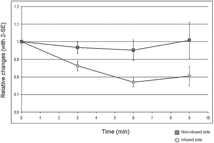
Although baseline MCA FVs were higher on the infusion side, they decreased to below the values of the noninfused side during the infusion of cold saline (Table 2). FVs decreased on the noninfused side as well, but the relative decrease in systolic FVs was significantly greater on the infusion side compared with the noninfusion side (P = .009; Fig 2). The changes in diastolic FVs were not significant (P = .065). PIs increased on both sides (21% versus 14%), but the difference was not significant. The average cold saline temperature was 15.6 ± 1.7°C. The average room temperature was 22 ± 2.1°C. Capillary oxygen saturation as measured by pulse oxymetry remained >95% during the procedure.
Table 2:
Doppler characteristics, vitals, and laboratory values
| Baseline |
Maximum Change |
Relative Change (%) |
||||
|---|---|---|---|---|---|---|
| Infusion Side | No Infusion Side | Infusion Side | No Infusion Side | Infusion Side | No Infusion Side | |
| TCD (n = 6) | ||||||
| Systolic velocity | 101 ± 27 cm/s | 83 ± 24 cm/s | 73 ± 18 cm/s | 78 ± 21 cm/s | −26 ± 8a | −4 ± 27a |
| Diastolic velocity | 47 ± 23 cm/s | 42 ± 18 cm/s | 29 ± 16 cm/s | 38 ± 17 cm/s | −38 ± 16b | −6 ± 38b |
| PI | 0.849 ± 0.354 | 0.738 ± 0.209 | 1.091 ± 0.373 | 0.908 ± 0.288 | 21 ± 2 | 14 ± 23 |
| Vitals (n = 18) | Absolute changec | |||||
| Heart rate (beats/minute) | 71 ± 9 | 75 ± 24 | 10 ± 25 | |||
| Blood pressure (mm Hg) | Systemic 113 ± 16 | Systemic 120 ± 11 | Systemic 7 ± 12 | |||
| Diastolic 65 ± 10 | Diastolic 72 ± 13 | Diastolic 11 ± 21 | ||||
| Laboratory values (n = 9) | ||||||
| ΔA-V oxygen content (mL O2/100 mL) | 5.1 ± 2.1 | 5.3 ± 1.4 | 0.2 ± 1.0 | |||
| Hematocrit, local (%) | 35.8 ± 4.7 | 34.4 ± 5.1 | −1.3 ± 0.9 | |||
| Hematocrit, systemic (%) | 37.9 ± 6.0 | 36.0 ± 5.4 | −1.2 ± 1.1 | |||
| Catecholamines (n = 5) | ||||||
| Epinephrine (pg/mL) | 0 | 3 ± 6c | ||||
| Norepinephrine (pg/mL) | 167 ± 30 | 166 ± 45c | ||||
Note:— P = .009;
not significant, P = .065;
not significant.
Fig 2.
Systolic MCA blood flow velocity on the side of the infusion and the noninfused side. Relative changes (2 SEs).
Part 2
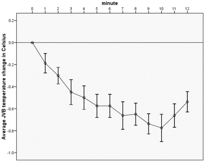
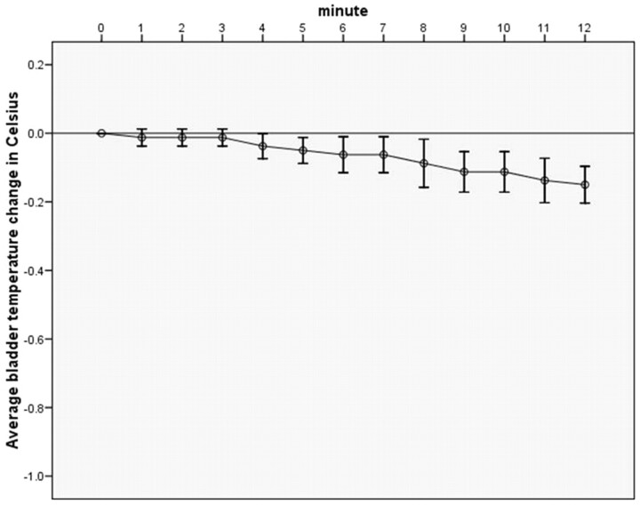
Nine patients were enrolled into the second part of the study. The mean JVB temperature (baseline, 35.7 ± 0.7°C) dropped by 0.84 ± 0.13°C in a mean time of 8.2 ± 1.9 minutes (Fig 3) and the bladder temperature (baseline, 35.6 ± 0.7°C) dropped by 0.15 ± 0.08°C (Fig 4). The JVB temperature decrease was significantly greater when compared with the decrease in bladder temperature (P = .0006). The changes in systemic hematocrit and arteriovenous oxygen difference were not significant (Table 2). Other laboratory values (blood count, coagulation, electrolytes, epinephrine, and norepinephrine) and vitals remained in the normal range. The average cold saline temperature was 7.4 ± 2.4°C. Urinary output during study procedure was measured in the last 3 patients (mean output, 317 ± 120 mL).
Fig 3.
Jugular venous bulb temperature changes (°C; SD) during endovascular cold saline infusion into the ICA.
Fig 4.
Bladder/esophageal temperature changes (°C; SD) during endovascular cold saline infusion into the ICA.
Other Parameters
Vital signs did not change significantly during the study procedure. Consciousness and verbal response in the 11 conscious patients remained unaltered during the study procedure. In 3 awake patients either partial (face) or systemic shivering was observed shortly after the start of the cold infusion. However, this shivering stopped after a few minutes and remained unnoticed by 1 of the 3 patients. No pain or discomfort was reported. Neurologic examination revealed no focal deficits in any of the 18 patients. Neither Doppler spectra nor angiographic control revealed signs of microemboli or vascular spasm.
Discussion
The present study shows that endovascular intracarotid infusion of cold normal saline at a flow-rate of 33 mL/min led to a rapid decrease in JVBT and an ipsilateral drop of MCA blood FV. To the best of our knowledge, this is the first report that describes the hemodynamic effects, feasibility, and safety of selective brain cooling in humans by means of endovascular intracarotid infusion of cold saline.
The most commonly described goal of hypothermia as a neuroprotective measure is to reduce metabolic rate. It is known that the physiologic parameters of metabolic rate and CBF are coupled to temperature over a wide range of temperatures, uncoupling only at <20°C.32,33 Studies in animals and humans investigating systemic hypothermia and selective brain cooling have confirmed the validity of the concept of this physiologic coupling.32–36 In our study, oxygen extraction remained unchanged after selective brain cooling. This may be because cooling duration in our pilot study was only 10 minutes; feasibility and safety were the main goals of this study. The linkage between temperature and metabolism might be demonstrated with longer cooling durations and deeper brain cooling. Eventual demonstration of the temperature-dependent change in brain metabolism would support the hypothesis that the local cold infusion is acting as a neuroprotective agent by inducing a hypometabolic state.
JVBT was used as our measure of brain temperature because the use of invasive parenchymal temperature probes cannot be justified in elective or acute ischemic stroke patients. However, considering the phantom model results, the rapidly achieved temperature equilibrium between blood in the capillary network and brain matter (heat conduction), and potentially mixed composition of the JV blood from contributions of both hemispheres (“contamination” with warm blood), it appears likely that the actual brain temperature is lower than the measured JV blood temperature, which in itself was significantly lower than the body temperature measured in our cohort. According to the phantom model results the perfusate temperature was ∼35.2°C when saline input temperature was ∼7°C (Fig 1), as it was in our clinical study, part 2. Considering the experimental baseline temperature was 38°C, the difference would be 2.8°C. Thus, we assume that the brain temperature (including the possibility of contributions from the contralateral hemisphere) may have decreased by between 0.8°C and 2.8°C. Apart from theoretic models that support the feasibility of endovascular brain cooling,25–27 Kleiber's rule, which describes the relationship between the BMR of animals with their body weight as a function of BMR = 3.52 × weight0.74, may give further insights.37 Applied to an 8-kg rhesus monkey or a 10-kg dog, the BMRs relative to a 70-kg human body are 1.7 and 1.6, respectively (Table 1). When adjusted for these factors, the cold infusion (4–13°C) rates used in the animal experiments projected to human scales would range between ∼20 and 44 mL/min, which would lead to a significant brain temperature decrease.22–24 Furthermore, the exponential shape of our JVB temperature curve can be found in several animal experiments where actual brain temperatures were measured,18,22,24 giving insights into speed and degree of cooling when infusion parameters are changed, for instance to colder saline temperatures than were used in the present pilot study. MR imaging-based thermometry is a promising alternative38 for noninvasive on-line monitoring of brain temperature during clinical investigations such as ours, which may become a reality with further technologic advancement, also allowing endovascular procedures in an MR environment.
The initial (baseline) difference in systolic velocities we found between the hemispheres before cold infusion might be explained by hemodilution created by the infusion of nonhematologic material, which has been shown to increase CBF.39–41 With regard to changes in flow after cold infusion, our study setup did not allow the calculation of CBF. TCD FVs were measured as a surrogate. A lack of knowledge about whether there were any changes in MCA diameter precluded quantitative CBF assessment. However, FV data are still useful with respect to hypothesis generation. While the constrictive effect of cold on peripheral vessels is widely accepted (eg, vasculature of the skin during cold exposure), there is controversy with regard to the effect of cold on the diameter of larger vessels supplying the brain. Studies on ex vivo specimens from ICAs of animals and MCAs from human autopsies revealed vascular relaxation during cold exposure.42,43 Relaxation of the MCA during TCD insonation would result in a decrease in MCA FV, which we observed in this study. A concurrent increase in peripheral vascular resistance (increase in PI) suggests that there was constriction of arterioles and precapillaries. Our findings led to 2 possible physiologic scenarios: first, that peripheral vasoconstriction occurred as an autoregulatory response to reduced metabolism, or second, that our observed changes occurred as a direct effect of cold on the vasculature.44–46 It is also possible that both mechanisms are involved. Longer cooling times with additional data on oxygen extraction may help sort out the underlying mechanism. One limitation on the technique could be the volume of normal saline and the hemodilution effect, though it appears from clinical experience in cardiac arrest and acute ischemic stroke that the body is able to cope with volume loads of >2000 mL/h.47–49 Finally, we observed transient shivering in 3 patients. It remains unclear whether this was caused by brain cooling or the slight reduction in body temperature. Although overall discomfort during short-term selective cooling was mild (transient systemic shivering in 2 of 11 patients) and no pain was reported, it is possible that during longer term selective cooling greater discomfort may occur. A warming blanket may then not suffice and pharmacologic means may be necessary to counteract the shivering.
Despite these limitations of our pilot study, we argue that selective endovascular cooling is feasible and safe. Dissociation between JVB and bladder temperature occurred almost immediately and the brain cooled within minutes (Figs 3 and 4). This method has the potential to add to our armamentarium for treatment of acute cerebrovascular disease, especially as adjunct hypothermic neuroprotection in acute ischemic stroke patients undergoing local recanalization therapy. Apart from antiinflammatory and antiexcitatory effects of hypothermia,1,3,4,19 other mechanisms, such as decrease in metabolism and blood flow,33,35,36 and suppression of blood-brain barrier disruption50 may provide neuroprotection in a crucial early period and may help prevent complications from reperfusion. Passive thermal conduction may also help lower the temperature in a region of compromised flow even before the artery is opened (MCA infarction) because the infusate would cool the entire anterior circulation. Furthermore, selective brain cooling would be achieved via the same endovascular catheter as used for clinical treatment. Finally, selective brain cooling during local recanalization therapy could serve as a bridging procedure to long-term systemic cooling. The logistics for endovascular procedures are not widely developed. However, because of the increasing availability of endovascular centers, the systematic investigation of this promising alternative treatment method is needed.12 Potential advantages over systemic cooling, especially for acute stroke patients undergoing endovascular treatment, include speed of cooling of the organ of interest (brain) and adjunct neuroprotection during recanalization. Lowering brain temperature beyond that of the present study appears feasible and is limited only by infusion temperature and speed of infusion.25,26 Assessment of other blood markers (eg, catecholamines and antidiuretic hormone) may give further insights into the physiologic mechanisms that are triggered during local cold stress.44,51 Finally, local cooling might also be applied during neuroendovascular procedures such as carotid angioplasty and stent placement, where thromboembolic events may place the brain at ischemic risk by creating a more ischemia-tolerant state (pre- and intraischemic neuroprotection).52
Conclusions
The results of this clinical pilot study suggest that selective endovascular brain cooling by means of intra-arterial infusion of cold saline is feasible and safe in the setting of a neuroendovascular suite. Endovascular brain cooling may constitute a rapid, selective, and safe method for hypothermic neuroprotection in patients with acute ischemic stroke, especially in conjunction with local recanalization therapy, or in patients undergoing neuroendovascular procedures, where thromboembolic events are major threats. The underlying neuroprotective mechanism (temperature-dependent effect on brain metabolism and CBF) requires confirmation in a larger study with more direct measurements of CBF and cerebral metabolism. Further clinical studies are necessary (and underway) to assess the safety and feasibility of longer term endovascular brain cooling and its physiologic effects in acute ischemic stroke patients.
Supplementary Material
Acknowledgments
We are grateful for the technical support by the Horace W. Goldsmith Doppler Laboratory and the Interventional Neuroradiology Angiography team.
Abbreviations
- ABC
automated blood count
- AComA
anterior communicating artery
- AVM
arteriovenous malformation
- BA
basilar artery
- BMR
basal metabolic rate
- CBF
cerebral blood flow
- ΔA-V
difference between arterial and venous oxygen content
- FV
flow velocity
- IA
intra-arterial
- ICA
internal carotid artery
- IC-CSI
intracarotid cold saline infusion
- JV
jugular vein
- JVB
jugular venous bulb
- JVBT
jugular venous bulb temperature
- MCA
middle cerebral artery
- MES
microemboli signals
- N/A
not available
- O2-Sat
oxygen saturation
- PI
pulsatility index
- PICA
posterior inferior cerebral artery
- TCD
transcranial Doppler
Footnotes
This study was supported by the New York State Office of Science, Technology, and Academic Research (NYSTAR) and the Dana Foundation (Clinical Neuroscience Research Grant).
Previously presented in part at: 16th European Stroke Conference, Glasgow, Scotland, May 29–June 1, 2007.
Indicates article with supplemental on-line table.
References
- 1. Morikawa E, Ginsberg MD, Dietrich WD, et al. The significance of brain temperature in focal cerebral ischemia: histopathological consequences of middle cerebral artery occlusion in the rat. J Cereb Blood Flow Metab 1992;12:380–89 [DOI] [PubMed] [Google Scholar]
- 2. van der Worp HB, Sena ES, Donnan GA, et al. Hypothermia in animal models of acute ischaemic stroke: a systematic review and meta-analysis. Brain 2007;130:3063–74 [DOI] [PubMed] [Google Scholar]
- 3. Yanamoto H, Hong SC, Soleau S, et al. Mild postischemic hypothermia limits cerebral injury following transient focal ischemia in rat neocortex. Brain Res 1996;718:207–11 [DOI] [PubMed] [Google Scholar]
- 4. Ishikawa M, Sekizuka E, Sato S, et al. Effects of moderate hypothermia on leukocyte-endothelium interaction in the rat pial microvasculature after transient middle cerebral artery occlusion. Stroke 1999;30:1679–86 [DOI] [PubMed] [Google Scholar]
- 5. Hypothermia after Cardiac Arrest Study Group. Mild therapeutic hypothermia to improve the neurologic outcome after cardiac arrest. N Engl J Med 2002; 346: 549– 56 [DOI] [PubMed] [Google Scholar]
- 6. Bernard SA, Gray TW, Buist MD, et al. Treatment of comatose survivors of out-of-hospital cardiac arrest with induced hypothermia. N Engl J Med 2002;346:557–63 [DOI] [PubMed] [Google Scholar]
- 7. Randomised trial of normothermic versus hypothermic coronary bypass surgery: The Warm Heart Investigators. Lancet 1994; 343: 559– 63 [PubMed] [Google Scholar]
- 8. Grigore AM, Mathew J, Grocott HP, et al. ; Neurological Outcome Research Group; CARE Investigators of the Duke Heart Center. Cardiothoracic Anesthesia Research Endeavors.Prospective randomized trial of normothermic versus hypothermic cardiopulmonary bypass on cognitive function after coronary artery bypass graft surgery.Anesthesiology 2001;95: 1110– 19 [DOI] [PubMed] [Google Scholar]
- 9. Lyden PD, Allgren RL, Ng K, et al. Intravascular cooling in the treatment of stroke (ICTuS): early clinical experience. J Stroke Cerebrovasc Dis 2005;14:107–14 [DOI] [PubMed] [Google Scholar]
- 10. De Georgia MA, Krieger DW, Abou-Chebl A, et al. Cooling for acute ischemic brain damage (COOL AID): a feasibility trial of endovascular cooling. Neurology 2004;63:312–17 [DOI] [PubMed] [Google Scholar]
- 11. Georgiadis D, Schwarz S, Kollmar R, et al. Endovascular cooling for moderate hypothermia in patients with acute stroke: first results of a novel approach. Stroke 2001;32:2550–53 [DOI] [PubMed] [Google Scholar]
- 12. Cloft HJ, Rabinstein A, Lanzino G, et al. Intra-arterial stroke therapy: an assessment of demand and available work force. AJNR Am J Neuroradiol 2009;30:453–58 [DOI] [PMC free article] [PubMed] [Google Scholar]
- 13. Choi JH, Bateman BT, Mangla S, et al. Endovascular recanalization therapy in acute ischemic stroke. Stroke 2006;37:419–24 [DOI] [PubMed] [Google Scholar]
- 14. Furlan A, Higashida R, Wechsler L, et al. Intra-arterial prourokinase for acute ischemic stroke: PROACT II study-a randomized controlled trial. Prolyse in acute cerebral thromboembolism. JAMA 1999;282:2003–11 [DOI] [PubMed] [Google Scholar]
- 15. Smith WS, Sung G, Saver J, et al. ; for the Multi MERCI Investigators. Mechanical thrombectomy for acute ischemic stroke: final results of the Multi MERCI trial. Stroke 2008;39:1205–12 [DOI] [PubMed] [Google Scholar]
- 16. The Penumbra Pivotal Stroke Trial Investigators. The Penumbra Pivotal Stroke Trial: safety and effectiveness of a new generation of mechanical devices for clot removal in intracranial large vessel occlusive disease. Stroke 2009; 40: 2761– 68 [DOI] [PubMed] [Google Scholar]
- 17. Hallenbeck JM, Dutka AJ. Background review and current concepts of reperfusion injury. Arch Neurol 1990;47:1245–54 [DOI] [PubMed] [Google Scholar]
- 18. Ohta T, Sakaguchi I, Dong LW, et al. Selective cooling of brain using profound hemodilution in dogs. Neurosurgery 1992;31:1049–55 [DOI] [PubMed] [Google Scholar]
- 19. Ding Y, Li J, Rafols JA, et al. Prereperfusion saline infusion into ischemic territory reduces inflammatory injury after transient middle cerebral artery occlusion in rats. Stroke 2002;33:2492–98 [DOI] [PubMed] [Google Scholar]
- 20. Luan X, Li J, McAllister JP, II, et al. Regional brain cooling induced by vascular saline infusion into ischemic territory reduces brain inflammation in stroke. Acta Neuropathol 2004;107:227–34 [DOI] [PubMed] [Google Scholar]
- 21. Zhao WH, Ji XM, Ling F, et al. Local mild hypothermia induced by intra-arterial cold saline infusion prolongs the time window of onset of reperfusion injury after transient focal ischemia in rats. Neurol Res 2009;31:43–51 [DOI] [PubMed] [Google Scholar]
- 22. Jiang JY, Xu W, Yang PF, et al. Marked protection by selective cerebral profound hypothermia after complete cerebral ischemia in primates. J Neurotrauma 2006;23:1847–56 [DOI] [PubMed] [Google Scholar]
- 23. Furuse M, Preul MC, Kinoshita Y, et al. Rapid induction of brain hypothermia by endovascular intra-arterial perfusion. Neurol Res 2007;29:53–57 [DOI] [PubMed] [Google Scholar]
- 24. Nishihara K, Furuse M, Kinoshita Y, et al. Differential brain cooling induced by transarterial perfusion of cooled crystalloid solution in canines. Neurol Res 2009;31:251–57 [DOI] [PubMed] [Google Scholar]
- 25. Konstas AA, Neimark MA, Laine AF, et al. A theoretical model of selective cooling using intracarotid cold saline infusion in the human brain. J Appl Physiol 2007;102:1329–40 [DOI] [PubMed] [Google Scholar]
- 26. Neimark MA, Konstas AA, Choi JH, et al. The role of intracarotid cold saline infusion on a theoretical brain model incorporating the circle of Willis and cerebral venous return. Conf Proc IEEE Eng Med Biol Soc 2007;1:1140–43 [DOI] [PubMed] [Google Scholar]
- 27. Slotboom J, Kiefer C, Brekenfeld C, et al. Locally induced hypothermia for treatment of acute ischaemic stroke: a physical feasibility study. Neuroradiology 2004;46:923–34 [DOI] [PubMed] [Google Scholar]
- 28. Choi JH, Rundek T, Marshall RS, et al. Cold saline infusion into the carotid artery decreases ipsilateral middle cerebral artery blood flow [abstract]. Cerebrovasc Dis 2007;23(suppl 2):46 [Google Scholar]
- 29. Choi JH, Neimark MA, Konstas AA, et al. Selective brain cooling with endovascular intra-carotid infusion of cold saline [abstract]. Ann Neurol 2008;64(suppl 12):14 [Google Scholar]
- 30. Basic identification criteria of Doppler microembolic signals: Consensus Committee of the Ninth International Cerebral Hemodynamic Symposium. Stroke 1995; 26: 1123 [PubMed] [Google Scholar]
- 31. Grosset DG, Straiton J, du Trevou M, et al. Prediction of symptomatic vasospasm after subarachnoid hemorrhage by rapidly increasing transcranial Doppler velocity and cerebral blood flow changes. Stroke 1992;23:674–79 [DOI] [PubMed] [Google Scholar]
- 32. Schwartz AE, Stone JG, Pile-Spellman J, et al. Selective cerebral hypothermia by means of transfemoral internal carotid artery catheterization. Radiology 1996;201:571–72 [DOI] [PubMed] [Google Scholar]
- 33. Walter B, Bauer R, Kuhnen G, et al. Coupling of cerebral blood flow and oxygen metabolism in infant pigs during selective brain hypothermia. J Cereb Blood Flow Metabol 2000;20:1215–24 [DOI] [PubMed] [Google Scholar]
- 34. Sustanskii AL, Yablonskiy DA. Theoretical model of temperature regulation in the brain during changes in functional activity. Proc Natl Acad Sci U S A 2006;103:12144–49 [DOI] [PMC free article] [PubMed] [Google Scholar]
- 35. Rosomoff HH, Holoday DA. Cerebral blood flow and cerebral oxygen consumption during hypothermia. Am J Physiol 1954;179:85–88 [DOI] [PubMed] [Google Scholar]
- 36. Michenfelder JD, Milde JH, Katusic ZS. Postischemic canine cerebral blood flow is coupled to cerebral metabolic rate. J Cereb Blood Flow Metab 1991;11:611–16 [DOI] [PubMed] [Google Scholar]
- 37. Kleiber M. Body size and metabolism. Hilgardia 1932;6:315–51 [Google Scholar]
- 38. Corbett R, Laptook A, Weatherall P. Noninvasive measurements of human brain temperature using volume-localized proton magnetic resonance spectroscopy. J Cereb Blood Flow Metab 1997;17:363–69 [DOI] [PubMed] [Google Scholar]
- 39. Henriksen L, Paulson OB, Smith RJ. Cerebral blood flow following normovolemic hemodilution in patients with high hematocrit. Ann Neurol 1981;9:454–57 [DOI] [PubMed] [Google Scholar]
- 40. Tu YK, Liu HM. Effects of isovolemic hemodilution on hemodynamics, cerebral perfusion, and cerebral vascular reactivity. Stroke 1996;27:441–45 [DOI] [PubMed] [Google Scholar]
- 41. Bruder N, Cohen B, Pellissier D, et al. The effect of hemodilution on cerebral blood flow velocity in anesthetized patients. Anesth Analg 1998;86:320–32 [DOI] [PubMed] [Google Scholar]
- 42. Mustafa S, Thulesius O. Cooling-induced carotid artery dilatation: an experimental study in isolated vessels. Stroke 2002;33:256–60 [DOI] [PubMed] [Google Scholar]
- 43. Sagher O, Huang DL, Webb RC. Induction of hypercontractibility in human cerebral arteries by rewarming following hypothermia: a possible role for tyrosine kinase. J Neurosurg 1997;87:431–35 [DOI] [PubMed] [Google Scholar]
- 44. Frank SM, Higgins MS, Fleisher LA, et al. Adrenergic, respiratory, and cardiovascular effects of core cooling in humans. Am J Physiol 1997;272:557–62 [DOI] [PubMed] [Google Scholar]
- 45. Boulant JA, Dean JB. Temperature receptors in the central nervous system. Annu Rev Physiol 1986;48:639–54 [DOI] [PubMed] [Google Scholar]
- 46. Raichle ME, Hartman BK, Eichling JO, et al. Central noradrenergic regulation of cerebral blood flow and vascular permeability. Proc Natl Acad Sci U S A 1975;72:3726–30 [DOI] [PMC free article] [PubMed] [Google Scholar]
- 47. Bernard S, Buist M, Monteiro O, et al. Induced hypothermia using large volume, ice-cold intravenous fluid in comatose survivors of out-of-hospital cardiac arrest: a preliminary report. Resuscitation 2003;56:9–13 [DOI] [PubMed] [Google Scholar]
- 48. Polderman KH, Rijnsburger ER, Peerdeman SM, et al. Induction of hypothermia in patients with various types of neurologic injury with use of large volumes of ice-cold intravenous fluid. Crit Care Med 2005;33:2844–45 [DOI] [PubMed] [Google Scholar]
- 49. Kollmar R, Schellinger PD, Steigleder T, et al. Ice-cold saline for the induction of mild hypothermia in patients with acute ischemic stroke: a pilot study. Stroke 2009;40:1907–09 [DOI] [PubMed] [Google Scholar]
- 50. Karibe H, Zarow GJ, Graham SH, et al. Mild intraischemic hypothermia reduces postischemic hyperperfusion, delayed postischemic hypoperfusion, blood-brain barrier disruption, brain edema, and neuronal damage volume after temporary focal cerebral ischemia in rats. J Cereb Blood Flow Metab 1994;14:620–27 [DOI] [PubMed] [Google Scholar]
- 51. Peppiatt CM, Howarth C, Mobbs P, et al. Bidirectional control of CNS capillary diameter by pericytes. Nature 2006;443:700–04 [DOI] [PMC free article] [PubMed] [Google Scholar]
- 52. Drew KL, Buck CL, Barnes BM, et al. Central nervous system regulation of mammalian hibernation: implications for metabolic suppression and ischemia tolerance. J Neurochem 2007;102:1713–26 [DOI] [PMC free article] [PubMed] [Google Scholar]
Associated Data
This section collects any data citations, data availability statements, or supplementary materials included in this article.


