Abstract
BACKGROUND AND PURPOSE:
AICA aneurysms are rare and a challenge to treat surgically. We present our experience of the angiographic results and the clinical outcomes for 9 AICA aneurysms treated by EVT.
MATERIALS AND METHODS:
Between 1997 and 2009, EVT was attempted for 9 AICA aneurysms. Six patients presented with SAH, and 3 aneurysms were found incidentally. The location of the aneurysms was the proximal AICA in 7 and the distal AICA in 2. Five aneurysms originated from an AICA-PICA variant. Clinical outcomes and procedural complications were evaluated, and angiography was performed 6, 12, and 24 months after embolization to confirm recanalization of the coiled aneurysm.
RESULTS:
EVT was technically successful in 7 patients (78%). Surgical trapping was performed in 1 patient after failure of EVT, and another aneurysm occluded spontaneously, along with the parent artery during EVT. In 7 patients, the AICAs had good patency on postoperative angiography. Stent-assisted coiling was performed in 3 patients. Follow-up angiographies were performed in 7 patients and showed no evidence of recanalization or progressive occlusion with further thrombosis except in 1 patient. There was no evidence of aneurysm rupture during the follow-up period, and 8 patients were able to perform all usual activities (mRS score, 0–1).
CONCLUSIONS:
EVT may provide a feasible and safe option as an alternative, though a microsurgical option is initially considered for the management of AICA aneurysms. Further follow-up and more experience are also necessary.
AICA aneurysms are extremely rare and account for less than 1.5% of all intracranial aneurysms.1–7 These aneurysms are a therapeutic challenge, with high surgical risk.
Since the first surgery of an AICA aneurysm by Schwartz,8 dozens of case reports and some articles have been published concerning surgical treatment of AICA aneurysms.9–21 Although various surgical approaches have been introduced to access these aneurysms, surgical treatment is often limited due to the complexity of the adjacent neurovascular structure, the presence of brain stem perforators, the narrow and deep surgical field, and the necessity of an experienced surgeon.
Although EVT has been reported for the treatment of AICA aneurysms, the most common procedure is parent artery occlusion.22–24 Recently Kusaka et al25 reported coil embolization of an AICA aneurysm with preservation of the parent artery.
We present our experience in the treatment and the clinical outcome of AICA aneurysms using EVT.
Materials and Methods
Patients
This retrospective study was approved by the institutional review board, and informed consent was obtained from the patients or their families before the procedure. The data base of all the patients who underwent endovascular treatment for cerebral aneurysms was reviewed from 1997 to 2009 at 2 tertiary hospitals. Nine patients with 9 AICA aneurysms were enrolled in this study, and the mean age of the patients (4 men and 5 women) was 56.7 years (range, 28–73 years; Table).
Characteristics of patients with AICA aneurysms
| No | Sex/ Age (yr) | Presentation | HH | Location | Size (mm) | Associated Condition | Tx | Result | Cx | mRS | Follow-Up Angiography (mo) | AICA | F/U (mo) |
|---|---|---|---|---|---|---|---|---|---|---|---|---|---|
| 1 | F/67 | SAH | III | Proximal | 4 × 3 × 3 | AICA-PICA variant | Coil/EVD | Partial | None | 1 | IO (24) | P | 36 |
| 2 | M/28 | SAH | I | Proximal | 8 × 5 × 5 | Moyamoya disease | Coil | Partial | None | 1 | NA | P | – |
| 3 | M/41 | Incidental | Proximal | 9 × 7 × 7 | AVM | Coil | Complete | None | 1 | CO (24) | P | 120 | |
| 4a | M/46 | SAH | IV | Distal | 5 × 4 × 3 | Surgical trapping | Cerebellar infarct | 2 | NA | O | – | ||
| 5a | F/73 | SAH | III | Distal | 3 × 2.5 × 3 | Spontaneous occlusion | TIA | 0 | CO (24) | O | 24 | ||
| 6 | F/71 | SAH | IV | Proximal | 4 × 3 × 3 | AICA-PICA variant | Coil/EVD | Complete | None | 0 | CO (24) | P | 42 |
| 7 | M/66 | Incidental | Proximal | 3 × 3 × 3 | AICA-PICA variant | Coil/stent (4 × 20) | Complete | None | 0 | CO (20) | P | 20 | |
| 8 | F/72 | Incidental | Proximal | 8 × 7 × 7 | AICA-PICA variant | Coil stent (4 × 15) | Partial | None | 0 | IO (12) | P | 30 | |
| 9 | F/46 | SAH | IV | Proximal | 15 × 10 × 10 | AICA-PICA variant | Coil/stent (4 × 14)/EVD | Complete | None | 1 | CO (10) | P | 10 |
Endovascularly failed case.
Six patients presented with SAH, and 3 aneurysms were found incidentally. According to the HH, grades III-IV were reported in 5 patients and grade I, in 1. All the involved aneurysms were of the saccular type, varying from 3 to 15 mm in the largest diameter.
AICA aneurysms are classified as proximal (from the BA-AICA junction to the AICA bifurcation or a combined AICA-PICA origin, premeatal segment) and distal (from the end of the meatal loop to the distal AICA, postmeatal segment) with reference to the seventh and eighth cranial nerve complex.6,26 According to this classification, the location of aneurysms was as follows: the proximal AICA in 7 and the distal AICA in 2. Five aneurysms were associated with the AICA-PICA variant, and 2 aneurysms, with Moyamoya disease or arteriovenous malformation.
In all patients, a diagnostic cerebral angiogram was obtained before the endovascular procedure to evaluate the associated vascular anatomy of the posterior circulation and define the underlying vascular disease. The endovascular treatment option was decided on the basis of discussion between a neurosurgeon and interventional neuroradiologist regarding the patient's condition, the anticipated clinical outcome, and the surgical difficulty associated with the size and location of the aneurysm.
Endovascular Procedure
EVT was attempted in all patients with the patients under general anesthesia. In 6 patients with SAH, EVT was attempted immediately after diagnostic angiography, and in 3 patients, electively. All patients had heparinization (activated clotting time, >300) immediately after or during EVT, and heparinization was maintained for 24 hours after the procedure. In cases of stent-assisted coiling, a dual antiplatelet regimen (clopidogrel, 75 mg/day, and aspirin, 325 mg/day) was started 3 days before the EVT. In 1 patient (patient 9), clopidogrel, 300 mg, and aspirin, 500 mg, were administered orally 1 day after the procedure and heparinization was maintained as above.
For EVT of AICA aneurysms, a 5F or 6F guiding catheter system (Envoy; Codman Neurovascular, Raynham, Massachusetts) was introduced via the femoral artery. Through the vertebral artery, a microcatheter (Prowler 14 microcatheter, Codman Neurovascular; Excelsior SL-10 microcatheter, Boston Scientific, Natick, Massachusetts) was guided carefully into the aneurysm sac or via the proximal segment of the AICA. For stent-assisted coiling, a self-expandable intracranial stent (Neuroform stent, Boston Scientific; Enterprise stent, Codman Neurovascular) was placed in the BA or the AICA to protect the parent artery from coil protrusion. Afterward, a detachable coil was delivered into the aneurysm sac via a microcatheter, and this process was repeated until coil embolization was achieved adequately. A closure device (Perclose; Abbott Vascular Devices, Redwood City, California) was used to seal off the femoral artery puncture. All treated patients were maintained with heparin for 24 hours after the procedure; and clopidogrel, 75 mg/day, and aspirin, 150 mg/day, were administered for 6 months in cases of stent implantation. After the procedure, EVD was performed if the development of hydrocephalus was detected.
Clinical Outcome and Follow-Up
All patients were evaluated by postoperative angiography to confirm the status of aneurysm coiling. Angiographic findings were categorized as either complete occlusion (no filling of contrast agent in the aneurysm sac) or partial occlusion (residual filling of contrast agent in the remnant aneurysm sac). Periprocedural and postoperative complications, such as parent artery occlusion/dissection, rebleeding, thromboembolism, and retroperitoneal hematoma, were also evaluated.
A follow-up angiography was performed 6, 12, and 24 months after EVT to confirm recanalization and regrowth of the coiled aneurysm and the patency of the AICA. These angiographies were reviewed and classified as changed compared with the previous postoperative and initial angiographies by 2 senior neuroradiologists (D.I.K., T.-S.C). Clinical outcomes were assessed according to the mRS, 6, 12, and 24 months after discharge.
Results
EVT was attempted in 9 patients but was successful in 7 patients (Table). In 1 patient, surgical trapping was performed after failure of EVT. Another aneurysm had occluded spontaneously along with the parent artery during trials of catheterizing the AICA. There was no evidence of postoperative rebleeding or infarction in the 7 patients with EVT, and the patient with surgical trapping had postoperative complications of cerebellar infarction and cranial nerve palsy. In the patient with spontaneous occlusion of the AICA and its aneurysm, transient ischemic symptoms developed and she recovered after being discharged. At discharge, 8 patients recovered well without neurologic deficits (mRS score, 0–1) and 1 patient with surgical trapping had an mRS score of 2.
In the 7 patients with EVT, complete occlusion was obtained in 4 aneurysms and 3 aneurysms had partial occlusion to preserve the AICA. Stent-assisted coiling was used in 3 aneurysms. In these 7 patients, AICAs had good patency on the postoperative angiography. EVD was performed in 3 patients because of progressive hydrocephalus.
The follow-up period was 10–120 months (mean, 39.9 months). Follow-up angiographies were performed in 7 patients and showed no instance of recanalization or progressive occlusion with further thrombosis except in 1 patient (patient 8). There was no evidence of aneurysm rupture, in-stent stenosis, or AICA occlusion during the follow-up period. All 8 patients carried out all their usual activities without symptoms (mRS scores, 0–1) except 1 (patient 4).
Case 1
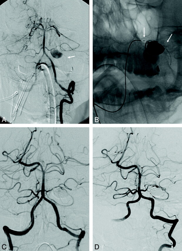
A 46-year-old woman (patient 9) presented with SAH and stuporous mental status. Diagnostic angiography revealed a large saccular aneurysm located just after the bifurcation of the left AICA-PICA variant (Fig 1A). EVT was chosen due to the patient's condition and surgical risk, and informed consent was obtained. With the patient under general anesthesia, catheterization of the left AICA aneurysm and the distal AICA was performed with 2 microcatheters and a Synchro microwire (Boston Scientific) via the right femoral artery access. To avoid the possibility of an extensive infarction of the cerebellum and brain stem, we placed an Enterprise stent (14 mm, Codman Neurovascular) in the parent artery immediately after coil embolization to preserve the AICA-PICA variant (Fig 1B). Control angiography 20 minutes after stent deployment confirmed good patency of the stented artery with antegrade flow to the left AICA-PICA territory (Fig 1C). The patient was delivered to the intensive care unit with heparinization and antiplatelet therapy with aspirin, and clopidogrel was started orally 1 day later. Her postoperative course was uneventful without neurologic deficits, and EVD was performed 4 days after EVT due to progressive hydrocephalus. At 10 months, the follow-up angiography showed no interval change of the coiled aneurysm with good patency of the AICA-PICA variant (Fig 1D).
Fig 1.

A 46-year-old woman with a proximal AICA aneurysm. A, Left VA angiogram reveals a large saccular aneurysm just after the bifurcation of the left AICA-PICA variant, which is located at the proximal PICA branch (white arrow). B, Fluoroscopic image shows the deployment of the Enterprise stent (white arrows) after coil embolization of the ruptured aneurysm. C, The right VA angiogram shows complete occlusion of the aneurysm with preservation of the parent artery immediately after embolization. D, Follow-up angiogram depicts good patency of the left AICA, with complete occlusion 10 months after the procedure.
Case 2
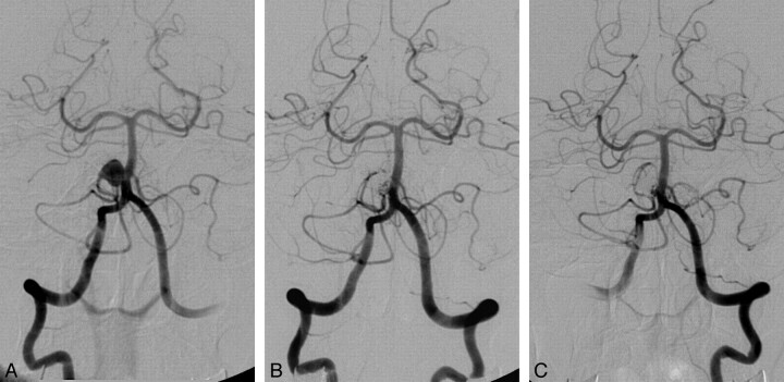
A 72-year-old woman (patient 8) was admitted with an incidentally found proximal AICA aneurysm on brain MR imaging. Cerebral angiography revealed a wide-neck aneurysm from the proximal AICA-PICA variant (Fig 2A). With the patient under general anesthesia, two 5F Envoy guiding catheters were placed in both VAs via the bilateral femoral arteries. After catheterization of the aneurysm and parent artery was performed by using 2 microcatheters, a Neuroform stent (15 mm) was placed in the BA. Coil embolization was performed partially to preserve the common trunk (Fig 2B). Dual antiplatelet therapy was maintained before and after the procedure. At 12 months, a follow-up angiography revealed aneurysm neck filling and minimal coil compaction while preserving the AICA-PICA variant (Fig 2C).
Fig 2.
A 72-year-old woman with a proximal AICA aneurysm, which originated from the AICA-PICA variant. A, Right VA angiogram reveals a wide-neck saccular aneurysm on the proximal right AICA-PICA variant. B, The right VA angiogram confirms partial occlusion of the aneurysm with preservation of the AICA-PICA variant immediately after embolization. C, The follow-up angiogram shows residual neck filling of the aneurysm with minimal coil compaction 12 months later.
Discussion
The AICA arises from the first or middle third of the BA and has a single origin bilaterally in 72% of patients and a common trunk with the PICA in >30%.27,28 It is distributed to the pons, foramen of Luschka, middle cerebellar peduncle, and petrosal surface of the cerebellum. Near the seventh and eighth cranial nerve complex, it frequently divides into the rostral and caudal trunk. The AICA gives off the internal auditory artery, recurrent perforating arteries, and subarcuate artery as well as perforators to the brain stem and choroidal branches to the tela and choroid plexus.
AICA aneurysms are an extremely rare pathology and have been reported in only 2 of 6368 aneurysms by Locksley7 and in only 34 AICA aneurysms (1.3%) of >3500 saccular aneurysms by Gonzalez et al.6 Most aneurysms arise from the proximal segment of the AICA and rarely from the distal one. The former can be treated by clip ligation at the neck, and the latter may be trapped or occluded surgically or endovascularly.6,21,24,25,29–31 The etiology of the AICA aneurysm is controversial: hemodynamic stress, embryonic vulnerability, flow-related vascular pathology, and arterial dissection by local trauma or nonspecific inflammation.29,32–39 Our study showed that 2 aneurysms were accompanied by AVMs and Moyamoya disease.
In this study, EVT showed 78% partial or complete occlusion, with a good clinical outcome in the treatment of AICA aneurysms. Moreover, all treated aneurysms were proximal; aneurysms in this location have been regarded as candidates for surgery in previous studies. To date, there have been reported only 17 AICA aneurysms treated by EVT, all of which were in the distal AICA and were treated by parent artery occlusion, except in 2 cases.22–25,29,30,40–47 The reasons for EVT in proximal AICA aneurysms may be as follows: 1) development of technologies regarding endovascular devices, 2) all treated aneurysms not fusiform but saccular, and 3) accessibility to anatomic configuration by an endovascular device. Because all the treated AICA aneurysms were saccular, it was possible to preserve the parent artery with endosaccular embolization, and stent-assisted coiling was also applied in cases of the wide-neck AICA aneurysms. Unlike our cases, some AICA aneurysms were reported to be fusiform or dissecting, which were treated by parent artery occlusion.23,30
The high rate of postoperative complications is a major concern, though surgical therapy has been a mainstay in the management of AICA aneurysms since the first surgical treatment of these aneurysms by Gonzalez6 and Schwartz.8 Morbidity and mortality related to SAH could be explained by the surrounding neurovascular complexity, narrow surgical field, rarity and deep location of AICA aneurysms, and lack of surgeon expertise. However, our study showed no postoperative neurologic complication in 7 cases treated by EVT. Moreover in the literature review of 15 cases treated by parent artery occlusion, most of the patients also recovered well with minimal neurologic deficit.22–25,30,42,43,45–49 In view of the condition and clinical outcome of the patients, EVT may be considered a treatment option instead of surgery, despite fewer than 30 cases reported, including ours.
In association with the prognosis, the patency of the AICA origin during surgical clipping is a major issue,6 as in EVT. Two cases of a ruptured dissecting aneurysm treated by endosaccular coil embolization, preserving the AICA, have been reported.25,45 In particular, 5 AICA aneurysms in this study developed from the AICA-PICA variant, which had a common trunk from the BA or the distal VA and supplied blood to their territories as well as the brain stem. Of 9 AICA aneurysms, 7 were also treated with preservation of the AICA to avoid infarction by arterial occlusion. Although all the treated aneurysms were saccular, it was very useful to apply a stent-assisted technique in cases of wide-neck aneurysms. However dual antiplatelet therapy is necessary to prevent thromboembolism after stent placement and may increase the risk of intracranial hemorrhage or rebleeding from a ruptured aneurysm.50 Because a ruptured AICA aneurysm is often accompanied by intraventricular hemorrhages, an intracranial procedure such as EVD should be carefully performed after dual antiplatelet therapy. In this study, 1 patient with stent-assisted coiling required EVD, which was performed without intracranial hemorrhage.
Although technologies related to EVT are developing rapidly, there are some restrictions to EVT for the treatment of AICA aneurysms. After failure of EVT, 1 aneurysm was treated by surgical trapping and 1 by spontaneous occlusion. Spontaneous occlusion of the AICA occurred following forced manipulation by a Synchro microwire, which was due to acute thrombosis or arterial dissection after intimal injury. From the latter, one can infer the causes of failure of EVT: 1) acute angled configuration of the AICA orifice from the BA (patients 4 and 5), and 2) the very small caliber of the AICA (patient 5) combined with a distal location of the aneurysm. The anatomic configuration and adequate caliber of the vessel are mandatory for accessibility of the microcatheter delivery system. Most of the reported cases of EVT satisfied these requirements, and the surgical approach was used in endovascularly failed cases.6
On the basis of these conditions, a treatment strategy should be carefully decided between EVT and surgery. For the past 13 years, we have considered it most important to choose a better therapeutic option from the standpoint of the patient, though AICA aneurysms may be more easily treated by EVT than before, along with advances in materials and devices relevant to EVT. Stent-assisted coiling cannot always be applied to all AICA aneurysms or the AICA itself, and thromboembolic complications should be monitored after this procedure.
There are some limitations in our study. First, the number of the enrolled patients was too small to assess the safety and feasibility of EVT. However, the incidence of AICA aneurysm is extremely rare, and therapeutics of this pathology are minimal as well. Second, recanalization of the coiled aneurysm may be encountered inevitably in the long-term follow-up period, despite good follow-up results in our study. Three AICA aneurysms were incompletely embolized to preserve the AICA, and in the follow-up period, findings of these aneurysms were same as the initial angiographic results or showed minimal coil compaction. Choi et al45 reported a rebleeding case after EVT of a distal AICA aneurysm with parent artery preservation. Therefore, close monitoring of the patients treated by coils is essential. Finally, in-stent stenosis or acute in-stent thrombosis of the stent may occur in the small-diameter AICA. In general, the diameter of the AICA is <2 mm, and use of a self-expandable stent on this small vessel is limited. Turk et al51 reported the use of the same stent in smaller cerebral vessels. Above all, it is important to maintain dual antiplatelet therapy thoroughly before and after the procedure.
Conclusions
In the treatment of AICA aneurysms, EVT provides another viable option to surgery when the microsurgical option is not tolerable. Until now, not every AICA aneurysm was treated with stents and coils. Further follow-up and more experience are also necessary to determine long-term results for EVT of AICA aneurysms.
Abbreviations
- AICA
anterior inferior cerebellar artery
- AVM
arteriovenous malformation
- BA
basilar artery
- CO
complete occlusion
- Cx
procedural complication
- EVD
external ventricular drainage
- EVT
endovascular therapy
- F/U
follow-up period
- HH
Hunt and Hess scale
- IO
incomplete occlusion
- mRS
modified Rankin Scale
- NA
not available
- O
occlusion
- P
preservation
- PICA
posterior inferior cerebellar artery
- SAH
subarachnoid hemorrhage
- TIA
transient ischemic accident
- Tx
treatment
- VA
vertebral artery
Footnotes
This work was supported by a faculty research grant of Yonsei University College of Medicine for 2009 (6–2009–0081).
References
- 1. Drake CG. The treatment of aneurysms of the posterior circulation. Clin Neurosurg 1979;26:96–144 [DOI] [PubMed] [Google Scholar]
- 2. Drake CG. Surgical treatment of ruptured aneurysms of the basilar artery: experience with 14 cases. J Neurosurg 1965;23:457–73 [DOI] [PubMed] [Google Scholar]
- 3. Drake CG. The surgical treatment of aneurysms of the basilar artery. J Neurosurg 1968;29:436–46 [DOI] [PubMed] [Google Scholar]
- 4. Drake CG. The surgical treatment of vertebral-basilar aneurysms. Clin Neurosurg 1969;16:114–69 [DOI] [PubMed] [Google Scholar]
- 5.. Drake CG, Peerless SJ, Hernesniemi JA. Surgery of Vertebrobasilar Aneurysms: London, Ontario Experience on 1767 Patients. New York: Springer-Verlag; 1995 [Google Scholar]
- 6. Gonzalez LF, Alexander MJ, McDougall CG, et al. Anteroinferior cerebellar artery aneurysms: surgical approaches and outcomes—a review of 34 cases. Neurosurgery 2004;55:1025–35 [DOI] [PubMed] [Google Scholar]
- 7. Locksley HB. Natural history of subarachnoid hemorrhage, intracranial aneurysms and arteriovenous malformations: based on 6368 cases in the cooperative study. J Neurosurg 1966;25:219–39 [DOI] [PubMed] [Google Scholar]
- 8. Schwartz HG. Arterial aneurysm of the posterior fossa. J Neurosurg 1948;5:312–16 [DOI] [PubMed] [Google Scholar]
- 9. Porter RJ, Eyster EF. Aneurysm in the anterior inferior cerebellar artery at the internal acoustic meatus: report of a case. Surg Neurol 1973;1:27–18 [PubMed] [Google Scholar]
- 10. Johnson JH, Jr, Kline DG. Anterior inferior cerebellar artery aneurysms: case report. J Neurosurg 1978;48:455–60 [DOI] [PubMed] [Google Scholar]
- 11. Mori K, Miyazaki H, Ono H. Aneurysm of the anterior inferior cerebellar artery at the internal auditory meatus. Surg Neurol 1978;10:297–300 [PubMed] [Google Scholar]
- 12. Zlotnik EI, Sklyut JA, Smejanovich AF, et al. Saccular aneurysm of the anterior inferior cerebellar-internal auditory artery: case report. J Neurosurg 1982;57:829–32 [DOI] [PubMed] [Google Scholar]
- 13. Nishimoto A, Fujimoto S, Tsuchimoto S, et al. Anterior inferior cerebellar artery aneurysm: report of three cases. J Neurosurg 1983;59:697–702 [DOI] [PubMed] [Google Scholar]
- 14. Nakagawa K, Sakaki S, Kimura H, et al. Aneurysm of the anterior inferior cerebellar artery at the internal auditory meatus. Surg Neurol 1984;21:231–35 [DOI] [PubMed] [Google Scholar]
- 15. Uede T, Matsumura S, Ohtaki M, et al. Aneurysm of the anterior inferior cerebellar artery at the internal auditory meatus: report of two cases [in Japanese]. No Shinkei Geka 1986;14:1263–68 [PubMed] [Google Scholar]
- 16. Fukuya T, Kishikawa T, Ikeda J, et al. Aneurysms of the peripheral portion of the anterior inferior cerebellar artery: report of two cases. Neuroradiology 1987;29:493–96 [DOI] [PubMed] [Google Scholar]
- 17. Oana K, Murakami T, Beppu T, et al. Aneurysm of the distal anterior inferior cerebellar artery unrelated to the cerebellopontine angle: case report. Neurosurgery 1991;28:899–903 [DOI] [PubMed] [Google Scholar]
- 18. Yokoyama S, Kadota K, Asakura T, et al. Aneurysm of the distal anterior inferior cerebellar artery: case report. Neurol Med Chir (Tokyo) 1995;35:587–90 [DOI] [PubMed] [Google Scholar]
- 19. Mizushima H, Kobayashi N, Yoshiharu S, et al. Aneurysm of the distal anterior inferior cerebellar artery at the medial branch: a case report and review of the literature. Surg Neurol 1999;52:137–42 [DOI] [PubMed] [Google Scholar]
- 20. Bambakidis NC, Manjila S, Dashti S, et al. Management of anterior inferior cerebellar artery aneurysms: an illustrative case and review of literature. Neurosurg Focus 2009;26:E6. [DOI] [PubMed] [Google Scholar]
- 21. Sun Y, Wrede KH, Chen Z, et al. Ruptured intrameatal AICA aneurysms: a report of two cases and review of the literature. Acta Neurochir (Wien) 2009;151:1525–30 [DOI] [PubMed] [Google Scholar]
- 22. Suzuki K, Meguro K, Wada M, et al. Embolization of a ruptured aneurysm of the distal anterior inferior cerebellar artery: case report and review of the literature. Surg Neurol 1999;51:509–12 [DOI] [PubMed] [Google Scholar]
- 23. Mitsos AP, Corkill RA, Lalloo S, et al. Idiopathic aneurysms of distal cerebellar arteries: endovascular treatment after rupture. Neuroradiology 2008;50:161–70 [DOI] [PubMed] [Google Scholar]
- 24. Peluso JP, van Rooij WJ, Sluzewski M, et al. Distal aneurysms of cerebellar arteries: incidence, clinical presentation, and outcome of endovascular parent vessel occlusion. AJNR Am J Neuroradiol 2007;28:1573–78 [DOI] [PMC free article] [PubMed] [Google Scholar]
- 25. Kusaka N, Maruo T, Nishiguchi M, et al. Embolization for aneurismal dilatation associated with ruptured dissecting anterior inferior cerebellar artery aneurysm with preservation of the parent artery: case report [in Japanese]. No Shinkei Geka 2006;34:729–34 [PubMed] [Google Scholar]
- 26. Pritz MB. Aneurysms of the anterior inferior cerebellar artery. Acta Neurochir (Wien) 1993;120:12–19 [DOI] [PubMed] [Google Scholar]
- 27. Baba T, Matsushima T, Fukui M, et al. Relationship between angiographical manifestations and operative findings in 100 cases of hemifacial spasm [in Japanese]. No shinkei geka 1988;16:1355. [PubMed] [Google Scholar]
- 28. Baskaya MK, Coscarella E, Jea A, et al. Aneurysm of the anterior inferior cerebellar artery-posterior inferior cerebellar artery variant: case report with anatomical description in the cadaver. Neurosurgery 2006;58:E388, discussion E88 [DOI] [PubMed] [Google Scholar]
- 29. Zager EL, Shaver EG, Hurst RW, et al. Distal anterior inferior cerebellar artery aneurysms: report of four cases. J Neurosurg 2002;97:692–96 [DOI] [PubMed] [Google Scholar]
- 30. Fukushima S, Hirohata M, Okamoto Y, et al. Anterior inferior cerebellar artery dissecting aneurysm in a juvenile: case report. Neurol Med Chir (Tokyo) 2009;49:81–84 [DOI] [PubMed] [Google Scholar]
- 31. Takeuchi S, Takasato Y, Masaoka H, et al. Trapping of ruptured dissecting aneurysm of distal anterior inferior cerebellar artery: case report [in Japanese]. Brain Nerve 2009;61:203–07 [PubMed] [Google Scholar]
- 32. Graves VB, Strother CM, Partington CR, et al. Flow dynamics of lateral carotid artery aneurysms and their effects on coils and balloons: an experimental study in dogs. AJNR Am J Neuroradiol 1992;13:189–96 [PMC free article] [PubMed] [Google Scholar]
- 33. Strother CM, Graves VB, Rappe A. Aneurysm hemodynamics: an experimental study. AJNR Am J Neuroradiol 1992;13:1089–95 [PMC free article] [PubMed] [Google Scholar]
- 34. Khayata MH, Aymard A, Casasco A, et al. Selective endovascular techniques in the treatment of cerebral mycotic aneurysms: report of three cases. J Neurosurg 1993;78:661–65 [DOI] [PubMed] [Google Scholar]
- 35. Cockrill HH, Jr, Jimenez JP, Goree JA. Traumatic false aneurysm of the superior cerebellar artery simulating posterior fossa tumor. J Neurosurg 1977;46:377–80 [DOI] [PubMed] [Google Scholar]
- 36. Heros RC. Posterior inferior cerebellar artery. J Neurosurg 2002;97:747–48, discussion 48 [DOI] [PubMed] [Google Scholar]
- 37. Horiuchi T, Tanaka Y, Hongo K, et al. Characteristics of distal posteroinferior cerebellar artery aneurysms. Neurosurgery 2003;53:589–95, discussion 95–96 [DOI] [PubMed] [Google Scholar]
- 38. Biondi A. Trunkal intracranial aneurysms: dissecting and fusiform aneurysms. Neuroimaging Clin N Am 2006;16:453–65, viii [DOI] [PubMed] [Google Scholar]
- 39. Akyuz M, Tuncer R. Multiple anterior inferior cerebellar artery aneurysms associated with an arteriovenous malformation: case report. Surg Neurol 2005;64(suppl 2):S106–08 [DOI] [PubMed] [Google Scholar]
- 40. Kaech D, de Tribolet N, Lasjaunias P. Anterior inferior cerebellar artery aneurysm, carotid bifurcation aneurysm, and dural arteriovenous malformation of the tentorium in the same patient. Neurosurgery 1987;21:575–82 [DOI] [PubMed] [Google Scholar]
- 41. Saito A, Ezura M, Takahashi A, et al. An arterial dissection of the distal anterior inferior cerebellar artery treated by endovascular therapy [in Japanese]. No Shinkei Geka 2000;28:269–74 [PubMed] [Google Scholar]
- 42. Fujimura N, Abe T, Hirohata M, et al. Bilateral anomalous posterior inferior cerebellar artery-anterior inferior cerebellar artery anastomotic arteries associated with a ruptured cerebral aneurysm: case report. Neurol Med Chir (Tokyo) 2003;43:396–98 [DOI] [PubMed] [Google Scholar]
- 43. Maekawa M, Awaya S, Fukuda S, et al. A ruptured choroidal artery aneurysm of the anterior inferior cerebellar artery obliterated via the endovascular approach: case report [in Japanese]. No Shinkei Geka 2003;31:523–27 [PubMed] [Google Scholar]
- 44. Benes L, Kappus C, Sure U, et al. Treatment of a partially thrombosed giant aneurysm of the vertebral artery by aneurysm trapping and direct vertebral artery-posterior inferior cerebellar artery end-to-end anastomosis: technical case report. Neurosurgery 2006;59:ONSE166–67, discussion ONSE66–67 [DOI] [PubMed] [Google Scholar]
- 45. Choi CH, Cho WH, Choi BK, et al. Rerupture following endovascular treatment for dissecting aneurysm of distal anterior inferior cerebellar artery with parent artery preservation: retreatment by parent artery occlusion with Guglielmi detachable coils. Acta Neurochir (Wien) 2006;148:363–66, discussion 66 [DOI] [PubMed] [Google Scholar]
- 46. Takao T, Fukuda M, Kawaguchi T, et al. Ruptured intracranial aneurysm following gamma knife surgery for acoustic neuroma. Acta Neurochir (Wien) 2006;148:1317–18, discussion 18 [DOI] [PubMed] [Google Scholar]
- 47. Tsutsumi M, Kazekawa K, Aikawa H, et al. Development of unusual collateral channel from the posterior meningeal artery after endovascular proximal occlusion of the posterior inferior cerebellar artery. Neurol Med Chir (Tokyo) 2007;47:503–05 [DOI] [PubMed] [Google Scholar]
- 48. Saito R, Tominaga T, Ezura M, et al. Distal anterior inferior cerebellar artery aneurysms: report of three cases and literature review [in Japanese]. No Shinkei Geka 2001;29:709–14 [PubMed] [Google Scholar]
- 49. Hue YH, Yi HJ, Kim YJ. Extravasation during aneurysm embolization without neurologic consequences: lessons learned from complications of pseudoaneurysm coiling—report of 2 cases. J Korean Neurosurg Soc 2008;44:178–81. Epub 2008 Sep 30 [DOI] [PMC free article] [PubMed] [Google Scholar]
- 50. Tumialan LM, Zhang YJ, Cawley CM, et al. Intracranial hemorrhage associated with stent-assisted coil embolization of cerebral aneurysms: a cautionary report. J Neurosurg 2008;108:1122–29 [DOI] [PubMed] [Google Scholar]
- 51. Turk AS, Niemann DB, Ahmed A, et al. Use of self-expanding stents in distal small cerebral vessels. AJNR Am J Neuroradiol 2007;28:533–36 [PMC free article] [PubMed] [Google Scholar]


