Abstract
Steroid hormones, including glucocorticoids and androgens, exert a wide variety of effects in the body across almost all tissues. The steroid A-ring 5β-reductase (AKR1D1) is expressed in human liver and testes, and three splice variants have been identified (AKR1D1-001, AKR1D1-002, AKR1D1-006). Amongst these, AKR1D1-002 is the best described; it modulates steroid hormone availability and catalyses an important step in bile acid biosynthesis. However, specific activity and expression of AKR1D1-001 and AKR1D1-006 are unknown. Expression of AKR1D1 variants were measured in human liver biopsies and hepatoma cell lines by qPCR. Their three-dimensional (3D) structures were predicted using in silico approaches. AKR1D1 variants were overexpressed in HEK293 cells, and successful overexpression confirmed by qPCR and Western blotting. Cells were treated with either cortisol, dexamethasone, prednisolone, testosterone or androstenedione, and steroid hormone clearance was measured by mass spectrometry. Glucocorticoid and androgen receptor activation were determined by luciferase reporter assays. AKR1D1-002 and AKR1D1-001 are expressed in human liver, and only AKR1D1-006 is expressed in human testes. Following overexpression, AKR1D1-001 and AKR1D1-006 protein levels were lower than AKR1D1-002, but significantly increased following treatment with the proteasomal inhibitor, MG-132. AKR1D1-002 efficiently metabolised glucocorticoids and androgens and decreased receptor activation. AKR1D1-001 and AKR1D1-006 poorly metabolised dexamethasone, but neither protein metabolised cortisol, prednisolone, testosterone or androstenedione. We have demonstrated the differential expression and role of AKR1D1 variants in steroid hormone clearance and receptor activation in vitro. AKR1D1-002 is the predominant functional protein in steroidogenic and metabolic tissues. In addition, AKR1D1-001 and AKR1D1-006 may have a limited, steroid-specific role in the regulation of dexamethasone action.
Keywords: steroids, dexamethasone, cortisol, testosterone, liver
Introduction
Steroid hormones, including glucocorticoids, androgens and oestrogens, are fat-soluble molecules biosynthesised from cholesterol that play a crucial role in development, differentiation and metabolism (Simons 2008). Glucocorticoids, produced by the adrenal cortex, are released in response to stress and, following binding to their cognate receptor, the glucocorticoid receptor (GR), regulate anti-inflammatory and metabolic processes. Androgens are predominantly produced by the male testes, but also by the adrenal glands and the ovaries in females. Upon binding to the androgen receptor (AR), they have multiple actions, including the initiation of adrenarche and stimulation and control of secondary sexual characteristics. Following biosynthesis and delivery into the circulation, steroid hormones can be reduced, oxidised or hydroxylated by a variety of enzymes, including the 11β-hydroxysteroid dehydrogenases (11β-HSD) and the A-ring reductases (5α-reductases, (5αR) and 5β-reductase (AKR1D1)) in a tissue-specific manner. Dysregulation of glucocorticoid and androgen steroid metabolism have been associated with adverse metabolic features, including insulin resistance, hypertension, glucose intolerance and hepatic triacylglycerol (TG) accumulation (Pitteloud et al. 2005, Wang 2005, Tomlinson et al. 2008, Nasiri et al. 2015).
The steroid 5β-reductase is encoded by the AKR1D1 (aldo-keto reductase 1D1) gene, and is highly expressed in the liver, where it inactivates steroid hormones, including glucocorticoids and androgens, and catalyses a fundamental step in bile acid biosynthesis (Chen et al. 2011, Jin et al. 2014). AKR1D1 utilises NADPH as the hydride donor and catalyses a stereospecific irreversible double bond reduction between the C4 and C5 positions of the A-ring of steroids. 5β-reduction of steroids is unique in steroid enzymology as it introduces a 90° bend and creates an A/B cis-ring junction, resulting in the formation of steroids with different properties from either the α,β-unsaturated or 5α-reduced steroids (which possess a largely planar steroidal structure with an A/B trans ring-junction) (Jin et al. 2014). The human AKR1D1 protein is highly homologous with other members of the AKR1 family, including the AKR1C subfamily (which includes the hydroxysteroid dehydrogenases), the AKR1A subfamily (aldehyde reductases) and the AKR1B subfamily (aldose reductases) (Jez et al. 1997). The human gene for AKR1D1 consists of nine exons, and three splice variants have been identified, all of which are predicted to lead to functional proteins: AKR1D1-001 (NM_001190906), AKR1D1-006 (NM_001190907), and AKR1D1-002 (NM_005989) (Barski et al. 2013). AKR1D1-002 encodes a 326 amino acid 5β-reductase enzyme that includes all 9 exons. AKR1D1-001 lacks exon 5 and is translated into a 285 amino acid protein, whilst AKR1D1-006 omits exon 8 and translates into a 290 amino acid protein (Barski et al. 2013) (Fig. 1). No sex-specific differences in hepatic AKR1D1 expression have been reported (Amai et al. 2020). Loss-of-function mutations in the AKR1D1 gene have been revealed in patients with 5β-reductase deficiency, and are associated with decreased 5β-reduced urinary corticosteroids and impaired bile acid biosynthesis (Palermo et al. 2008).
Figure 1.
(A) AKR1D1 splice variants, showing the full form of AKR1D1-002, resulting in a 326 amino acid (aa) protein, as well as AKR1D1-001 and AKR1D1-006 that lack exons 5 and 8, resulting in 285 and 290 amino acid proteins, respectively.
Amongst all AKR1D1 splice variants (AKR1D1-SVs), AKR1D1-002 is the best characterised and represents the full-length WT protein. In both cellular and cell-free system models, AKR1D1-002 metabolises endogenous glucocorticoids, including cortisol and cortisone, to their 5β-reduced metabolites, 5β-dihydrocortisol and 5β-dihydrocortisone, respectively. The 5β-reduced metabolites are then converted, in a non-rate limiting step, to their inactive tetrahydro-metabolites (5β-tetrahydrocortisol and 5β-tetrahydrocortisone) by the hepatic 3α-hydroxysteroid dehydrogenases, AKR1C1-C4, with downstream effects on steroid receptor activation and target gene transcription (Chen et al. 2011, Nikolaou et al. 2019a). Crucially, AKR1D1-002 is also implicated in drug metabolism, as it metabolises the synthetic glucocorticoids prednisolone and dexamethasone, with downstream decreases in glucocorticoid receptor activation (Nikolaou et al. 2020).
The relative expression levels of AKR1D1-SVs in human tissues have not been identified. Similarly, it is entirely unexplored whether the truncated variants lead to functional proteins, and whether they have a role in glucocorticoid, androgen and drug metabolism. The aim of our study was therefore to define the ability of AKR1D1-SVs to regulate endogenous and synthetic steroid availability in appropriate human cellular models, as well as to determine their expression levels in human cells.
Materials and methods
Genotype-tissue expression data (GTEx)
AKR1D1-SV expression data (ENSG00000122787.14) were extracted from the Genotype-Tissue Expression (GTEx) Project (https://www.gtexportal.org). GTEx was supported by the Common Fund (https://commonfund.nih.gov/GTEx) of the Office of the Director of the National Institutes of Health, and by NCI, NHGRI, NHLBI, NIDA, NIMH, and NINDS. The data used for the analyses described in this manuscript were obtained from dbGaP Accession phs000424.v8.p2 on 24/04/2020. Limits of detection for data analysis were not provided.
Cell culture and human liver tissue
Liver biopsy samples originated from the Oxford Gastrointestinal Illness Biobank (REC reference 16/YH/0247). HepG2 cells (male donor, Cat#HB-8065) and HEK293 cells (female donor, Cat#CRL-11268) were purchased from ATCC. Huh7 cells (male donor) were purchased from the Japanese Cancer Research Resources Bank (Cat#JCRB0403). All cell lines were cultured in DMEM (Thermo Fisher Scientific), containing 4.5 g/L glucose, and supplemented with 10% foetal bovine serum, 1% penicillin/streptomycin and 1% non-essential amino acids (Thermo Fisher Scientific). Cells were grown at 37°C in a 5% CO2 setting.
Dexamethasone, cortisol, prednisolone, testosterone, androstenedione and MG-132 were purchased from Sigma-Aldrich. For all cell treatments, HEK293 cells were cultured in serum-free and phenol red-free media containing 4.5 g/L glucose (Sigma-Aldrich), 1% penicillin/streptomycin, 1% non-essential amino acids and 1% l-glutamine (Sigma-Aldrich).
Transfection studies
For overexpression transfection studies, 1 × 10⁵ cells/well were plated in 24-well Cell Bind plates (CORNING) 24 h prior to transfection with either empty pCMV6 vector (#PCMV6XL4), pCMV6+AKR1D1-002 (#SC116410), pCMV6+AKR1D1-001 (#RC231126) or pCMV6+AKR1D1-006 (#RC231133) construct variants (Origene Technologies, Rockville, USA). In total 0.5 µg DNA construct and 1 µL X-tremeGENE DNA Transfection Reagent (Roche) were diluted in 50 µL OPTIMEM serum-free media (Invitrogen). The mixture was vortexed and incubated at room temperature for 20 min; 50 µL was added to each well and cells incubated at 37°C for 48 h before treatment. To investigate transfection efficiency, co-transfection experiments using 0.1 µg Green Fluorescent Protein (GFP) construct and 0.5 µg from each AKR1D1 transcript variant were performed, as described previously.
For cell treatments, HEK293 cells were cultured in serum-free and phenol red-free media containing 4.5 g/L glucose, and either steroid hormone (glucocorticoids: 500 nM; androgens: 200 nM) for 24 h, post-transfection. For proteasome inhibition studies, cells were cultured in serum-free media, and 20 µM MG-132 were added 3 and 6 h prior to harvesting.
RNA extraction and gene expression
Total RNA was extracted from cells using the Tri-Reagent system (Sigma-Aldrich) and RNA concentrations were determined spectrophotometrically at OD260 on a Nanodrop spectrophotometer (ThermoFisher Scientific). Genomic DNA was removed using a commercially available DNAse Treatment and Removal Reagent kit, according to the manufacturer’s protocol (#AM1906, ThermoFisher Scientific). RT was performed in a 20 μL volume; 1 μg of total RNA was incubated with 10× RT Buffer, 100 mM dNTP Mix, 10x RT Random Primers, 50 U/μL MultiScribe Reverse Transcriptase and 20 U/μL RNase Inhibitor (ThermoFisher Scientific). The reaction was performed under the following conditions: 25°C for 10 min, 37°C for 120 min and then terminated by heating to 85°C for 5 min.
All quantitative PCR (qPCR) experiments were conducted using an ABI 7900HT sequence detection system (Perkin-Elmer Applied Biosystems). Reactions were performed in 6 μL volumes on 384-well plates in reaction buffer containing 3 μL of 2× Kapa Probe Fast qPCR Master Mix (Sigma-Aldrich), in triplicate. All probes were supplied by Thermo Fisher Scientific as pre-designed FAM dye-labelled TaqMan Gene Expression Assays (AKR1D1 spanning exons 3–4: Hs00973526_g1; AKR1D1 spanning exons 5–6: Hs00973528_gH; AKR1D1 spanning exons 7–8: Hs00975611_m1). The reaction conditions were 95°C for 3 min, then 40 cycles of 95°C for 3 s and 60°C for 20 s. The Ct (dCt) of each sample was measured using the following calculation (where E is reaction efficiency – determined from a standard curve): dCt = E(min Ct-sample Ct), using a 1/40 sample dilution. The standard curve was generated from a pool of all cDNAs – including a negative control, containing sample diluent (10 mM TRIS:HCl) only – and used as the calibrator for all samples. The relative expression ratio was calculated using the following equation: Ratio= dCt(target)/dCt(ref) and expression values were corrected to the geometric mean of three reference genes: 18SrRNA (Hs03003631_g1), TBP (Hs00427620_m1) and ACTB (Hs01060665_g1) (Pfaffl 2001).
All AKR1D1 Taqman assays had similar efficiency, and for experiments investigating the relative expression of the different AKR1D1-SVs, this was corrected by running one sample from AKR1D1-002 overexpressed cells, which was detected by all three AKR1D1 Taqman assays. In this set of experiments, minimum dCt values were defined by the negative controls and maximum dCt values were defined by the AKR1D1-002 overexpressed cells. Relative mRNA expression of AKR1D1-001 and AKR1D1-006 variants were determined as a percentage of total AKR1D1 expression, which was set at 100% (Supplementary Fig. 1, see section on supplementary materials given at the end of this article).
Glucocorticoid measurements
Cell media, cortisol, prednisolone and dexamethasone concentrations were measured using quantitative liquid chromatography–mass spectrometry (LC-MS/MS) in selected ion-monitoring analysis using previously published methods (Owen et al. 2005, Hawley et al. 2018). The lower limit of quantitation was 5.2 nmol/L for prednisolone, 0.25 nmol/L for dexamethasone, and 22 nmol/L for cortisol.
Androgen measurements
A cocktail (100 µL) containing 1.5 ng D2-testosterone and 15 ng D7-androstenedione in water was added to each sample after which the steroids were extracted using three volumes of tert-Butyl methyl ether (MTBE). Samples were mixed by vortexing and incubated at −80°C for 1 h. The organic layer was subsequently transferred to a clean test tube and dried under a stream of nitrogen at 45°C. The dried steroid residue was reconstituted in 50% MeOH in water (150 μL) and stored at −20°C prior to analysis.
Androgens were quantified by UPLC-MS/MS. Briefly, steroids were separated using a high strength silica (HSS) T3 column (2.1 mm × 50 mm, 1.8 µm) (Waters, Milford, USA) coupled to an ACQUITY UPLC (Waters). A 5 min linear gradient from 55% A (1% formic acid) to 75% B (100% methanol) was used to separate the steroids at a constant flow rate of 0.6 mL/min and a column temperature of 50°C. The injection volume was 10 µL. Steroids were quantified in multiple reaction monitoring (MRM) using a Xevo TQ-S triple quadrupole mass spectrometer (Waters) containing an electrospray probe set to positive ionisation mode (ESI+). The following settings were used: capillary voltage of 4.0 kV, source temperature 150°C, desolvation temperature 500°C, desolvation gas 800 L/hand cone gas 150 L/h. Data collection and analysis were performed using MassLynx 4.1 (Waters Corporation).
Luciferase reporter assay
To determine GR activation, HEK293 cells were plated in 24-well Cell Bind plates (CORNING) and co-transfected with either empty pCMV6 vector, pCMV6+AKR1D1-002, pCMV6+AKR1D1-001 or pCMV6+006 construct variants, followed by treatments with serum-free and phenol-red free cell media containing each glucocorticoid for a further 24 h. Cell media aliquots (500 µL) were collected and stored at −20°C. In another set of experiments, HEK293 cells were transiently transfected with a glucocorticoid responsive element (GRE) reporter – a mixture of an inducible GRE-responsive firefly luciferase construct and a constitutively expressing renilla luciferase construct (#CCS-006L, QIAgen). Forty-eight hours post-transfection, cell media was replaced with the steroid-containing media aliquots described previously, and cells were incubated for 24 h.
Similarly, to determine AR activation, HEK293 cells were plated in 24-well Cell Bind plates (CORNING) and co-transfected with either empty pCMV6 vector, pCMV6+AKR1D1-002, pCMV6+AKR1D1-001 or pCMV6+006 construct variants, followed by treatments with serum-free and phenol-red free cell media containing testosterone or androstenedione for a further 24 h. Cell media aliquots (500 µL) were collected and stored at −20°C. In another set of experiments, HEK293 cells were transiently co-transfected with a pcDNA3.1+AR construct and an androgen responsive element (ARE) reporter – a mixture of an inducible ARE-responsive firefly luciferase construct and a constitutively expressing renilla luciferase construct (#CCS-1019L, QIAgen). Forty-eight hours post-transfection, cell media was replaced with the steroid-containing media aliquots described above, and cells were incubated for 24 h.
Cell lysates were harvested in passive lysis buffer, and reporter activity was measured using the Luciferase Assay System (Promega) and an EnSpire Multimode plater reader (PerkinElmer). The data were presented as the % ratio of firefly to renilla luciferase activity (Fluc/Rluc).
Protein extraction and immunoblotting
Cells were lysed using RIPA buffer (Sigma-Aldrich), supplemented with protease and phosphatase inhibitor cocktails (both 1/100) (Thermo Fisher Scientific). Protein concentrations were determined using the Bio-Rad protein assay (Bio-Rad Laboratories Inc.), according to the manufacturer’s instructions. Primary human AKR1D1 (dilution 1/250 – HPA057002, Atlas Antibodies AB, Bromma, Sweden), β-tubulin (#15115, monoclonal) (Cell Signalling), β-actin (#3700, monoclonal) (Cell Signalling), and secondary antibodies (P044801-2, polyclonal) from Dako (Agilent) were used at a dilution 1/1000 (primary) and 1/2000 (secondary), respectively, unless stated otherwise. Bands were visualised with Bio Rad Clarity Western ECL (Watford, Hertfordshire, UK) and ChemiDocXS imager (Bio Rad). Western blots were quantified by densitometry analysis using ImageJ (NIH, http://rsb.info.nih.gov/ij), normalised to β-tubulin and β-actin to correct for variability in gel loading.
Statistics
Data are presented as mean ± s.e., unless otherwise stated. Normal distribution was confirmed using Shapiro–Wilk test. Two-tailed, paired t-tests were used to compare single treatments to control. For comparisons between control and different treatments, statistical analysis was performed using one-way ANOVA with Dunnett corrections. Statistical analysis on qPCR data was performed on mean relative expression ratio values (Ratio= ΔCt(target)/ΔCt (Pfaffl 2001)). Data analysis was performed using GraphPad Prism version 9.0.0 for MacOS (GraphPad Software, www.graphpad.com), and considered statistically significant at P < 0.05, unless otherwise stated.
Results
AKR1D1-SVs are differentially expressed in human liver and testes
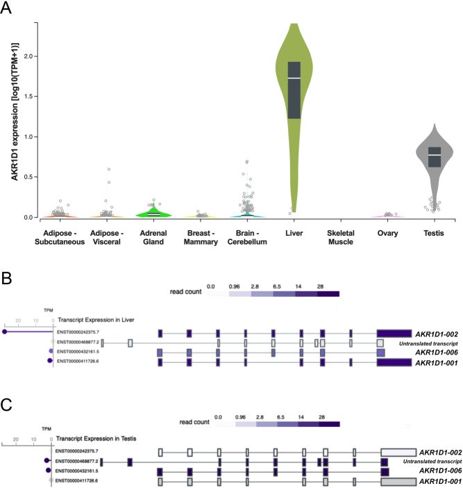
To define the transcription levels of AKR1D1 in human tissues, data were initially extracted from publicly available databases (https://www.gtexportal.org). Consistent with previous studies, AKR1D1 is predominantly expressed in the liver (median transcripts per million (TPM) = 51.66; n = 226), although significant levels of expression have also been detected in human testes (median TPM = 4.87; n = 381) (Fig. 2A). Low levels of AKR1D1 have also been detected in human adrenal glands (median TPM = 0.07; n = 258) and mammary breast (median TPM = 0.02; n = 459). No transcription levels were, however, detected in other metabolic or steroidogenic tissues, including adipose (s.c. (n = 663) or visceral (n = 541)), skeletal muscle, ovary (n = 180) or brain (n = 241) (Fig. 2A). Specifically with regards to AKR1D1-SVs, database analysis revealed AKR1D1-002 as the predominant transcript expressed in the liver, with lower levels of AKR1D1-001 expression, and almost no detectable AKR1D1-006 expression (Fig. 2B).
Figure 2.
(A) AKR1D1 transcript levels in human adipose, adrenal gland, breast, brain, liver, skeletal muscle, ovary and testicular tissues.$ (B) Transcript expression levels of AKR1D1 splice variants in human liver.$ (C) Transcript expression levels of AKR1D1 splice variants in human testes.$ $Data extracted from https://www.gtexportal.org (GTEx). GTEx-extracted expression values are shown as transcripts per million (TPM). Box plots are shown as median and 25th and 75th percentiles. Points displayed are outliers 1.5 times above or below the interquartile range.
Additional database analysis exploring testicular AKR1D1 expression demonstrated that the AKR1D1-006 splice variant was the main transcript present, with no expression of either AKR1D1-002 or AKR1D1-001 (Fig. 2C). Interestingly, a fourth transcript variant (ENST00000468877.2) was also detected in human testes, however, this variant is not translated due to the lack of an opening reading frame within its sequence.
AKR1D1-SVs are variably expressed in human liver and are differentially targeted for proteasomal degradation
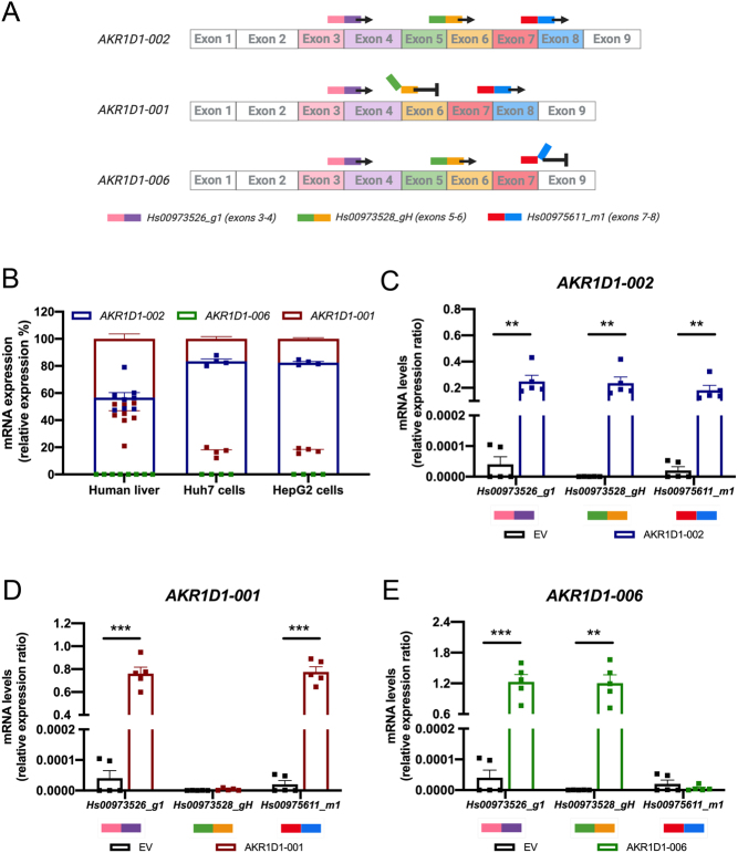
AKR1D1 variant-specific expression was confirmed using multiple pre-designed Taqman gene expression assay systems. First, an assay spanning the 3–4 exon–exon junction in the AKR1D1 mRNA was used to identify all variants. A second Taqman assay spanning the 5–6 exon–exon junction was able to identify AKR1D1-002 and -006, but not -001 transcripts. Finally, a third assay was used spanning the 7–8 exon–exon junction that was able to identify AKR1D1-002 and -001, but not -006 transcripts (Fig. 3A).
Figure 3.
(A) Schematic representation demonstrating the mechanism of TaqMan qPCR targeting the different AKR1D1 splice variants using specific exon–exon junction primers. (B) Relative mRNA expression levels of AKR1D1-001, AKR1D1-002 and AKR1D1-006 splice variants in male and female human liver biopsies (n = 8), Huh7 (n = 4) and HepG2 (n = 4) hepatoma cell lines. mRNA overexpression levels of (C) AKR1D1-002, (D) AKR1D1-001, and (E) AKR1D1-006 in HEK293 cells, confirmed using multiple exon–exon junction Taqman primer assays (n = 5). mRNA expression data are presented as mean ± s.e., performed in duplicate.
qPCR analysis in liver biopsies from male and female donors (n = 8) revealed that AKR1D1-002 is the most highly expressed splice variant in human liver (56.7 ± 3.6%), followed by AKR1D1-001 (43.3 ± 3.6%), and a complete lack of AKR1D1-006 expression. Similarly, in HepG2 and Huh7 cells, AKR1D1-002 was the predominant splice variant (HepG2: 82.5 ± 0.9%; Huh7: 83.5 ± 1.6%), with lower levels of AKR1D1-001 (HepG2: 17.5 ± 0.8%; Huh7: 16.5 ± 1.6%), and no AKR1D1-006 expression (Fig. 3B).
To define the functional role of AKR1D1 variants in the regulation of steroid hormone metabolism in vitro, HEK293 cells were transfected with either empty pCMV6 (EV), AKR1D1-002, AKR1D1-001, or AKR1D1-006 containing vectors for 48 h. Co-transfection experiments using GFP revealed no differences in transfection efficiency amongst the different DNA constructs (Supplementary Fig. 2A and B), and successful overexpression of each variant was confirmed by qPCR, using the expression system assays described above (Fig. 3C, Dand E).
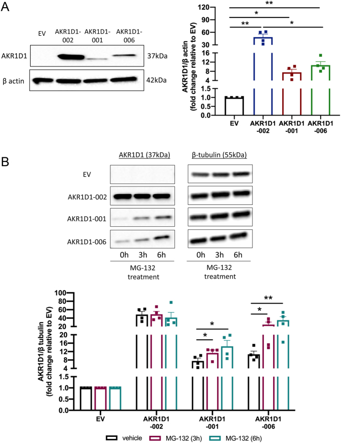
AKR1D1 overexpression was further confirmed at protein level by Western blotting. Although protein expression levels of all variants were detected – and despite the higher mRNA levels of AKR1D1-001 and -006, compared to AKR1D1-002 (Supplementary Fig. 2C, D and E) – protein expression levels of both truncated isoforms were significantly lower, compared to the full WT protein, indicative of rapid intracellular proteasomal degradation (Fig. 4A).
Figure 4.
(A) Protein expression levels of AKR1D1-002, AKR1D1-001 and AKR1D1-006, following overexpression in HEK293 cells, as measured by Western blotting. (B) Protein expression levels of AKR1D1-002, AKR1D1-001 and AKR1D1-006 following overexpression in HEK293 cells for 48 h, and subsequent treatment with either DMSO (vehicle, black bars) or 20 µM MG-132 (proteasomal inhibitor) for 3 h (maroon bars) and 6 h (teal bars). Representative Western blotting images are shown from one biological replicate, however, formal quantification was performed in n = 4 replicates. Data are presented as mean ± s.e. *P < 0.05, **P < 0.01, compared to empty vector (EV) transfected controls.
To investigate the hypothesis that truncated AKR1D1-SVs undergo proteasomal degradation, HEK293 cells were transfected with either EV, AKR1D1-002, AKR1D1-001, or AKR1D1-006 containing vectors and treated with the proteasome inhibitor, MG-132 (20 μM) for 3 or 6 h, as previously described (Chui et al. 2019). Confirming our hypothesis, MG-132 treatment significantly increased protein levels of both AKR1D1-001 and AKR1D1-006 in a time-dependent manner (Fig. 4B).
Truncated AKR1D1-SVs demonstrate distinct protein structures
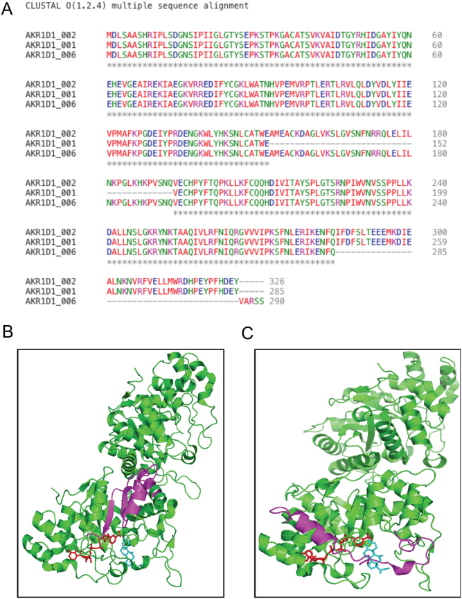
Following AKR1D1-SVs overexpression in in vitro human cell systems, multiple amino acid sequence alignments were conducted for the three variants (Clustal Omega) (Goujon et al. 2010, Sievers et al. 2011). The sequence alignments revealed that the AKR1D1-001 protein misses the amino acids 153–193, whilst the AKR1D1-006 protein lacks amino acids 286–326 and has an additional 5 amino acids at the C-terminus (Val, Ala, Arg, Ser, Ser) (Fig. 5A). Following that, in silico protein structure modelling was performed to predict the 3D structures of the truncated variants, using cortisone and NADP+ as substrate and co-factor, respectively (www.PyMol.org). Prediction modelling on AKR1D1-001 revealed that the deleted 153–193 amino acid region could disrupt the interaction between the nicotinamide head of the co-factor and likely hydride transfer to the A-ring of the steroid. In addition, the residues S166 and N170, which bind the carboxamide side chain of the nicotinamide ring, would be absent from the structure (Fig. 5B). In contrast, prediction modelling for AKR1D1-006 demonstrated that the absence of exon 8 plus the addition of five amino acids at the C-terminus lead to the loss of the C-terminal flexible loop (amino acids 286–326), which borders the steroid channel, as well as loss of the last helix in the structure, and is predicted to have decreased affinity for steroid substrates (Fig. 5C).
Figure 5.
(A) Clustal Omega multiple alignment of the amino acid sequences of the human AKR1D1 splice variants -002, -001 and -006. (B) AKR1D1-001.NADP+.Cortisone Ternary Complex (PDB 3CMF). Magneta shows deletion of residues 153-193; NADP+ (red) and cortisone (blue). There are two monomers per asymmetric unit. (C) AKR1D1-006.NADP+.Cortisone Ternary Complex (PDB 3CMF). Magneta shows deletion of exon 8 (residues 285–326); NADP+ (red) and cortisone (blue). There are two monomers per asymmetric unit. Colour coding in figure A represents amino acid physicochemical properties: RED: Small (small + hydrophobic (aromatic)); BLUE: Acidic; MAGENTA: Basic – H; GREEN: Hydroxyl + sulfhydryl + amine + G; ✶ positions which have a single, fully conserved residue. B and C made in PyMol.
Truncated AKR1D1-SVs differentially regulate glucocorticoid metabolism in vitro
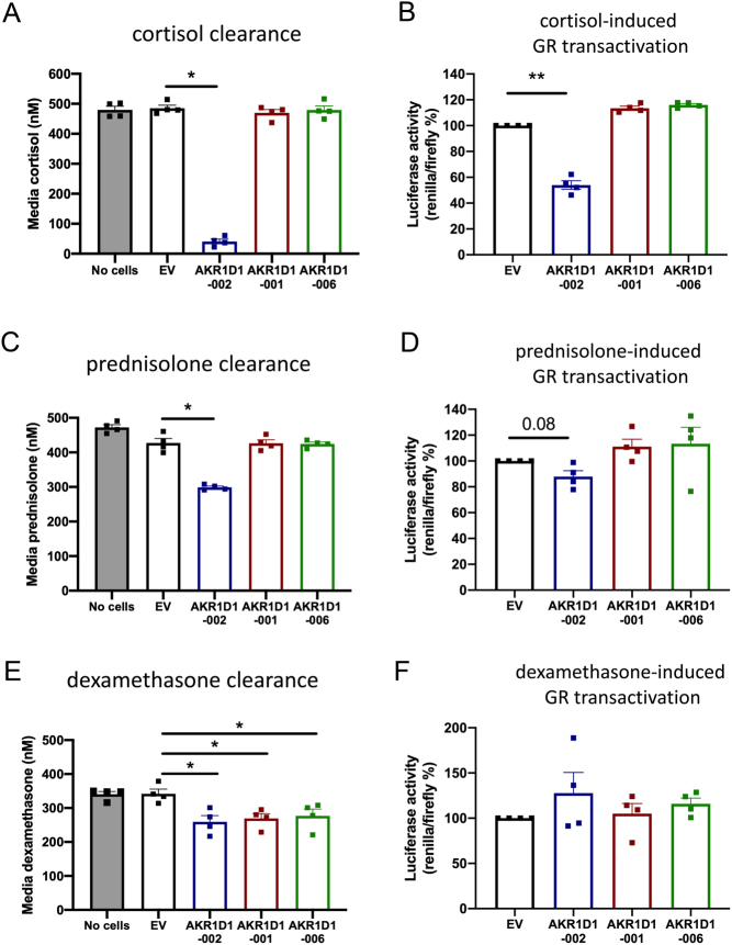
To investigate the functional role of truncated AKR1D1 variants in glucocorticoid metabolism, HEK293 cells were transfected with either EV, AKR1D1-002, AKR1D1-001, or AKR1D1-006 containing vectors and treated with cortisol, dexamethasone or prednisolone. Amongst the three variants, only AKR1D1-002 was able to metabolise cortisol and prednisolone, with concomitant decreases in GR activation (Fig. 6A, B, C and D). While, all AKR1D1-SVs reduced cell media dexamethasone concentrations, no differences in GR activation were observed (Fig. 6E and F). There was no change in cell media concentrations or GR activation with any of the glucocorticoid treatments in the presence of EV.
Figure 6.
(A) Cortisol, (C) prednisolone, and (E) dexamethasone clearance in AKR1D1-002 (blue bar), AKR1D1-001 (red bar), AKR1D1-006 (green bar) or empty vector (EV, black bar) transfected HEK293 cells (after 24 h of steroid treatment, 500 nM). (B, D and F) Glucocorticoid (GR) receptor transactivation in AKR1D1-002 (blue bar), AKR1D1-001 (red bar), AKR1D1-006 (green bar) overexpressing and empty vector (EV, black bar) transfected HEK293 cells after 24 h of (B) cortisol, (D) prednisolone, and (F) dexamethasone treatment (500 nM). Data are presented as mean ± s.e. of n = 4 experiments, performed in duplicate, *P < 0.05, **P < 0.01, compared to empty vector (EV) transfected controls.
Truncated AKR1D1-SVs differentially regulate androgen metabolism in vitro
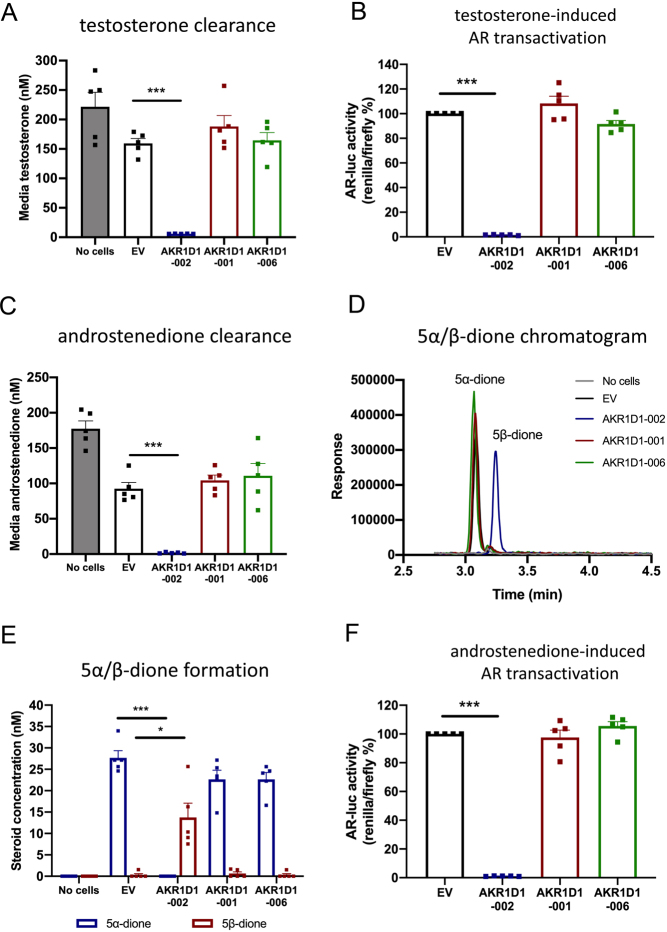
AKR1D1 is reported to have a crucial role in the regulation of androgen availability (Charbonneau & The 2001). In our study, testosterone was completely metabolised after 24 h of treatment in AKR1D1-002-transfected HEK293 cells. However, no reduction in media testosterone levels was observed in cells expressing either AKR1D1-001 or AKR1D1-006. Consistent with this data, AKR1D1-002 overexpression significantly reduced testosterone-mediated AR activation compared to EV-transfected cells, but neither AKR1D1-001 nor AKR1D1-006 had any impact on activation of the receptor (Fig. 7B).
Figure 7.
(A) Testosterone clearance in AKR1D1-002 (blue bar), AKR1D1-001 (red bar), AKR1D1-006 (green bar) or empty vector- (EV, black bar) transfected HEK293 cells after 24 h of testosterone treatment (200 nM). (B) Testosterone-induced androgen receptor (AR) activation in AKR1D1-002 (blue bar), AKR1D1-001 (red bar), AKR1D1-006 (green bar) overexpressing and empty vector (EV, black bar) transfected HEK293 cells after 24 h of testosterone treatment (200 nM). (C) Androstenedione clearance in AKR1D1-002 (blue bar), AKR1D1-001 (red bar), AKR1D1-006 (green bar) or empty vector- (EV, black bar) transfected HEK293 cells after 24 h of androstenedione treatment (200 nM). (D) Representative UHPLC-MS/MS chromatogram of 5α- and 5β-androstanedione in empty vector (EV, black line), AKR1D1-002 (blue line), AKR1D1-001 (red line) and AKR1D1-006- (green line) transfected HEK293 cells after 24 h of androstenedione treatment (200 nM). (E) Media 5α- and 5β-androstanedione levels in empty vector (EV, black line), AKR1D1-002 (blue line), AKR1D1-001 (red line) and AKR1D1-006- (green line) transfected HEK293 cells after 24 h of androstenedione treatment (200 nM). (F) Androstenedione-induced androgen receptor (AR) activation in AKR1D1-002 (blue bar), AKR1D1-001 (red bar), AKR1D1-006 (green bar) overexpressing and empty vector (EV, black bar) transfected HEK293 cells after 24 h of androstenedione treatment (200 nM). Data are presented as mean ± s.e. of n = 5 experiments, performed in duplicate, *P < 0.05, **P < 0.01, ***P < 0.001 compared to empty vector (EV) transfected controls.
Similarly, when cells were treated with androstenedione, AKR1D1-002 overexpression resulted in complete media androstenedione clearance, with a parallel increase in 5β-androstanedione formation (5β-reduced form of androstenedione), compared to EV-transfected cells (Fig. 7C, D and E). No differences in either androstenedione clearance or 5β-androstanedione formation were observed following AKR1D1-001 and AKR1D1-006 overexpression (Fig. 7C, D and E). Interestingly, in all androstenedione-treated cell models (with the exception of AKR1D1-002), we also detected low levels of 5α-androstanedione, indicative of endogenous SRD5A activity (Fig. 7C, D and E). Finally, and in line with this data, AKR1D1-002 significantly reduced androstenedione-mediated AR activation, compared to EV-transfected cells, but neither AKR1D1-001 nor AKR1D1-006 had any impact on receptor activation (Fig. 7F).
Discussion
This study provides the first evidence of the functional role of the three AKR1D1-SVs in glucocorticoid and androgen clearance. We show that AKR1D1-001 and AKR1D1-002 are expressed in human liver biopsies and liver cell lines, whilst AKR1D1-006 is the predominant variant, and expressed only, in human testes. We demonstrate that, similar to AKR1D1-002, the truncated AKR1D1-001 and AKR1D1-006 proteins metabolise dexamethasone (albeit poorly) in vitro, but neither of the truncated AKR1D1 proteins metabolise cortisol or prednisolone. Similarly, we show that, amongst the three variants, only AKR1D1-002 metabolises the androgen testosterone or the androgen precursor androstenedione. Finally, we show that truncated AKR1D1-SVs undergo rapid intracellular proteasomal degradation.
Examination of the predicted structures of the truncated AKR1D1-SVs suggest an effect on function. The exon 5 omitted in AKR1D1-001 does not cause a frameshift in the protein; a previous study from Barski et al. (2013) suggested that, as residues in the middle of the protein sequence are missing, this protein may be structurally compromised, leading to improper folding. Notably, the loss of exon 5 will also interfere with the reaction trajectory because S166 and N167 are absent and these stabilise the carboxamide side-chain of the nicotinamide ring (Penning et al. 2019). In contrast, AKR1D1-006, which omits exon 8, does cause a frameshift in the protein and misses residues that close over the steroid channel; thus, it is predicted to decrease affinity for steroid substrates (Barski et al. 2013). These differences in protein structure are shown in cartoon form in Fig. 5B. It is possible that the differences we observed in steroid clearance between the long AKR1D1-002 and the shorter AKR1D1-001/AKR1D1-006 SVs may reflect the structural disruption in the truncated proteins (thus a lower affinity for steroid hormones), or potentially decreased protein stability.
Protein stability has recently been explored in disease-related AKR1D1 mutations (Drury et al. 2010). Missense mutations in AKR1D1 have been associated with inherited 5β-reductase and bile acid deficiency, including Leu106Phe, Pro133Arg, Pro198Leu, Gly223Glu, and Arg261Cys1. Following expression in HEK293 cells, these AKR1D1 mutants showed significantly lower protein expression levels than WT AKR1D1, despite equal mRNA expression. Analysis of protein degradation rate using a protein synthesis inhibitor, cycloheximide, suggested the mutations impaired protein folding and stability (Drury et al. 2010). Importantly, the mutants retained some 5β-reductase activity (via detection of 5β-reduced testosterone) despite 100-fold lower expression, indicating the disease phenotypes may not be caused by defects in enzymatic properties, but rather by reduced expression of active AKR1D1 protein.
Similarly, in our study, and despite high mRNA expression, protein levels of both AKR1D1-001 and AKR1D1-006 were significantly lower than those of AKR1D1-002, and proteasomal inhibition treatment partially restored truncated AKR1D1 protein levels. As the ubiquitin-proteasome pathway regulates digestion of misfolded or damaged polypeptides in the cell (Goldberg 2003), it is plausible that omitting exons 5 or 8 in the AKR1D1 transcripts results in improper post-translational protein folding, therefore degrading the truncated AKR1D1 proteins.
AKR1D1 is the first member of the 1D subfamily, along with all known mammalian 5β-reductases, including the rat (AKR1D2), the rabbit (AKR1D3) and the mouse (AKR1D4) homologs (Onishi et al. 1991, Faucher et al. 2008, Chen et al. 2019). Amongst these, two mouse AKR1D4-SVs, AKR1D4L and AKR1D4S, have been recently characterised. Both mouse transcripts were expressed in mouse hepatic and testicular tissues, and enzymatic kinetic assays revealed their ability to metabolise cortisol, progesterone and androstenedione to their 5β-reduced metabolites (Chen et al. 2019). Interestingly, they also displayed lower 3α-hydroxysteroid dehydrogenase activity; however, this was limited to C19 steroids only (Chen et al. 2019). In our study, neither of the truncated human AKR1D1-SVs metabolised cortisol, testosterone or androstenedione, adding evidence to the presence of distinct functional differences between the human and murine 5β-reductases.
Glucocorticoids are lipophilic molecules that undergo a variety of metabolic conversions to increase their water solubility and enable efficient renal excretion; however, synthetic glucocorticoid clearance has only been examined in a limited number of studies. CYP3A isoforms drive dexamethasone clearance through the formation of 6-hydroxylated metabolites in vitro (Gentile et al. 1996, Tomlinson et al. 1997a,b). In a clinical study, urinary steroid metabolome analysis of samples from healthy male volunteers, following prednisolone administration, identified 20 different prednisolone metabolites, including 11-hydroxylated, 20-reduced and 5α/β-reduced products (Matabosch et al. 2015). Supporting these findings, we have recently demonstrated the ability of AKR1D1-002 to clear prednisolone and dexamethasone, with concomitant decreases in hepatic GR activation (Nikolaou et al. 2019a, 2020). We now show that, in addition to AKR1D1-002, AKR1D1-001 and AKR1D1-006 metabolise dexamethasone (albeit poorly) in vitro, but this poor clearance did not result in any significant differences in GR activation. This finding was not surprising, as levels of dexamethasone remained at high levels in our cell system, and the higher potency of dexamethasone, compared to prednisolone and cortisol, is well described (Nebesio et al. 2016). It is of interest that, despite the higher protein levels of AKR1D1-002, all three variants resulted in a similar decrease in dexamethasone availability, raising the possibility for the truncated isoforms to have an even more potent role in dexamethasone clearance. Considering, however, the reduced protein stability of AKR1D1-001 and AKR1D1-006, the role of these truncated AKR1D1-SVs in dexamethasone clearance is likely to be minimal.
Up to 3% of the UK population are prescribed glucocorticoids therapeutically (predominantly prednisolone and dexamethasone), for the suppression of inflammation in chronic inflammatory diseases, including rheumatoid arthritis, inflammatory bowel disease and asthma (Barnes 1998, van Staa et al. 2000, van der Goes et al. 2014, Vandewalle et al. 2018). In addition, they are used in combination with anti-cancer agents to reduce the genotoxic side effects of chemotherapy treatment, including nausea and vomiting (Collins et al. 2007, Buxant et al. 2015). Prolonged use of synthetic glucocorticoids is, however, associated with adverse metabolic effects including obesity, insulin resistance, and non-alcoholic fatty liver disease (NAFLD) (Woods et al. 2015), and decreased AKR1D1 expression has recently been shown in patients with type 2 diabetes and NAFLD (Valanejad et al. 2018, Nikolaou et al. 2019b). Our study indicates that AKR1D1-002 is the predominant functional variant in human liver; alterations in AKR1D1-002 expression and activity may therefore contribute to the adverse metabolic impact of exogenous glucocorticoids. Nevertheless, the potential additional role of truncated AKR1D1-SVs cannot be excluded.
Testosterone is the predominant androgen in males, and is synthesised in the Leydig cells in the testis from the precursors dehydroepiandrosterone (DHEA) and androstenedione (Preslock 1980). Following release into the circulation, the majority of testosterone metabolism (90–95%) to produce inactive androgen metabolites occurs in the liver. AKR1D1-002 is the predominant protein expressed in human liver, with studies showing the ability of this variant to metabolise testosterone and androstenedione in both cell-free and in vitro cell systems (Kondo et al. 1994, Chen et al. 2011, Barnard et al. 2020). Consistent with these reports, we showed that AKR1D1-002 potently cleared testosterone and androstenedione, with downstream decreases in AR activation; nonetheless, neither AKR1D1-001 nor AKR1D1-006 metabolised either of the two androgens. Importantly, in the AKR1D1-002-transfected cells, we were also able to measure the formation of 5β-androstanedione; however, 5β-reduced products do not ionise well on mass spectrometry, and these measurements did not reflect the full effect of AKR1D1-002 on product generation. Finally, we demonstrated that, in the absence of AKR1D1-002, androstenedione-treated HEK293 cells possess low SRD5A activity, resulting in the formation of 5α-androstanedione. This data support previous work by Panter et al. (2005), who revealed endogenous expression and activity of SRD5A2 in the same cell line.
Chen et al. (2019) demonstrated low, but detectable levels of AKR1D4 (the murine homolog of AKR1D1) in male mouse testicular tissues, and we now show that AKR1D1 is expressed in human testes, with AKR1D1-006 the predominant transcript. Considering our data, which revealed no activity of AKR1D1-006 towards androgen metabolism, it is intriguing what the actual role of this protein may be. AKR1D1 has an additional, and rather potent, role in bile acid biosynthesis; it is, therefore, possible that this variant is involved in bile acid metabolism within the testes. Indeed, a recent study by Martinot et al. demonstrated detectable expression levels of bile acid synthesising enzymes and de novo bile acid biosynthesis in mouse testes (Martinot et al. 2017). Further studies are now warranted to clarify the exact role of AKR1D1-006 in human testicular cells.
Our study comes with limitations. Overexpression of AKR1D1 transcripts in HEK293 cells may not accurately reflect the physiological expression of the variants in vivo. In addition, despite the advantages this cell line offers regarding the presence of post-translational modification machineries, it remains a heterologous cell system and the high levels of gene expression may result in protein translation differences. Western blotting of human protein samples would help to determine the actual expression levels of AKR1D1 variants in the liver and the testes. Nonetheless, access to healthy human biopsies is limited, and currently, there are no specific AKR1D1 antibodies developed for either AKR1D1-001 or AKR1D1-006 proteins.
In conclusion, we have shown that of the three 5β-reductase (AKR1D1) transcript variants which translate into proteins, AKR1D1-002 and AKR1D1-001 are expressed in human liver, and only AKR1D1-006 is expressed in human testes. AKR1D1-002 metabolises endogenous and synthetic glucocorticoids as well as the androgen testosterone and its precursor androstenedione. AKR1D1-001 and AKR1D1-006 metabolise only dexamethasone, but both truncated proteins are intracellularly targeted for proteasome degradation. Additional questions now need to be addressed to dissect the broader role of these variants in translational clinical studies in vivo.
Supplementary Material
Declaration of interest
T M P is a consultant for Research Institute for Fragrance Materials, is a recipient of a sponsored research agreement from Forendo, and is founding director of Penzymes, LLC.
Funding
This work was supported by the Society for Endocrinology (Early Career Grant awarded to N N, Summer Studentship Grant awarded to N A); Medical Research Council, UK (programme grant awarded to J W T, ref. MR/P011462/1); NIHR Oxford Biomedical Research Centre (Principal investigator award to J W T), based at Oxford University Hospitals NHS Trust and University of Oxford; P30-ES013508 awarded to T M P by the National Institute of Environmental Health Sciences. The views expressed are those of the author(s) and not necessarily those of the NHS, the NIHR or the Department of Health or the National Institute of Environmental Health Sciences.
Author contribution statement
Conceptualisation, N N; Methodology, T M P, B K, K H S, N N; Investigation, N A, H G, E G, N J D, K M, S G, A A, T M P, N N; Writing – Original draft, N A, N N; Writing – Review and Editing, T M P, L L G, J W T, N N; Supervision, J W T, N N; Funding Acquisition, N A, J T W, N N.
References
- Amai K, Fukami T, Ichida H, Watanabe A, Nakano M, Watanabe K, Nakajima M.2020. Quantitative analysis of mRNA expression levels of aldo-keto reductase and short-chain dehydrogenase/reductase isoforms in human livers. Drug Metabolism and Pharmacokinetics 35 539–547. ( 10.1016/j.dmpk.2020.08.004) [DOI] [PubMed] [Google Scholar]
- Barnard L, Nikolaou N, Louw C, Schiffer L, Gibson H, Gilligan LC, Gangitano E, Snoep J, Arlt W, Tomlinson JWet al. 2020. The A-ring reduction of 11-ketotestosterone is efficiently catalysed by AKR1D1 and SR5A2 but not SRD5A1. Journal of Steroid Biochemistry and Molecular Biology 202 105724. ( 10.1016/j.jsbmb.2020.105724) [DOI] [PubMed] [Google Scholar]
- Barnes PJ.1998. Anti-inflammatory actions of glucocorticoids: molecular mechanisms. Clinical Science 94 557–572. ( 10.1042/cs0940557) [DOI] [PubMed] [Google Scholar]
- Barski OA, Mindnich R, Penning TM.2013. Alternative splicing in the aldo-keto reductase superfamily: implications for protein nomenclature. Chemico-Biological Interactions 202 153–158. ( 10.1016/j.cbi.2012.12.012) [DOI] [PMC free article] [PubMed] [Google Scholar]
- Buxant F, Kindt N, Laurent G, Noël JC, Saussez S.2015. Antiproliferative effect of dexamethasone in the MCF-7 breast cancer cell line. Molecular Medicine Reports 12 4051–4054. ( 10.3892/mmr.2015.3920) [DOI] [PMC free article] [PubMed] [Google Scholar]
- Charbonneau A, The VL.2001. Genomic organization of a human 5beta-reductase and its pseudogene and substrate selectivity of the expressed enzyme. Biochimica et Biophysica Acta 1517 228–235. ( 10.1016/S0167-4781(00)00278-5) [DOI] [PubMed] [Google Scholar]
- Chen M, Drury JE, Penning TM.2011. Substrate specificity and inhibitor analyses of human steroid 5β-reductase (AKR1D1). Steroids 76 484–490. ( 10.1016/j.steroids.2011.01.003) [DOI] [PMC free article] [PubMed] [Google Scholar]
- Chen M, Wangtrakuldee P, Zang T, Duan L, Gathercole LL, Tomlinson JW, Penning TM.2019. Human and murine steroid 5β-reductases (AKR1D1 and AKR1D4): insights into the role of the catalytic glutamic acid. Chemico-Biological Interactions 305 163–170. ( 10.1016/j.cbi.2019.03.025) [DOI] [PMC free article] [PubMed] [Google Scholar]
- Chui AJ, Okondo MC, Rao SD, Gai K, Griswold AR, Johnson DC, Ball DP, Taabazuing CY, Orth EL, Vittimberga BAet al. 2019. N-terminal degradation activates the NLRP1B inflammasome. Science 364 82–85. ( 10.1126/science.aau1208) [DOI] [PMC free article] [PubMed] [Google Scholar]
- Collins R, Fenwick E, Trowman R, Perard R, Norman G, Light K, Birtle A, Palmer S, Riemsma R.2007. A systematic review and economic model of the clinical effectiveness and cost-effectiveness of docetaxel in combination with prednisone or prednisolone for the treatment of hormone-refractory metastatic prostate cancer. Health Technology Assessment 11 iii–iv, xv. ( 10.3310/hta11020) [DOI] [PubMed] [Google Scholar]
- Drury JE, Mindnich R, Penning TM.2010. Characterization of disease-related 5beta-reductase (AKR1D1) mutations reveals their potential to cause bile acid deficiency. Journal of Biological Chemistry 285 24529–24537. ( 10.1074/jbc.M110.127779) [DOI] [PMC free article] [PubMed] [Google Scholar]
- Faucher F, Cantin L, Luu-The V, Labrie F, Breton R.2008. Crystal structures of human Δ4-3-ketosteroid 5β-reductase (AKR1D1) reveal the presence of an alternative binding site responsible for substrate inhibition. Biochemistry 47 13537–13546. ( 10.1021/bi801276h) [DOI] [PubMed] [Google Scholar]
- Gentile DM, Tomlinson ES, Maggs JL, Park BK, Back DJ.1996. Dexamethasone metabolism by human liver in vitro. Metabolite identification and inhibition of 6-hydroxylation. Journal of Pharmacology and Experimental Therapeutics 277 105–112. [PubMed] [Google Scholar]
- Goldberg AL.2003. Protein degradation and protection against misfolded or damaged proteins. Nature 426 895–899. ( 10.1038/nature02263) [DOI] [PubMed] [Google Scholar]
- Goujon M, McWilliam H, Li W, Valentin F, Squizzato S, Paern J, Lopez R.2010. A new bioinformatics analysis tools framework at EMBL-EBI. Nucleic Acids Research 38 W695–W699. ( 10.1093/nar/gkq313) [DOI] [PMC free article] [PubMed] [Google Scholar]
- Hawley JM, Owen LJ, Debono M, Newell-Price J, Keevil BG.2018. Development of a rapid liquid chromatography tandem mass spectrometry method for the quantitation of serum dexamethasone and its clinical verification. Annals of Clinical Biochemistry 55 665–672. ( 10.1177/0004563218766566) [DOI] [PubMed] [Google Scholar]
- Jez JM, Bennett MJ, Schlegel BP, Lewis M, Penning TM.1997. Comparative anatomy of the aldo-keto reductase superfamily. Biochemical Journal 326 625–636. ( 10.1042/bj3260625) [DOI] [PMC free article] [PubMed] [Google Scholar]
- Jin Y, Chen M, Penning TMM.2014. Rate of steroid double-bond reduction catalysed by the human steroid 5β-reductase (AKR1D1) is sensitive to steroid structure: implications for steroid metabolism and bile acid synthesis. Biochemical Journal 462 163–171. ( 10.1042/BJ20140220) [DOI] [PMC free article] [PubMed] [Google Scholar]
- Kondo KH, Kai MH, Setoguchi Y, Eggertsen G, Sjöblom P, Setoguchi T, Okuda KI, Björkhem I.1994. Cloning and expression of cDNA of human delta 4-3-oxosteroid 5 beta-reductase and substrate specificity of the expressed enzyme. European Journal of Biochemistry 219 357–363. ( 10.1111/j.1432-1033.1994.tb19947.x) [DOI] [PubMed] [Google Scholar]
- Martinot E, Baptissart M, Véga A, Sèdes L, Rouaisnel B, Vaz F, Saru JP, De Haze A, Baron S, Caira Fet al. 2017. Bile acid homeostasis controls CAR signaling pathways in mouse testis through FXRalpha. Scientific Reports 7 42182. ( 10.1038/srep42182) [DOI] [PMC free article] [PubMed] [Google Scholar]
- Matabosch X, Pozo OJ, Pérez-Mañá C, Papaseit E, Segura J, Ventura R.2015. Detection and characterization of prednisolone metabolites in human urine by LC-MS/MS. Journal of Mass Spectrometry 50 633–642. ( 10.1002/jms.3571) [DOI] [PubMed] [Google Scholar]
- Nasiri M, Nikolaou N, Parajes S, Krone NP, Valsamakis G, Mastorakos G, Hughes B, Taylor A, Bujalska IJ, Gathercole LLet al. 2015. 5α-Reductase type 2 regulates glucocorticoid action and metabolic phenotype in human hepatocytes. Endocrinology 156 2863–2871. ( 10.1210/en.2015-1149) [DOI] [PMC free article] [PubMed] [Google Scholar]
- Nebesio TD, Renbarger JL, Nabhan ZM, Ross SE, Slaven JE, Li L, Walvoord EC, Eugster EA.2016. Differential effects of hydrocortisone, prednisone, and dexamethasone on hormonal and pharmacokinetic profiles: a pilot study in children with congenital adrenal hyperplasia. International Journal of Pediatric Endocrinology 2016 17. ( 10.1186/s13633-016-0035-5) [DOI] [PMC free article] [PubMed] [Google Scholar]
- Nikolaou N, Gathercole LL, Kirkwood L, Dunford JE, Hughes BA, Gilligan LC, Oppermann U, Penning TM, Arlt W, Hodson Let al. 2019a. AKR1D1 regulates glucocorticoid availability and glucocorticoid receptor activation in human hepatoma cells. Journal of Steroid Biochemistry and Molecular Biology 189 218–227. ( 10.1016/j.jsbmb.2019.02.002) [DOI] [PMC free article] [PubMed] [Google Scholar]
- Nikolaou N, Gathercole LL, Marchand L, Althari S, Dempster NJ, Green CJ, van de Bunt M, McNeil C, Arvaniti A, Hughes BAet al. 2019b. AKR1D1 is a novel regulator of metabolic phenotype in human hepatocytes and is dysregulated in non-alcoholic fatty liver disease. Metabolism: Clinical and Experimental 99 67–80. ( 10.1016/j.metabol.2019.153947) [DOI] [PMC free article] [PubMed] [Google Scholar]
- Nikolaou N, Arvaniti A, Appanna N, Sharp A, Hughes BA, Digweed D, Whitaker MJ, Ross R, Arlt W, Penning TMet al. 2020. Glucocorticoids regulate AKR1D1 activity in human liver in vitro and in vivo. Journal of Endocrinology 245 207–218. ( 10.1530/JOE-19-0473) [DOI] [PMC free article] [PubMed] [Google Scholar]
- Onishi Y, Noshiro M, Shimosato T, Okuda K.1991. Molecular cloning and sequence analysis of cDNA encoding δ4-3-ketosteroid 5β-reductase of rat liver. FEBS Letters 283 215–218. ( 10.1016/0014-5793(91)80591-P) [DOI] [PubMed] [Google Scholar]
- Owen LJ, Gillingwater S, Keevil BG.2005. Prednisolone measurement in human serum using liquid chromatography tandem mass spectrometry. Annals of Clinical Biochemistry 42 105–111. ( 10.1258/0004563053492810) [DOI] [PubMed] [Google Scholar]
- Palermo M, Marazzi MG, Hughes BA, Stewart PM, Clayton PT, Shackleton CHL.2008. Human Δ4-3-oxosteroid 5β-reductase (AKR1D1) deficiency and steroid metabolism. Steroids 73 417–423. ( 10.1016/j.steroids.2007.12.001) [DOI] [PubMed] [Google Scholar]
- Panter BU, Jose J, Hartmann RW.2005. 5α-Reductase in human embryonic kidney cell line HEK293: evidence for type II enzyme expression and activity. Molecular and Cellular Biochemistry 270 201–208. ( 10.1007/s11010-005-4508-8) [DOI] [PubMed] [Google Scholar]
- Penning TM, Wangtrakuldee P, Auchus RJ.2019. Structural and functional biology of aldo-keto reductase steroid-transforming enzymes. Endocrine Reviews 40 447–475. ( 10.1210/er.2018-00089) [DOI] [PMC free article] [PubMed] [Google Scholar]
- Pfaffl MW.2001. A new mathematical model for relative quantification in real-time RT-PCR. Nucleic Acids Research 29 e45. ( 10.1093/nar/29.9.e45) [DOI] [PMC free article] [PubMed] [Google Scholar]
- Pitteloud N, Mootha VK, Dwyer AA, Hardin M, Lee H, Eriksson KF, Tripathy D, Yialamas M, Groop L, Elahi Det al. 2005. Relationship between testosterone levels, insulin sensitivity, and mitochondrial function in men. Diabetes Care 28 1636–1642. ( 10.2337/diacare.28.7.1636) [DOI] [PubMed] [Google Scholar]
- Preslock JP.1980. Steroidogenesis in the mammalian testis. Endocrine Reviews 1 132–139. ( 10.1210/edrv-1-2-132) [DOI] [PubMed] [Google Scholar]
- Sievers F, Wilm A, Dineen D, Gibson TJ, Karplus K, Li W, Lopez R, McWilliam H, Remmert M, Söding Jet al. 2011. Fast, scalable generation of high-quality protein multiple sequence alignments using Clustal Omega. Molecular Systems Biology 7 539. ( 10.1038/msb.2011.75) [DOI] [PMC free article] [PubMed] [Google Scholar]
- Simons SS.2008. What goes on behind closed doors: physiological versus pharmacological steroid hormone actions. BioEssays 30 744–756. ( 10.1002/bies.20792) [DOI] [PMC free article] [PubMed] [Google Scholar]
- Tomlinson ES, Maggs JL, Park BK, Back DJ.1997a. Dexamethasone metabolism in vitro: species differences. Journal of Steroid Biochemistry and Molecular Biology 62 345–352. ( 10.1016/S0960-0760(97)00038-1) [DOI] [PubMed] [Google Scholar]
- Tomlinson ES, Lewis DFV, Maggs JL, Kroemer HK, Park BK, Back DJ.1997b. In vitro metabolism of dexamethasone (DEX) in human liver and kidney: the involvement of CYP3A4 and CYP17 (17,20 lyase) and molecular modelling studies. Biochemical Pharmacology 54 605–611. ( 10.1016/S0006-2952(97)00166-4) [DOI] [PubMed] [Google Scholar]
- Tomlinson JW, Finney J, Gay C, Hughes BA, Hughes SV, Stewart PM.2008. Impaired glucose tolerance and insulin resistance are associated with increased adipose 11β-hydroxysteroid dehydrogenase type 1 expression and elevated hepatic 5α-reductase activity. Diabetes 57 2652–2660. ( 10.2337/db08-0495) [DOI] [PMC free article] [PubMed] [Google Scholar]
- Valanejad L, Ghareeb M, Shiffka S, Nadolny C, Chen Y, Guo L, Verma R, You S, Akhlaghi F, Deng R.2018. Dysregulation of Δ4-3-oxosteroid 5β-reductase in diabetic patients: implications and mechanisms. Molecular and Cellular Endocrinology 470 127–141. ( 10.1016/j.mce.2017.10.005) [DOI] [PMC free article] [PubMed] [Google Scholar]
- van der Goes MC, Jacobs JW, Bijlsma JW.2014. The value of glucocorticoid co-therapy in different rheumatic diseases – positive and adverse effects. Arthritis Research and Therapy 16 (Supplement 2) S2. ( 10.1186/ar4686) [DOI] [PMC free article] [PubMed] [Google Scholar]
- van Staa TP, Leufkens HGM, Abenhaim L, Begaud B, Zhang B, Cooper C.2000. Use of oral corticosteroids in the United Kingdom. QJM 93 105–111. ( 10.1093/qjmed/93.2.105) [DOI] [PubMed] [Google Scholar]
- Vandewalle J, Luypaert A, De Bosscher K, Libert C.2018. Therapeutic mechanisms of glucocorticoids. Trends in Endocrinology and Metabolism 29 42–54. ( 10.1016/j.tem.2017.10.010) [DOI] [PubMed] [Google Scholar]
- Wang M.2005. The role of glucocorticoid action in the pathophysiology of the metabolic syndrome. Nutrition and Metabolism 2 3. ( 10.1186/1743-7075-2-3) [DOI] [PMC free article] [PubMed] [Google Scholar]
- Woods CP, Hazlehurst JM, Tomlinson JW.2015. Glucocorticoids and non-alcoholic fatty liver disease. Journal of Steroid Biochemistry and Molecular Biology 154 94–103. ( 10.1016/j.jsbmb.2015.07.020) [DOI] [PubMed] [Google Scholar]
Associated Data
This section collects any data citations, data availability statements, or supplementary materials included in this article.


 This work is licensed under a
