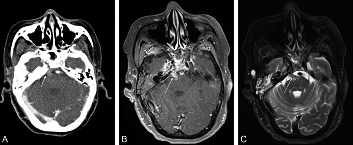
We report a case of perineural tumor spread (PTS) involving the abducens nerve in a 61-year-old Asian woman. The patient was previously treated for nasopharyngeal carcinoma and achieved a full clinical response. Two months following completion of chemotherapy, the patient developed a right abducens nerve palsy. She underwent both CT and MR imaging.
Enlargement and thickening of the right abducens nerve was noted on both CT and MR imaging compatible with PTS along the sixth nerve. Retrograde tumor spread was noted to extend into the right aspect of the pons (Fig 1).
Fig 1.
Axial CT and MR images in a 61-year-old woman treated previously for nasopharyngeal carcinoma. A, Contrast-enhanced CT image shows abnormal thickening and mild enhancement along the right sixth cranial nerve (arrow). B, T1-weighted fat-saturated gadolinium-enhanced axial image through the level of the pons more clearly demonstrates the band of abnormal enhancement and thickening extending from the clivus into the right aspect of the pons (arrow). C, Fat-saturated axial T2-weighted image also shows the lesion to be of heterogeneous internal signal intensity with clear extension into the pons (arrow).
PTS is a well-known phenomenon that occurs predominantly in malignancies of the head and neck, whereby tumor extends along the nerves away from the primary site of malignancy. Such tumor spread occurs in 2.5%–5% of patients with head and neck cancer, and most often progresses in a retrograde direction toward the central nervous system.1,2 Malignancies most often associated with PTS include squamous cell carcinoma, adenoid cystic carcinoma, and lymphoma, but any head and neck malignancy can spread via this mechanism.2 The second and third divisions of the trigeminal nerve and the facial nerve are the most commonly involved cranial nerves. Such disease can produce clinical symptoms of pain, paresthesia, numbness, formication, and motor weakness.2,3 PTS may also be asymptomatic and may produce very subtle findings on imaging and, therefore, can be easily missed by radiologists. The detection of such findings is critical because the presence of PTS leads to a poorer prognosis and will alter the course of therapy. Failure to diagnose PTS may have morbid implications if the mode of therapy fails to address the disease along the affected nerves.2 Our case is unique in that there had been no previous reports of isolated PTS along the abducens nerve in the literature, to our knowledge.
Sixth nerve palsy is the most common of the isolated ocular motor nerve palsies.4 The condition often presents with binocular horizontal diplopia being worse in the gaze direction of the paretic lateral rectus muscle. Examination reveals ipsilateral abduction deficit and a primary position esotropia that is worse in gazing toward the paretic muscle.8 When patients with cancer present with such findings, one would not usually consider PTS. However, radiologists should be aware of the possibility of perineural spread along the sixth nerve because the detection is associated with a poorer prognosis and increased risk for recurrence. PTS is generally considered an incurable disease. Surgical excision and adjuvant radiation therapy are the available treatment options. The mechanism of spread from the primary malignancy to the abducens nerve in our case was likely a manifestation of disease that had initially infiltrated the cavernous sinus with subsequent tracking along the sixth nerve.
In conclusion, one can argue that the search for PTS is one of the most important tasks of radiologists when examining a patient with head and neck carcinoma. The discovery of PTS drastically hinders the chance of surgical cure and adversely affects long-term prognosis. This report highlights an unusual pattern of PTS along the abducens nerve.
References
- 1. Maroldi R, Farina D, Borghesi A, et al. Perineural tumor spread. Neuroimaging Clin N Am 2008; 18: 413–29 [DOI] [PubMed] [Google Scholar]
- 2. Nemec ST, Herneth AM, Czerny C. Perineural tumor spread in malignant head and neck tumors. Top Magn Reson Imaging 2007; 18: 467–71 [DOI] [PubMed] [Google Scholar]
- 3. Warden KF, Parmar H, Trobe JD. Perineural spread of cancer along the three trigeminal divisions. J Neuroophthalmol 2009; 29: 300–07 [DOI] [PubMed] [Google Scholar]
- 4. Ayberk G, Ozveren MF, Yildirim T, et al. Review of a series with abducens nerve palsy. Turk Neurosurg 2008; 18: 355–73 [PubMed] [Google Scholar]


