Abstract
BACKGROUND AND PURPOSE:
A number of flow-diverting devices have become available for endovascular occlusion of cerebral aneurysms. This article reports immediate and midterm results in recently ruptured aneurysms treated with the PED.
MATERIALS AND METHODS:
A prospective registry was established at 3 Australian neurointerventional sites to collect data on ruptured and unruptured aneurysms treated with PED during a 12-month period from August 2009. From this data base of 65 patients, 11 cases of recent aneurysmal SAH were examined. Relevant data including antiplatelet therapy, technical issues, complications, and imaging findings during at least a 6-month period of follow-up were collected and analyzed.
RESULTS:
Eleven patients had acutely ruptured aneurysms with SAH. Clinical follow-up was available on all cases with imaging follow-up at 6 months in 9 patients. Two patients died from rebleeding during the acute illness. There was no other procedural or delayed significant symptomatic morbidity. Eight aneurysms were occluded with a single case of residual body filling.
CONCLUSIONS:
PED should be used in SAH with caution, reserved for suitable patients concomitantly treated with endosaccular coiling if possible.
Endovascular treatment has become a major technique for ruptured and unruptured aneurysms since publication of the International Subarachnoid Aneurysm Trial1 in 2002. Additional advances with the use of complex coils,2 balloon remodelling,3,4 “bioactive” coils,5–7 and stent-assisted coiling8–11 have occurred, facilitating treatment of wide-neck aneurysms. More recently, a number of flow-diverting devices have become available and are under evaluation.12–16
Published midterm results17–19 have demonstrated that treatment of wide-neck aneurysms with PED (Chestnut Medical Technologies, Menlo Park, California) reconstruction of the parent vessel is achieved safely. Acute treatment of difficult lesions such as blister aneurysms by using a stent or stent-assisted coiling, in the setting of SAH, has also been reported.20 The purpose of this report was to analyze early clinical experience with the PED in the setting of SAH.
Materials and Methods
Patient Population
This series represents a subset of a prospective case registry of all patients with lesions suitable for PED treated at 3 Australian neurointerventional centers between August 2009 and August 2010. Each case was reviewed by a multidisciplinary team; before general release of the PED, individual application for use of the PED on compassionate grounds was sought from hospital administration and the Therapeutic Goods and Services Administration. Written informed consent was obtained. The PED was only used in patients in whom other endovascular or surgical options were thought to carry higher morbidity and in 3 patients in whom endovascular therapy had failed in the same admission to control the aneurysm. Data were collected prospectively with respect to aneurysm morphology, symptoms, previous treatment, antiplatelet and anticoagulation regimen, and technical and clinical complications. Follow-up for at least 6 months evaluated occlusion, mass effect, delayed complications, ongoing antiplatelet therapy, and in-stent stenosis.
Antiplatelet and Anticoagulation Schedule
A variety of protocols was used depending on operator and clinical circumstances (Table). Most (7 cases) had a loading dose of 300–600 mg of clopidogrel and 300 mg of aspirin at the time of the procedure. One patient was on long-term clopidogrel for cardiac disease. One center had access to a point-of-care platelet inhibition unit and could prescribe additional clopidogrel in patients with measured inadequate platelet inhibition. Dual antiplatelet medication was prescribed for 6 months in the anterior circulation and for ≤12 months in the posterior circulation. Patients were monitored for clopidogrel compliance by direct questioning. Aspirin therapy was intended to be lifelong. All patients had procedural intravenous heparin with an activated clotting time of >200. Nine of 11 patients had heparin infusion for at least 24 hours postprocedure (activated partial thromboplastin time, ×2 normal).
Patient, aneurysm, and treatment summary characteristics
| Patient | Sex/ Age (yr) | WFNS Grade at Time of PED | Days after SAH at Time of PED Placement | Aneurysm Site | Type | Sac Size (mm) | Previous Treatment | No. of PEDs | Additional Coils at Time of PED | Clopidogrel/Aspirin (mg) Loading | mRSa | Aneurysm Occluded (at 6 mo) |
|---|---|---|---|---|---|---|---|---|---|---|---|---|
| 1 | M/48 | 1 | 24 | Basilar trunk | B | 2 | Nil | 2 | No | 600/300 | 0 | Yes |
| 2 | F/56 | 2 | 2 | Superior hypophyseal | S | 21 | Nil | 3 | No | 600/300 | 6 | Died |
| 3 | M/64 | 1 | 4 | Basilar tip/P1 | S | 12 | Nil | 1 | Yes | 600/300 | 0 | Yes |
| 4 | F/49 | 1 | 21 | Superior hypophyseal | S | 10 | Nil | 1 | Yes | 300/300 | 0 | Yes |
| 5 | F/52 | 2 | 11 | P1/P2 | B | 12 | Coils and stent (day 2) | 1 | Yes | 300/300 | 2 | Filling |
| 6 | M/51 | 3 | 26 | Vertebral | B | 2 | Nil | 1 | No | 450/150 | 2 | Yes |
| 7 | M/56 | 2 | 1 | Basilar trunk | F | 21 | Nil | 2 | Yes | Long-term daily 75/100 | 1 | Yes |
| 8 | F/50 | 1 | 16 | PcomA | F | 12 | Nil | 2 | No | 75/150 5-Day loading | 0 | Yes |
| 9 | F/37 | 5 | 1 | Dorsal paraclinoid ICA | F | 34 | Nil | 2 | No | 300/300 | 6 | Died |
| 10 | F/64 | 4 | 8b | Dorsal paraclinoid ICA | B | 2 | Coils and stent (day 1) | 1 | Yes | 75/150 7 Days | 4 | Yes |
| 11 | F/41 | 1 | 21 | P1/P2 | B | 6 | Coils (day 1) | 1 | No | 75/150 5-Day loading | 0 | Yes |
Note:—B indicates blister; F, fusiform; S, saccular.
mRS at 1 month posttreatment.
Bleed day 1 and second bleed on day 8.
Procedure
Therapy was undertaken with the patient under general anesthesia. All PEDs were deployed through a Marksman (ev3, Irvine, California) 2.8F microcatheter. Additional PEDs were deployed at the discretion of the operator and were placed if there was an ongoing jet or inadequate neck coverage. Ideally, stasis and a contrast/blood layer were observed at the cessation of the procedure. Where possible, coils were deployed in the aneurysm before PED deployment or after “jailing” a microcatheter.
Discharge and Follow-Up
Clinical follow-up was performed at 1 and 6 months, in addition to independent evaluation by a neurosurgeon. Patients were also seen more regularly if they had complex problems. A 6-month control angiogram was performed and reviewed by 2 interventional neuroradiologists. Further angiography was performed if the aneurysm was open or in-construct narrowing was present.
Study End Points
The primary end point was development of complications leading to patient morbimortality from the time of PED placement to 6 months. All clinical incidents (TIA, stroke, SAH, or mass effect) in the first 6 months posttreatment were documented. The secondary end point was the angiographic appearance at 6 months with assessment of aneurysm closure and parent vessel stenosis. Extended follow-up was undertaken beyond 6 months in cases with nonocclusion of aneurysm or in-stent stenosis.
Results
Patient and Aneurysm Characteristics
Data on 65 patients were collected between August 2009 and August 2010. From this cohort, 11 cases of acute SAH were identified. Six patients were treated between day 1 and 14 post-SAH. Five others were treated between day 15 and 26. Details of the clinical status, aneurysm morphology, and therapy of patients are summarized in the Table. There were 7 women and 4 men with age range of 41–69 years.
Aneurysm morphology, treatment, and outcome are documented in the Table. There were 8 fusiform and 3 saccular aneurysms in the cohort of 11 cases. Of the fusiform aneurysms, 5 were blister or posterior circulation dissecting blisterlike type (1 ICA, 1 basilar trunk, 1 vertebral artery, and 2 P1/P2). These showed typical features of small irregular lesions with poorly defined wide necks and angiographic evidence of growth and morphologic instability with time. Three of these had failed previous endovascular treatment (2 stent/coil, 1 coils only) during the same admission.
Treatment and Procedural Outcomes
Six patients received coils as well as PEDs during their cumulative treatment. Of the 5 who received only a PED in their treatment, 2 had dissecting blisterlike lesions with no sac to hold the coils and 1 was treated in a delayed fashion (16 days postictus). Twenty-one-millimeter (patient 2) and 34-mm (patient 9) aneurysms were treated acutely with PED only. Patient 2 was intended to have PED/coiling (Fig 1). Both died of aneurysm rerupture during their acute admission.
Fig 1.
A 56-year-old patient 2 with a poorly opened PED requiring angioplasty. All images (unless stated) are left ICA angiograms in the lateral projection. Images A−E are day 2 after SAH. A, Subtracted image of 21-mm superior hypophyseal aneurysm. B, Nonsubtracted image demonstrates the PED in the ICA with a delivery catheter tip (black arrowhead) and jailed microcatheter tip (white arrowhead) in the aneurysm with the intention of coiling the aneurysm after PED deployment. C, Subtracted image after 2 PEDs were implanted. Despite sequential removal of both microcatheters, there is ongoing marked stenosis of the ICA with no aneurysm filling. D, Nonsubtracted image demonstrates an angioplasty balloon within the PED. Note contrast stasis in the sac. E, Subtraction image postangioplasty documents a normal-caliber ICA with a PED now fully open. Minimal dependent filling of the aneurysm (small black arrows) without discernible inflow jet is seen. Because the jailed microcatheter was removed before angioplasty, coiling of the sac is not possible. The patient did well but developed symptomatic vasospasm and right upper limb weakness and dysphasia day 8 post-SAH. Because she was being prepared for angiography with a view to angioplasty, the aneurysm reruptured and she became obtunded, requiring intubation. CT (not shown) demonstrated increased SAH, and emergency angiography was immediately performed. F, Anteroposterior subtracted DSA of the left ICA demonstrates severe M1 vasospasm. G and H, Lateral unsubtracted DSA (G) and lateral subtracted DSA (H) demonstrate aneurysm refilling with a new rupture point locule (black arrow). Balloon angioplasty and deployment of an additional PED (not shown) was performed, but the patient remained in poor condition and died 3 days later.
Acute Procedural Technical and Clinical Complications
A poorly opened PED requiring angioplasty occurred in 1 patient, patient 2 (Fig 1); 2 PEDs were deployed with an Echelon-10 microcatheter (ev3) jailed in the aneurysm sac with the expressed intent of coiling the sac after PED deployment. However, after deployment of 2 PEDs, there was poor flow in the carotid siphon, causing concerns of raised intrasaccular pressure, despite no demonstrable aneurysm filling. The microcatheters were sequentially removed to determine if they were the cause of diminished flow. This proved not to be the case, and angioplasty of the PED was performed, which restored flow in the parent vessel. The coiling opportunity had been lost because the jailed catheter had been removed. Minimal clot on the surface of the PED was demonstrated with no evidence of distal embolism. The clot was lysed with 3 mg of intra-arterial abciximab. The patient awoke intact and unchanged in condition.
Two patients experienced acute aneurysm rerupture and died. One of these, patient 2, developed upper limb weakness on day 8 and elevated transcranial Doppler velocities. The patient was emergently prepared for angiography but had a rapid decline in consciousness on the way to the angiography suite. CT demonstrated additional SAH, and severe vasospasm of the M1 segment (distal to the treated aneurysm) was documented on angiography (Fig 1), potentially pressurizing the sac. At this time, balloon angioplasty of the M1 was performed, and a third PED was deployed. The patient died 3 days later.
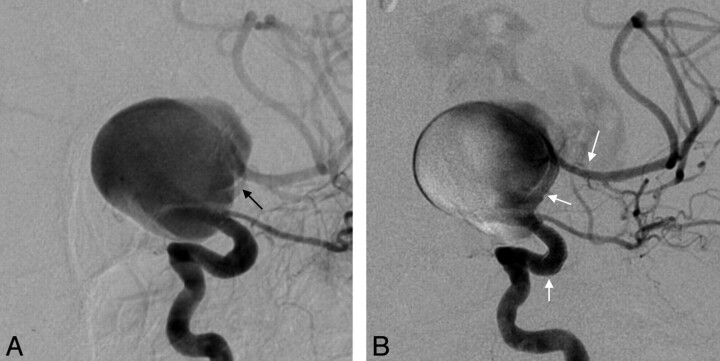
Patient 9, WFNS grade 5, had a fusiform 34-mm paraclinoid carotid aneurysm with no normal parent vessel lumen at the aneurysm interface (Fig 2). On close inspection, there was some smooth narrowing of the parent vessel distal to the neck, possibly due to extrinsic compression. Two PEDs were telescoped across the aneurysm, and the aneurysm re-ruptured immediately after deployment of the second device. Active treatment was withdrawn, and the patient died during the next 24 hours.
Fig 2.
Acute aneurysm rupture on PED deployment. Patient 9 presented with SAH, WFNS grade 5, and a 34-mm fusiform paraclinoid aneurysm treated on day 1 with 2 PEDs. A, Anteroposterior oblique left ICA DSA demonstrates the same narrowing of the terminal ICA (black arrow) distal to the fusiform aneurysm neck. B, Two PEDs have been deployed (white arrows outlining the proximal PED, point of overlap of the 2 PEDs in the previously narrowed portion of the distal ICA, and the distal PED in the proximal MCA M1 segment). Note the aneurysm is rerupturing with contrast extravasation superior to the dome. The patient died the same day.
PED migration requiring additional treatment occurred in a single case (patient 1) with a 2-mm dissecting blisterlike aneurysm in the distal basilar trunk. A single PED migrated caudally for 5 minutes, uncovering the neck and necessitating placement of a second PED with no consequence. Migration was related to inadequate distal purchase, unsheathing the PED rather than pushing the device out of the catheter, and the tendency for the device to seek a larger diameter (“melon seed”).
A cerebellar bleed related to anticoagulation and a lacunar infarct occurred in the same patient (patient 3) who had a basilar tip/P1 fusiform aneurysm treated with coils and PED. The patient was confused on day 1 postprocedure. CT demonstrated a left cerebellar tonsillar 2-cm hematoma and a small 5-mm anterior right thalamic lacune. The patient settled within 48 hours with no morbidity and was discharged home 13 days post-SAH with an mRS score of 0 at 1 month.
Three patients failed initial endovascular therapy, which necessitated PED deployment as a salvage procedure during their admission for SAH.
Patient 5 presented with WFNS 4 with a fusiform dissecting blisterlike aneurysm of the left P1/P2 segment aneurysm (Fig 3), originally treated with a Solitare stent (ev3) and coils. Due to the nature of the aneurysm, MRA was performed on day 11, demonstrating a large recurrence. The patient was now WFNS grade 2 and was immediately treated with additional coils and PED placement.
Fig 3.
Nonoccluded blister aneurysm previously treated with stent/coil combination. Patient 5 presented with SAH and a blister aneurysm of the left P1/P0 segment, initially treated with a Solitare stent and coils. All images are subtracted DSA of the left vertebral artery in an anteroposterior transfacial projection. A, A 6-mm fusiform blister-type aneurysm projects inferiorly. Note the dilated basilar trunk. B, An aneurysm secured with coils passes through a nondetected Solitare stent. The black arrowhead designates the nondetached proximal marker of the stent. C, Enlarging fusiform blister aneurysm day 11 after MRA (not shown), performed the same, day demonstrates recurrence of the aneurysm. A PED has been deployed in the left P1 segment, across the aneurysm and into the basilar trunk. A microcatheter is jailed in the sac (small black arrow) precoiling. D, Postcoiling angiogram shows a secured aneurysm and good filling of left posterior cerebral artery. The patient developed vasospasm subsequently, which was treated with balloon angioplasty of the ICA and middle cerebral arteries bilaterally (not shown), and made a good recovery. E, Six-month angiogram shows some body filling contained within the coils. Note that the proximal margin of the PED (small white arrow) is proximal to the proximal marker of the stent (black arrowhead). The distal marker of the stent within the P1/P2 segment junction is also shown (black arrowhead). The patient's clopidogrel was stopped as a consequence of the change, but aspirin was continued. F, Ten-month angiogram shows a stable appearance. No action has been taken thus far, and a follow-up angiogram in 12 months is scheduled because there is a reluctance to overlap the PED in the basilar tip and trunk.
Patient 11 presented with a WFNS grade 1 SAH and a 7-mm dissecting blisterlike aneurysm at the left P1/P2 junction. The aneurysm was treated on day 1 with coil occlusion, resulting in a residual 1-mm neck. Due to the etiology of the aneurysm, a check angiogram was obtained at day 7 demonstrating an acute 6-mm recurrence. A decision was made to wait before treating the recurrence with PED. This occurred without incident on day 21 post-SAH.
Patient 10 presented with a WFNS grade 3 SAH due to a blister aneurysm of the right dorsal paraclinoid ICA. This was treated on the day 1 with 2 coils and a Solitaire stent. A check angiogram was obtained at day 7 to exclude early regrowth, and the aneurysm remained secure. On day 8, the patient rebled and deteriorated to WFNS grade 4. Angiography confirmed a 2-mm recurrence of the aneurysm proximal and superior to the existing coils. A microcatheter was placed in the recurrent aneurysm through the stent, and 2 additional coils were deployed. This procedure was followed immediately by a PED because the patient was already receiving clopidogrel and aspirin due to the previously inserted stent. DSA at 1 month demonstrated aneurysm occlusion, but the patient did not improve and had a poor outcome (mRS 4).
No patient had a TIA, stroke, new-onset mass effect, or delayed aneurysm rupture during outpatient follow-up.
Aneurysm Closure at Imaging Follow-Up
Nine patients were available for follow-up (2 deaths). Eight had DSA (7 at 6 months and 1 at 1 month) with 1 patient (patient 6) undergoing MRA at 6 and 12 months due to chronic renal failure. Eight aneurysms were occluded with a single case of residual body filling (Fig 3) in patient 5 who had a pre-existing stent in situ.
In-Stent Stenosis
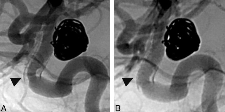
In 1 case, patient 4, treated with coils and a PED, 50% in-construct stenosis was detected at 6 months. Dual antiplatelet agents were maintained, and the narrowing decreased to 25% at 10-month DSA (Fig 4) and <20% after 14 months.
Fig 4.
In-construct stenosis. Patient 4 lives in a remote location and has a 10-mm superior hypophyseal aneurysm treated with coils and PED 21 days post-SAH. ICA angiography in lateral projection. A, Unsubtracted image at 6 months demonstrates aneurysm occlusion with dense coil packing. Fifty percent focal stenosis within the proximal margin of the PED (black arrowhead) is seen with a longer segment of neointimal hyperplasia throughout the construct. B, Unsubtracted image at 10 months documents that focal stenosis is reduced as is the extent of neointimal hyperplasia (black arrowhead).
Discussion
Endovascular treatment of ruptured aneurysms in the setting of SAH is most safely performed with coils. Wide-neck, blister, and dissecting blisterlike aneurysms are difficult to treat with both open and endovascular techniques. A balloon-assisted technique may not be possible in all small blister or dissecting blisterlike aneurysms, fusiform aneurysms, and some wide-neck aneurysms in which the native vessel is incorporated into the neck of the aneurysm. Treatment of acutely ruptured aneurysms with stent placement/coils is problematic because of the perceived requirement for concomitant antiplatelet medication, particularly clopidogrel, during the time of acute SAH. External ventricular drainage and the potential for aneurysm rehemorrhage (particularly if the aneurysm sac is not tightly packed with coils) is a concern.
There are a number of articles outlining the treatment of acutely ruptured wide-neck21,22 or acute blister aneurysms23–25 with stents and/or coils, for which coil placement is difficult or impossible. Tahtinen et al21 treated 61 patients with wide-neck aneurysms (including 3 vertebral dissections) within 72 hours of the onset of SAH by using Neuroform stents (Boston Scientific, Natick, Massachusetts) and coils. Intravenous heparin and aspirin were given during the procedure, and 300 mg of clopidogrel was given postprocedure. In 10% of patients, stent deployment was not successful; 19.8% (12/61) had complications directly due to primary stent/coil treatment (7 thromboembolic events requiring abciximab and 4 aneurysm perforations) or due to early rebleeding of the aneurysm (1 patient). Thirty-day mortality was 20% with favorable Glasgow Outcome Score of 4 or 5 in 69%. Katsaridis et al22 reported a subcohort of 33 acutely ruptured aneurysms within a population of 54 cases treated intraprocedurally with Neuroform 2 stents (Boston Scientific) and coils with intraoperative heparin and postoperative clopidogrel, aspirin, and low-molecular-weight heparin. There were no procedure-related clinical complications, and 2 technical issues were encountered with no clinical sequelae. Good outcome (mRS score, 0–3) was seen in 64%, with 21% having a poor outcome and 15% resulting in death. Angiographic follow-up, however, for assessment of the durability of treatment was limited in the patients with SAH.
In acute carotid blister aneurysms or dissecting dissecting blisterlike posterior circulation aneurysms, treatment with coils alone is problematic. Lee et al25 reported 9 patients with blister aneurysm cases, 6 treated with Neuroform stents and coils initially; 4 of these subsequently had aneurysm recurrence requiring placement of an additional stent. Three others were primarily treated with covered stents. One patient in this small group died acutely due to vessel rupture during stent placement. The 8 survivors had a good outcome. Park et al24 reported acute blister aneurysm recurrence in all 4 cases in the setting of acute endovascular reconstructive therapy (3 coiling, 1 stent-coiling). Two of these had failed surgical treatment before endovascular therapy. The authors advocated endovascular trapping in these cases due to the high recurrence rate, but another group reported very poor outcome with acute vessel sacrifice in the setting of acute SAH, mainly due to vasospasm.26 Meckel et al20 reported 12 blister or dissecting blisterlike posterior circulation ruptured aneurysms treated acutely with stent-assisted coiling in 11 and double stent placement alone in 1. Two patients rebled, 1 of whom died. Three required retreatment, with 2 having parent vessel occlusion in the acute admission and 1 undergoing delayed vessel sacrifice. All 11 survivors had an excellent outcome and angiographic appearance, but primary endovascular treatment failed in 4/12.
The development of flow diverters to reconstruct vessels and occlude aneurysms has created much interest,27–31 and recent PED series17–19 showed occlusion rates of 93% and 94% at 6-month follow-up in large or wide-neck aneurysms, with low morbidity. The Pipeline for the Intracranial Treatment of Aneurysms trial19 studied 31 aneurysms with no recent history of SAH. Lylyk et al17 described a series of 63 aneurysms, without recent SAH, though 7 patients had had previous SAH treated remotely by endovascular means. Byrne et al32 reported the use of another flow diverter, Silk (Balt Extrusion, Montmorency, France), in 10 patients with previously ruptured aneurysms, but only 4 were treated within 30 days of hemorrhage. The application of these devices in the setting of acute SAH in aneurysms is not well-documented in the literature.
Kulcsar et al33 described 2 patients with aneurysms <2 mm presenting with acute SAH, primarily treated with the Silk stent at 10 and 24 days after the initial bleed. The first was a carotid blister aneurysm, and the patient was premedicated with 100 mg of aspirin and 75 mg of clopidogrel for 3 days. The second was a basilar tip aneurysm, and the patient was loaded with 300 mg of both aspirin and clopidogrel. Follow-up angiography demonstrated some in-stent narrowing in one and stenosis of the superior cerebellar artery origin in the other. Both had excellent outcomes and aneurysm obliteration.
Our population varied from that in other published series in that it consisted of 11 patients with acute SAH treated with PED: 6 in the first 2 weeks (including 4 within 72 hours of ictus) and 5 in the third or fourth week. Five were acute ICA blister or posterior circulation dissecting blisterlike lesions, and 3 of these recurred acutely after failing primary endovascular therapy of coils (1 patient) or a Solitare stent and coils (2 patients) during the same admission, with 1 patient representing with a second SAH. The mortality rate was 18.2% with 2 ruptures but with no procedure-related symptomatic significant morbidity in the other 9 patients.
The 6-month occlusion rate of (8/9) in our small series is consistent with that in previous reports,17–19 with the only nonoccluded aneurysm, initially treated with stent/coils, appearing stable at follow-up.
The literature has recently highlighted delayed rupture related to flow division in elective cases.34,35 Kulcsar et al34 documented 13 patients with delayed rupture following Silk stent placement. Two of these may have had acute SAH at presentation. In our series, 2 patients died from aneurysm rerupture in the setting of acute SAH. It is our practice to only use a stent in SAH if necessary and, wherever possible, to coil the aneurysm at the time of stent placement, because stent or PED deployment does not necessarily lower pressure in the fundus of the aneurysm. Although it may be easier to coil the aneurysm by using a jailed microcatheter after deploying the PED, it may be better to coil as much as possible first, with balloon assistance if practicable, deploy the PED, and then complete the coiling. This will maximize fundus protection in the acute setting. Alternatively, it could be argued that acute fundus protection with coils, followed by delayed PED at 1–2 months, could be undertaken in appropriate situations (such as the case described in Fig 1).
Cebral et al35 highlighted the possible role of parent vessel stenosis in aneurysm rupture post-PED, suggesting that coiling these aneurysms may also be required in that setting. In retrospect, patient 9 did have a degree of smooth narrowing in the ICA distal to the neck, which did improve a little in diameter post-PED, though the M1 was more gracile. Treatment of any type of this particular aneurysm will continue to be challenging in the setting of SAH.
Conclusions
PED reconstruction of acutely ruptured wide-neck aneurysms or dissecting blisterlike aneurysms without a well-defined sac is possible, but caution needs to be exercised in cases of acute SAH. Ancillary attenuated coiling of the sac is warranted in patients with acute SAH or in other high-risk situations (such as stenosis in the parent vessel), by using the PED as a coil scaffold rather than a flow-diverter for acute aneurysm treatment in appropriate cases. Staged treatment with PED, temporizing with balloon-assist coiling acutely, is an option. It may be better to avoid conventional stent-assisted coiling in the acute setting, if possible, as the occlusion rate with PED is lower if deployed inside a stent.
ABBREVIATIONS:
- mRS
modified Rankin Scale
- PcomA
posterior communicating artery
- PED
Pipeline Embolization Device
- WFNS
World Federation of Neurosurgery
Footnotes
Disclosures: Jason D. Wenderoth—Consultancy: ev3, Comments: proctoring for deployment of PED.
References
- 1. Molyneux A, Kerr R, Stratton I, et al. International Subarachnoid Aneurysm Trial (ISAT) of neurosurgical clipping versus endovascular coiling in 2143 patients with ruptured intracranial aneurysms: a randomised trial. Lancet 2002; 360: 1267– 74 [DOI] [PubMed] [Google Scholar]
- 2. Taschner CA, Thines L, El-Mahdy M, et al. GDC 360 degrees for the endovascular treatment of intracranial aneurysms: a matched-pair study analysing angiographic outcomes with GDC 3D coils in 38 patients. Neuroradiology 2009; 51: 45– 52 [DOI] [PubMed] [Google Scholar]
- 3. Moret J, Cognard C, Weill A, et al. Reconstruction technic in the treatment of wide-neck intracranial aneurysms: long-term angiographic and clinical results—apropos of 56 cases [in French]. J Neuroradiol 1997; 24: 30– 44 [PubMed] [Google Scholar]
- 4. Sluzewski M, van Rooij WJ, Beute GN, et al. Balloon-assisted coil embolization of intracranial aneurysms: incidence, complications, and angiography results. J Neurosurg 2006; 105: 396– 99 [DOI] [PubMed] [Google Scholar]
- 5. White PM, Lewis SC, Nahser H, et al. HydroCoil Endovascular Aneurysm Occlusion and Packing Study (HELPS trial): procedural safety and operator-assessed efficacy results. AJNR Am J Neuroradiol 2008; 29: 217– 23 [DOI] [PMC free article] [PubMed] [Google Scholar]
- 6. Geyik S, Ertugrul O, Yavuz K, et al. Comparison of bioactive coils and bare platinum coils for treatment of intracranial aneurysms: a matched-pair analysis. J Neurosurg 2010; 112: 709– 13 [DOI] [PubMed] [Google Scholar]
- 7. Katsaridis V, Papagiannaki C, Violaris C. Guglielmi detachable coils versus Matrix coils: a comparison of the immediate post-treatment results of the embolization of 364 cerebral aneurysms in 307 patients: a single-center, single-surgeon experience. AJNR Am J Neuroradiol 2006; 27: 1841– 48 [PMC free article] [PubMed] [Google Scholar]
- 8. Yavuz K, Geyik S, Saatci I, et al. WingSpan stent system in the endovascular treatment of intracranial aneurysms: clinical experience with midterm follow-up results. J Neurosurg 2008; 109: 445– 53 [DOI] [PubMed] [Google Scholar]
- 9. Weber W, Bendszus M, Kis B, et al. A new self-expanding nitinol stent (Enterprise) for the treatment of wide-necked intracranial aneurysms: initial clinical and angiographic results in 31 aneurysms. Neuroradiology 2007; 49: 555– 61 [DOI] [PubMed] [Google Scholar]
- 10. Lylyk P, Cohen JE, Ceratto R, et al. Combined endovascular treatment of dissecting vertebral artery aneurysms by using stents and coils. J Neurosurg 2001; 94: 427– 32 [DOI] [PubMed] [Google Scholar]
- 11. Fiorella D, Albuquerque FC, Deshmukh VR, et al. Usefulness of the Neuroform stent for the treatment of cerebral aneurysms: results at initial (3–6-mo) follow-up. Neurosurgery 2005; 56: 1191– 201, discussion 1201–02 [DOI] [PubMed] [Google Scholar]
- 12. Fiorella D, Kelly ME, Albuquerque FC, et al. Curative reconstruction of a giant midbasilar trunk aneurysm with the Pipeline embolization device. Neurosurgery 2009; 64: 212– 17, discussion 217 [DOI] [PubMed] [Google Scholar]
- 13. Fiorella D, Albuquerque F, Gonzalez F, et al. Reconstruction of the right anterior circulation with the Pipeline embolization device to achieve treatment of a progressively symptomatic, large carotid aneurysm. J NeuroInterv Surg 2010; 2: 31– 37 [DOI] [PubMed] [Google Scholar]
- 14. Kamran M, Yarnold J, Grunwald IQ, et al. Assessment of angiographic outcomes after flow diversion treatment of intracranial aneurysms: a new grading schema. Neuroradiology 2011; 53: 501– 08 [DOI] [PubMed] [Google Scholar]
- 15. Kulcsar Z, Ernemann U, Wetzel SG, et al. High-profile flow diverter (Silk) implantation in the basilar artery: efficacy in the treatment of aneurysms and the role of the perforators. Stroke 2010; 41: 1690– 96 [DOI] [PubMed] [Google Scholar]
- 16. Lubicz B, Collignon L, Raphaeli G, et al. Flow-diverter stent for the endovascular treatment of intracranial aneurysms: a prospective study in 29 patients with 34 aneurysms. Stroke 2010; 41: 2247– 53 [DOI] [PubMed] [Google Scholar]
- 17. Lylyk P, Miranda C, Ceratto R, et al. Curative endovascular reconstruction of cerebral aneurysms with the Pipeline embolization device: the Buenos Aires experience. Neurosurgery 2009; 64: 632– 42, discussion 642–43, quiz N636 [DOI] [PubMed] [Google Scholar]
- 18. Szikora I, Berentei Z, Kulcsar Z, et al. Treatment of intracranial aneurysms by functional reconstruction of the parent artery: the Budapest experience with the Pipeline embolization device. AJNR Am J Neuroradiol 2010; 31: 1139– 47 [DOI] [PMC free article] [PubMed] [Google Scholar]
- 19. Nelson PK, Lylyk P, Szikora I, et al. The Pipeline embolization device for the intracranial treatment of aneurysms trial. AJNR Am J Neuroradiol 2011; 32: 34– 40 [DOI] [PMC free article] [PubMed] [Google Scholar]
- 20. Meckel S, Singh TP, Undren P, et al. Endovascular treatment using predominantly stent-assisted coil embolization and antiplatelet and anticoagulation management of ruptured blood blister-like aneurysms. AJNR Am J Neuroradiol 2011; 32: 764– 71 [DOI] [PMC free article] [PubMed] [Google Scholar]
- 21. Tahtinen OI, Vanninen RL, Manninen HI, et al. Wide-necked intracranial aneurysms: treatment with stent-assisted coil embolization during acute (<72 hours) subarachnoid hemorrhage—experience in 61 consecutive patients. Radiology 2009; 253: 199– 208 [DOI] [PubMed] [Google Scholar]
- 22. Katsaridis V, Papagiannaki C, Violaris C. Embolization of acutely ruptured and unruptured wide-necked cerebral aneurysms using the Neuroform 2 stent without pretreatment with antiplatelets: a single center experience. AJNR Am J Neuroradiol 2006; 27: 1123– 28 [PMC free article] [PubMed] [Google Scholar]
- 23. Kim BM, Chung EC, Park SI, et al. Treatment of blood blister-like aneurysm of the internal carotid artery with stent-assisted coil embolization followed by stent-within-a-stent technique: case report. J Neurosurg 2007; 107: 1211– 13 [DOI] [PubMed] [Google Scholar]
- 24. Park JH, Park IS, Han DH, et al. Endovascular treatment of blood blister-like aneurysms of the internal carotid artery. J Neurosurg 2007; 106: 812– 19 [DOI] [PubMed] [Google Scholar]
- 25. Lee BH, Kim BM, Park MS, et al. Reconstructive endovascular treatment of ruptured blood blister-like aneurysms of the internal carotid artery. J Neurosurg 2009; 110: 431– 36 [DOI] [PubMed] [Google Scholar]
- 26. Meling TR, Sorteberg A, Bakke SJ, et al. Blood blister-like aneurysms of the internal carotid artery trunk causing subarachnoid hemorrhage: treatment and outcome. J Neurosurg 2008; 108: 662– 71 [DOI] [PubMed] [Google Scholar]
- 27. Fiorella D, Woo HH, Albuquerque FC, et al. Definitive reconstruction of circumferential, fusiform intracranial aneurysms with the Pipeline embolization device. Neurosurgery 2008; 62: 1115– 20, discussion 1120–21 [DOI] [PubMed] [Google Scholar]
- 28. Wanke I, Forsting M. Stents for intracranial wide-necked aneurysms: more than mechanical protection. Neuroradiology 2008; 50: 991– 98 [DOI] [PubMed] [Google Scholar]
- 29. Augsburger L, Farhat M, Reymond P, et al. Effect of flow diverter porosity on intraaneurysmal blood flow. Klin Neuroradiol 2009; 19: 204– 14 [DOI] [PubMed] [Google Scholar]
- 30. Kallmes DF, Ding YH, Dai D, et al. A new endoluminal, flow-disrupting device for treatment of saccular aneurysms. Stroke 2007; 38: 2346– 52 [DOI] [PubMed] [Google Scholar]
- 31. Kallmes DF, Ding YH, Dai D, et al. A second-generation, endoluminal, flow-disrupting device for treatment of saccular aneurysms. AJNR Am J Neuroradiol 2009; 30: 1153– 58 [DOI] [PMC free article] [PubMed] [Google Scholar]
- 32. Byrne JV, Beltechi R, Yarnold JA, et al. Early experience in the treatment of intra-cranial aneurysms by endovascular flow diversion: a multicentre prospective study. PLoS One 2010; 5: pii:e12492 [DOI] [PMC free article] [PubMed] [Google Scholar]
- 33. Kulcsar Z, Wetzel SG, Augsburger L, et al. Effect of flow diversion treatment on very small ruptured aneurysms. Neurosurgery 2010; 67: 789– 93 [DOI] [PubMed] [Google Scholar]
- 34. Kulcsar Z, Houdart E, Bonafe A, et al. Intra-aneurysmal thrombosis as a possible cause of delayed aneurysm rupture after flow-diversion treatment. AJNR Am J Neuroradiol 2011; 32: 20– 25 [DOI] [PMC free article] [PubMed] [Google Scholar]
- 35. Cebral JR, Mut F, Raschi M, et al. Aneurysm rupture following treatment with flow-diverting stents: computational hemodynamics analysis of treatment. AJNR Am J Neuroradiol 2011; 32: 27– 33 [DOI] [PMC free article] [PubMed] [Google Scholar]


