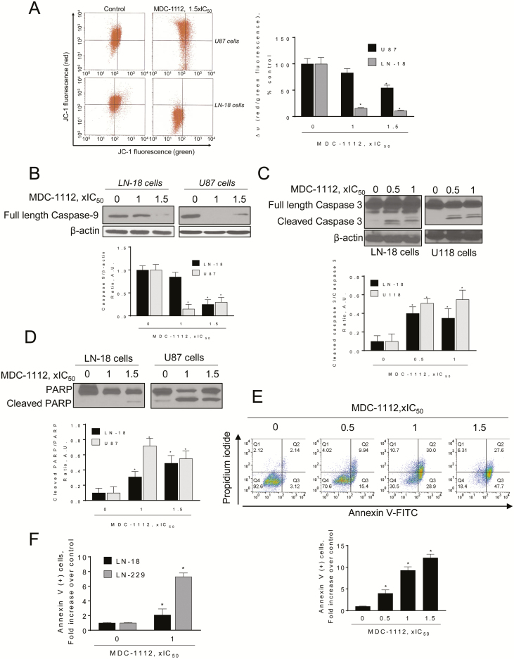
Figure 6.
MDC-1112 induces intrinsic apoptosis in GBM cells. (A) MDC-1112 collapses the mitochondrial membrane potential (ΔΨm) in a concentration-dependent manner. Cells were stained with JC-1 and analyzed with flow cytometry after treatment with MDC-1112 for 3 h. Data were quantified and results are shown as mean ± SEM; *P < 0.05 versus control. (B) Immunoblots for full-length caspase 9 in total cell protein extracts from LN-18 or U87 cells treated with MDC-1112, as indicated, for 24 h. Loading control: β-actin. Bands were quantified and results are shown as the ratio cleaved:β-actin; *P < 0.05 versus control. (C) Immunoblots for full length and cleaved caspase 3 in total cell protein extracts from LN-18 or U118 cells treated with MDC-1112, as indicated, for 24 h. Loading control: β-actin. Bands were quantified and results are shown as the ratio cleaved:full length protein; *P < 0.05 versus control. (D) Immunoblots for full length and cleaved PARP in total cell protein extracts from LN-18 or U87 cells treated with MDC-1112, as indicated, for 24 h. Bands were quantified and results are shown as the ratio cleaved/full length protein; *P < 0.05 versus control. (E) Cell death by apoptosis was determined by flow cytometry using the dual staining (Annexin V and PI) in U87 cells treated with increasing concentrations of MDC-1112 for 24 h. Results are expressed as fold-increase compared with the percentage of Annexin V (+) cells in the control group. (F) Cell death by apoptosis was determined by flow cytometry in LN-18 and LN-229 cells incubated without or with MDC-1112 1 × IC50 for 24 h. Results are expressed as fold-increase compared with the percentage of apoptotic cells in the control group.

