Abstract
The most effective and the standard treatment for bile duct stones (BDSs) is endoscopic retrograde cholangiopancreatography (ERCP). However, in 10% to 15% of patients with BDSs, the stones cannot be removed by conventional ERCP, which involves endoscopic sphincterotomy followed by balloon or basket extraction. Additional techniques or devices are often necessary to remove these difficult bile-duct stones, including endoscopic papillary large balloon dilatation to make a larger papillary opening and/or mechanical lithotripsy to fragment the stones. Advances in cholangioscopy have made possible electrohydraulic or laser lithotripsy under direct cholangioscopic visualization during ERCP. Cholangioscopy-guided lithotripsy could be another good option in the armamentarium of techniques for removing difficult BDSs. Here we review endoscopic techniques based on single-operator cholangioscopy for the management of difficult BDSs.
Keywords: Cholangiopancreatography, endoscopic retrograde, Choledocholithiasis, Lithotripsy, Cholangioscopy
INTRODUCTION
Bile duct stones (BDSs) are one of the most common biliary tract diseases, with a prevalence of 10% to 20% in patients with symptomatic gallbladder stones [1,2]. Patients with BDSs are at high risk of serious, potentially fatal complications, such as acute cholangitis, liver abscess, or acute gallstone pancreatitis. Endoscopic retrograde cholangiopancreatography (ERCP) is a standard procedure for the treatment of patients with BDSs. Approximately 90% of BDSs can be effectively removed with endoscopic sphincterotomy (EST) followed by stone extraction. However, the remaining 10% to 15%, which are considered difficult BDSs, often require additional techniques or devices for their removal [3]. These include cases in which access to the bile duct is challenging (e.g., periampullary diverticulum, altered anatomy after gastric bypass), and/or the size, shape, or location of the stones within the bile duct is difficult to determine for mechanical lithotripsy (ML) and extraction. ML and endoscopic papillary large balloon dilatation (EPLBD) are now the standard techniques for the removal of large BDSs. Cholangioscopy has been developed over the last decade and enables the management of BDSs by providing direct access to the bile duct. The second generation of digital single-operator cholangioscopy (SOC) is available and facilitates the management of difficult BDSs. This review highlights various aspects of difficult BDSs and SOC with a focus on recent strategies for SOC-guided lithotripsy.
DIFFICULT BILE-DUCT STONES
No clear consensus exists about the definition of difficult BDSs. Factors that contribute to the difficulty in removing BDSs can be divided into four major categories: (1) stone characteristics, such as stone size > 15 mm, multiple stones, hard stones, and strange shape of stones (e.g., barrel shape); (2) stone location, such as in the intrahepatic duct (IHD), above a stricture, or impacted in the bile duct/cystic duct; (3) anatomical alterations that make access to the papilla challenging, such as the presence of a periampullary diverticulum, Roux-en-Y gastric bypass anatomy, and Billroth II anatomy; and (4) patient factors, such as bleeding tendency due to the use of an antithrombotic agent, age > 65 years, and unstable vital signs (Table 1).
Table 1.
Difficult bile-duct stones
| Category | Conditions | Reasons |
|---|---|---|
| Characteristics of stone | Large stone (> 15 mm) Multiple stones (> 3 stones, size > 10 mm) Hard stone Strange-shaped stone |
Need for lithotripsy and difficulty in capturing the stone with a basket |
| Location of stone | Intrahepatic duct stone Stone above a stricture Impacted stone in the bile duct/cystic duct Mirizzi syndrome |
Difficulty in access |
| Anatomical situation | Altered anatomy Billroth II/Roux-en-Y gastric bypass anatomy Periampullary diverticulum |
Difficulty in biliary access and limitation of the endoscope/accessory |
| Patient factors | Old age/poor general condition Unstable vital signs Bleeding tendency Paradoxical response |
Risk of adverse events |
ENDOSCOPIC MANAGEMENT OF DIFFICULT BILE-DUCT STONES
The removal of difficult BDSs cannot be achieved with conventional ERCP, which involves EST followed by balloon or basket extraction. Thus, additional techniques including EPLBD, ML, and cholangioscopy-guided lithotripsy may be required.
Endoscopic papillary large balloon dilation
According to the 2019 guidelines of the European Society of Gastrointestinal Endoscopy (ESGE) [4], EST combined with EPLBD is recommended as the initial step in removing difficult BDSs. EPLBD was first described by Ersoz et al. [5] in 2003. It is performed with a dilation balloon of diameter > 12 mm. EPLBD can be used to dilate the biliary orifice using a large-diameter (typically 12 to 20 mm) balloon. Several studies on difficult BDSs have demonstrated the efficacy and safety of EPLBD. Additionally, EST + EPLBD reduces the need for ML by 30% to 50% compared with EST alone [6,7]. As a result, EPLBD after EST has become the standard technique for the management of difficult BDSs.
The adverse events (AEs) of EPLBD are mainly pancreatitis, bleeding, and perforation (the most serious AE). Distal common bile duct (CBD) stricture is an independent risk factor for perforation and is considered a relative contraindication to EPLBD. However, EST + EPLBD reduces the rate of AEs such as perforation. In a systematic review, the rate of AEs was significantly lower for EST + EPLBD than for EST alone (8.3% vs. 12.7%; odds ratio [OR], 1.60; p < 0.001) [8].
Mechanical lithotripsy
ML is an effective technique for crushing large or hard BDSs. The ESGE guidelines recommend ML when EST and EPLBD have failed or are inappropriate [4]. The generally used stone extraction basket cannot crush a hard stone because the basket wires are thin and weak. The ML basket captures the stone in a strong basket and then breaks it against a metal sheath. With the introduction and increased utilization of EPLBD, the use of ML has decreased. However, ML may be required if the stone is too large to remove after EPLBD, a stricture exists below the stone, or a traditional basket is impacted with a stone in the bile duct [9].
Although ML may require multiple sessions, it has been reported to be an effective and safe technique. The success rates range from 76% to 91%, and the overall rates of AEs range from 3% to 34% [10–14]. In a retrospective study by Lee et al. [12], stone impaction, stone size > 30 mm, and stone to CBD diameter ratio > 1 were significant predictors of ML failure. ML is associated with high rates of AEs, including cholangitis, hemorrhage, pancreatitis, and perforation [9]. The most common and feared AEs of ML are an impacted basket, a broken basket, traction wire fracture, or a broken handle [15].
Although the technical success rate of ML is approximately 90%, the procedure can be technically challenging depending on the size and location of the BDSs. An impacted stone is a risk factor for failure of ML, and a confluence stone is also technically challenging to manage. However, these difficulties and limitations have been overcome by the development of cholangioscopy-guided electrohydraulic lithotripsy (EHL) and cholangioscopy-guided laser lithotripsy (LL) [16–19].
CHOLANGIOSCOPY-GUIDED LITHOTRIPSY
Cholangioscopy system
Cholangioscopy is useful in the diagnosis and treatment of bile duct abnormalities, and was developed to overcome the limitations of ERCP. Three types of peroral cholangioscopy (POC) are currently available (Table 2) [20,21]:
Table 2.
Three types of cholangioscopy system
| “Mother–baby” dual-operator cholangioscopy | Single-operator cholangioscopy (SpyGlass DS system) | Direct peroral cholangioscopy (ultra-slim endoscope) | |
|---|---|---|---|
| Endoscopist | 2 | 1 | 1 |
| Additional endoscope system | Yes | Yes | No |
| Scope diameter, mm | 3.3–3.5 | 3.6 | 5–6 |
| Accessory channel, mm | 1.2 | 1.2 | 2 |
| Irrigation channel | No | Yes | No |
| Cost | High | High | Low |
| Image quality | High | Intermediate | High |
“Mother–baby” endoscope system: This system requires one operator to handle the duodenoscope and another to handle the cholangioscope.
SOC using the SpyGlass DS system: In this method, a digital and single-use cholangioscope is attached to the duodenoscope, allowing a single operator to manage the control of both scopes.
Direct POC using an ultra-slim endoscope: Direct access to the bile duct is a good option for the management of large BDSs because it offers direct visualization of the stone during therapy. However, because dual-operator cholangioscopy and direct cholangioscopy systems with an ultra-slim endoscope are inconvenient to use, expensive, and relatively thick, their application to the management of difficult BDSs has several limitations.
The mother–baby technique using classic cholangioscopes is limited by their fragility, cost, complex and difficult installation, two-way tip deflection only, and the need for two experienced endoscopists. Therefore, they are rarely used at present.
Single-operator cholangioscopy
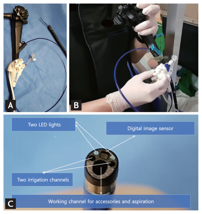
SOC, through the introduction of the SpyGlass DS Direct Visualization System (Boston Scientific, Marlborough, MA, USA), has led to a rapid increase in the use of cholangioscopy (cholangioscopy using the SpyGlass system is usually referred to as SOC). SpyGlass is a digital and single-use cholangioscope that overcomes several limitations of other types of cholangioscopes. Because this digital cholangioscope has a diameter of approximately 3.5 mm, it can be used to access the bile duct directly through the working channel of a duodenoscope. Because it is a ‘plug-and-play’ device, it is easy and quick to set up. This cholangioscope has both a 1.2 mm accessory channel and a 1.2 mm suction channel. Through the thin accessory channel, instruments, such as an EHL or LL probe, can be inserted and used for lithotripsy (Fig. 1).
Figure 1.
Single-operator cholangioscopy (SOC) using the SpyGlass DS system (Boston Scientific). (A) The SpyScope is inserted into the duodenoscope through its working channel. (B) A single endoscopic retrograde cholangiopancreatography endoscopist operates both the duodenoscope and the SOC (hence ‘single-operator’ cholangioscopy). (C) The SpyScope, the digital, single-use scope of the SpyGlass DS System. Its diameter is 3.6 mm. The diameter of the working channel is 1.2 mm. LED, light emitting diode.
The next generation of the SpyGlass DS System (SpyGlass DS 2.0) has recently been released. It has fourfold higher image resolution and optimized light emitting diode (LED) illumination, promising improved diagnostic and therapeutic capabilities.
Cholangioscopy-guided lithotripsy
Electrohydraulic lithotripsy
In this method, an EHL probe made of a coaxial bipolar device is passed into the working channel of the endoscope. EHL is based on the principle that, when a charge is applied, sparks generated underwater produce high-frequency hydraulic pressure waves. The energy is absorbed by the stones and results in their fragmentation. The probe is used with the probe tip 1 to 2 mm from the stone. Continuous saline irrigation is required to provide a medium for shock-wave energy transmission. Additionally, saline irrigation enables the maintenance of optimal vision during lithotripsy. Because direct bile-duct damage caused by shock waves can lead to bleeding or perforation, it is important to perform lithotripsy with direct visualization using cholangioscopy.
Laser lithotripsy
In LL, a laser light at a particular wavelength is focused on the surface of the stone to induce wave-mediated fragmentation. The first successful use of a pulsed laser for shock-wave lithotripsy of BDSs was reported in 1986 [22]. The technology has evolved since then, and other laser types, such as an neodymium:yttrium aluminum garnet (Nd:YAG) laser with an automatic stone recognition system and the frequency-doubled double-pulse Nd:YAG laser system, have been introduced [23,24]. The tip of the laser fiber has a green (or white) aiming beam, which is used to target the stone under direct vision. With the probe tip 1 to 2 mm from the stone and under continuous saline irrigation, laser bursts are delivered through the aqueous medium until stone fragmentation is deemed complete.
SOC-GUIDED LITHOTRIPSY
Indications for SOC-guided lithotripsy
No clear guidelines exist for SOC-guided lithotripsy, including those for indications, selection of cholangioscope type, and choice between EHL and LL [20]. The ESGE guidelines suggest that the type of cholangioscopy and lithotripsy should depend on local availability and experience [4]. Instead, the inclusion criteria of several studies on SOC-guided lithotripsy have suggested situations in which SOC-guided lithotripsy should be actively considered: stone size > 20 mm, multiple stones > 10 mm, stones proximal to a stricture, stones in the IHD, stones in difficult-to-access locations (cystic duct or IHD), impacted stones in the bile or cystic duct, lumen-occupying stone casts, and at least two failed attempts of stone clearance using conventional means.
Procedure, technique, and protocol
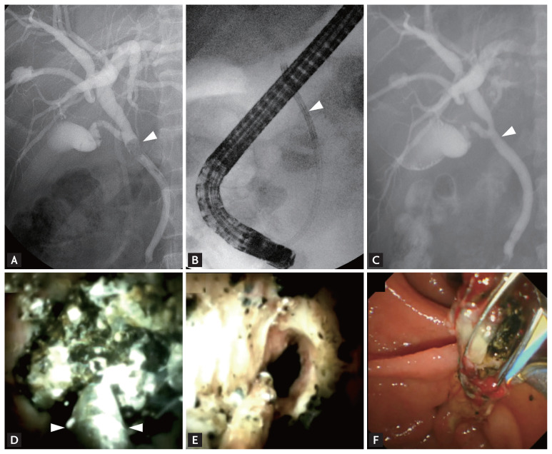
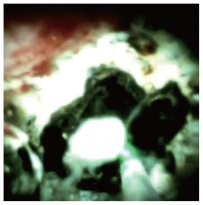
EST is performed before SOC. The choice of the SOC-guided lithotripsy method (EHL or LL) depends on system availability and the preferences of the endoscopist. When performing SOC, the SpyScope, which is a digital and single-use cholangioscope in the SpyGlass DS System, is inserted into the bile duct in a freehand or guidewire-guided method. The SpyScope is advanced into the CBD or cystic stump toward the stone of interest, with endoscopic vision and intermittent fluoroscopy. When deemed necessary, the SpyScope is advanced over the guidewire to the target site. After direct visualization of the stone, the guidewire is replaced by the EHL probe (Fig. 2). When using the holmium laser technology, the LL fiber is inserted into the 1.2 mm working channel of the SpyScope (Fig. 3). The energy of the laser fiber or EHL probe is delivered until stone fragmentation is deemed complete. The fragmented stones are removed using conventional extraction devices, which may include ML, at the discretion of the endoscopist. If complete ductal clearance is not achieved in a single session, one or more plastic biliary stents or a nasobiliary drain catheter is inserted until the next session.
Figure 2.
Single-operator cholangioscopy (SOC)-guided electrohydraulic lithotripsy (EHL). (A) Endoscopic retrograde cholangiopancreatography (ERCP) image showing an impacted bile duct stone (arrowhead). (B) Fluoroscopy image showing SOC (arrowhead) inserted into the bile duct. (C) Final ERCP image showing no residual filling defect after lithotripsy (arrowhead). (D) SOC image showing an impacted stone. EHL (arrowheads, EHL probe) is performed under endoscopic view. (E) SOC image showing the cleared bile duct after lithotripsy. (F) Fragmented bile duct stones are extracted using a multi-wire basket after lithotripsy.
Figure 3.
Single-operator cholangioscopy-guided laser lithotripsy. Green aiming beam from the laser fiber (probe) targeting and fragmenting the impacted stone.
Inserting an EHL or LL probe into the accessory channel of the SpyScope can be impossible because of strong resistance. This is because when the SpyScope is located inside the bile duct, an acute angulation is made by the twisted distal end of the duodenoscope and its elevator. After straightening the SpyScope by withdrawing to the duodenum, the probe can be more easily inserted. Thereafter, the SpyScope is re-inserted into the bile duct.
During SOC-guided EHL or LL, continuous saline irrigation is required to provide a medium for electric shock waves or laser bursts. However, in some situations, such as after full EST and EPLBD, it is impossible to fill the bile duct with saline. Various strategies can be attempted to overcome this situation—changing the patient’s position may be a good and easy solution. Increasing the irrigation pressure using a water pump can also be a good technical alternative. In addition, SOC-guided lithotripsy can be performed after occluding the bile duct with a balloon catheter. However, this procedure is complex and time consuming because it requires re-inserting the duodenoscope.
Efficacy of SOC-guided lithotripsy
Several studies have demonstrated the successful management of difficult biliary stones using cholangioscopy-guided LL and EHL, with success rates ranging from 67% to 100% [19,24–31].
In a meta-analysis of 49 studies, the overall stone clearance rate was 88% (95% confidence interval [CI], 85% to 91%) [28]. However, this study investigated all types of cholangioscopy-guided lithotripsy, including SOC-guided lithotripsy. Recent studies focusing on SOC-guided lithotripsy have reported better efficacy (Table 3) [19,26,27,29–34].
Table 3.
Efficacy of single-operator cholangioscopy-guided lithotripsy
| Study | Study design | Lithotripsy | No. of patients | Clearance, % | Clearance after 1 session, % | Mean no. of procedure | Mean size of stone, mm |
|---|---|---|---|---|---|---|---|
| Kurihara et al. (2016) [30] | Multicenter Prospective |
EHL + LL | 31 | 74.2 | 1.9 | 20.6 | |
| Bhandari et al. (2016) [19] | Single center | LL | 34 | 100 | 94.1 | 1.1 | |
| Navaneethan et al. (2016) [31] | Multicenter Retrospective |
LL | 31 | 97.2 | 86.1 | 1.1 | 14.9 |
| Wong et al. (2017) [26] | Single center | EHL + LL | 17 | 100 | 94.1 | 17 | |
| Brewer et al. (2018) [27] | Multicenter Retrospective |
EHL + LL | 407 | 97.3 | 77.4 | 1 | |
| Buxbaum at al. (2018) [37] | Single center Prospective |
LL | 42 | 93 | 1.9 | 18 | |
| Turowski et al. (2018) [32] | Multicenter Retrospective |
EHL + LL | 107 | 91.1 | 3 | ||
| Angsuwatcharakon et al. (2019) [33] | Multicenter Prospective |
LL | 32 | 100 | 100 | 1 | 19.5 |
| Bokemeyer et al. (2020) [29] | Multicenter Retrospective |
EHL + LL | 60 | 95 | 67 | 20 |
EHL, electrohydraulic lithotripsy; LL, laser lithotripsy.
In a recent systematic review and meta-analysis of 24 studies, Jin et al. [35] analyzed the efficacy and safety of SOC-guided lithotripsy in treating difficult BDSs. The rate of complete stone clearance was 94% (95% CI, 90.2% to 97.5%). The rate of single-session stone clearance was 71.1% (95% CI, 62.1% to 79.5%) in the pooled 2786 patients. The number of sessions needed for complete stone clearance was 1.26 (95% CI, 1.17% to 1.34%).
In a recent retrospective study, Bokemeyer et al. [29] reported that SOC-guided lithotripsy was successful in 95% of patients, with 15% needing at least two treatment sessions. They concluded that SOC-guided lithotripsy is an excellent rescue approach even in patients with difficult stones in whom previous conventional ERCP had failed. Additionally, they reported that SOC-guided lithotripsy reduced radiation exposure compared with the conventional ERCP method.
In a retrospective study of 407 patients who underwent POC for difficult biliary stones at 22 tertiary centers in the United States, United Kingdom, and Korea, technical success, defined as complete stone clearance, was achieved in 97.3% with a median number of lithotripsy sessions of 1 (range, 1 to 4 sessions) [27]. In a multivariate analysis, difficult anatomy and cannulation were associated with technical failure (adjusted OR, 5.18; 95% CI, 1.26 to 21.2; p = 0.02) and the duration of the procedure was a predictor of the need for more than one session to achieve complete stone clearance (adjusted OR, 1.02; 95% CI, 1.01 to 1.03; p < 0.001). The authors concluded that SOCguided lithotripsy is effective and safe in > 95% of patients with difficult BDSs.
In a prospective study by Wong et al. [26], complete biliary stone clearance with SOCguided LL was successful in 94% of patients (16/17) in a median of 1 endoscopic procedure (range, 1 to 3). The median duration of SOC-guided LL was 90 minutes (range, 46 to 164). This suggests that SOC with LL is effective for the management of difficult BDSs.
EHL vs. LL
We use both EHL and LL in our patients with biliary stones, according to endoscopist preference and equipment availability. A recent multicenter study compared EHL and LL by analyzing 407 patients [27]. This retrospective study compared the outcomes of 306 patients submitted to EHL and 101 patients treated with LL. The final clearance rate was similar for the two techniques (96.7% for EHL and 99% for LL). The ducts were cleared in a single session in 77.4% of patients. However, a trend favoring LL with respect to efficacy was observed in a single initial session (74.5% for EHL and 86.1% for LL, p = 0.20). The mean procedure time was significantly longer in the EHL group (73.9 minutes) than in the LL group (49.9 minutes, p < 0.001).
A recent systematic review showed that LL had a higher complete ductal clearance rate (95.1%) than EHL (88.4%, p < 0.001) [36]. Also, the AE rate was significantly higher with EHL (13.8%) than with LL (9.6%, p = 0.04). Thus, LL provides better clinical outcomes in difficult BDSs; however, it may depend on local expertise and the availability of each technique.
However, those studies did not explore the number of probes used and did not provide recommendations for choosing between EHL and LL. Further studies and guidelines that compare EHL and LL, including cost-effectiveness analyses, are needed.
EHL probes have a short life span, which is proportional to the power used during lithotripsy. In contrast, LL probes do not have this limitation but are more expensive. In addition, the generator/system is much more expensive. Probably, laser probes should be selected only in cases of very large, multiple, or hard stones, to overcome the life-span limitation of the EHL probe.
SOC-guided lithotripsy vs. direct POC-guided lithotripsy
Few studies have directly compared SOC-guided lithotripsy with direct POC-guided lithotripsy. In a systematic review of 49 studies, Korrapati et al. [28] analyzed the efficacy of POC for difficult BDSs. The overall estimated stone clearance rate was 88% (95% CI, 85% to 91%). Additionally, they identified a significant association between the type of POC used and the technical success rate, with SOC demonstrating a higher technical success rate than the other methods (p < 0.01). Direct POC involving the use of an ultra-slim endoscope by a single endoscopist is technically difficult. Moreover, because an ultra-slim endoscope is thicker than SpyGlass, cannulating a nondilated bile duct is more difficult using the former. The need for direct POC had decreased recently because of the widespread use of SpyGlass. However, because SpyGlass is extremely expensive, direct POC can be a good treatment option for difficult BDSs in centers where the use of SpyGlass is not possible.
SOC-guided lithotripsy vs. conventional ERCP (EPLBD and/or ML)
SOC-guided lithotripsy has been reported to have a better stone clearance rate than conventional ERCP (EPLBD and/or ML). However, there are advantages and disadvantages to consider when deciding which procedure to perform.
Buxbaum et al. [37] randomized patients with difficult BDSs at a 2:1 ratio to SOC-guided LL and conventional therapy alone. Complete clearance was achieved in 93% of patients treated with SOC-guided LL and in 67% of those treated with conventional therapy alone (p = 0.009). However, SOC-guided LL was associated with a longer procedure time (120.7 ± 40.2 minutes in the conventional group and 81.2 ± 49.3 minutes in the SOC-guided LL group, p = 0.0008). The authors concluded that SOC-guided LL has a significantly higher stone clearance rate and a lower need for surgery than conventional therapy alone, although it is associated with a longer procedure time.
The randomized controlled trial by Angsuwatcharakon et al. [33] compared SOC-guided LL and ML after a failed EPLBD. The complete stone removal rates in a single session were 100% and 63% with comparable AE rates (6% and 13%) in the SOC-guided LL and ML groups, respectively. In this study, the procedure time was not significantly different between the two groups (66 minutes vs. 83 minutes, p = 0.23), although the radiation exposure was significantly lower (20,989 mGy cm2 vs. 40,745 mGy cm2, p = 0.04) in the SOC-guided LL group. The rate of AEs was not different between the two groups (13% in the ML group vs. 6% in the LL group, p = 0.76).
Therefore, although ML is the standard procedure for difficult BDSs, SOC-guided lithotripsy is the more effective and safer treatment option and should be considered when available.
Role of SOC in the management of IHD stones
As mentioned earlier, SOC-guided lithotripsy is mainly used in the treatment of extrahepatic BDSs. However, few studies have investigated the efficacy of SOC in the management of IHD stones. The application of SOC in the removal of IHD stones is technically challenging. Usually, percutaneous transhepatic cholangioscopy (PTC) is the standard endoscopic method for the management of IHD stones in patients with a high operation risk, those who refuse surgery, and those with a previous hepatobiliary surgery or stones distributed in multiple parts of the liver. IHD stones are problematic to remove with ERCP because the IHD is difficult to access using the devices or accessories. This limitation can be overcome to some extent with SOC. Because the diameter of the cholangioscope is about 3.5 mm, access to the IHD is easier than with other types of cholangioscopy systems. With SOC, after finding the IHD site where the stone is located, the guidewire is placed, and the stones can be removed using a balloon and a basket over the guidewire. Although SOC is technically more difficult than PTC, it has the advantage of providing an excellent quality of life for patients because it requires simple patient preparation, takes a short time to complete, and does not require a transcutaneous route. Further studies and case reports on SOC-guided management of IHD stones are needed to develop appropriate guidelines.
Adverse events
Post-ERCP AEs include pancreatitis, bleeding, cholangitis, perforation, and cardiopulmonary complications. Generally, AEs after ERCP have been reported to occur in 7% of patients [38]. With respect to SOC, previous studies have reported rates of AEs ranging from 0% to 16.4% [26,27,32,34,39–44]. The profiles of SOC-related AEs are not different from those after conventional ERCP [44]. However, SOC-guided lithotripsy is more likely to have different or more frequent AEs than diagnostic SOC procedures because it has a longer procedure time and is intended for therapeutic use.
In a recent meta-analysis of 31 studies, the estimated overall AE rate was 7% (95% CI, 6% to 9%) [28]. The estimated rates of pancreatitis and cholangitis were 2% (95% CI, 2% to 3%) and 4% (95% CI, 3% to 5%), respectively. The estimated rate of severe AEs was 1% (95% CI, 1% to 2%). In several studies, including this one, cholangitis was the most frequently reported AE [24,25,34,43]. Sethi et al. [43] suggested that ERCP with cholangioscopy may be associated with higher total rates of AEs, especially cholangitis, despite antibiotic prophylaxis. The authors proposed that this risk could be explained by the increased pressure associated with water irrigation during the procedure. Pancreatitis is a rare AE, probably due to the high proportion of a preexistent EST state [34].
Overall, the available data suggest that SOC-guided lithotripsy is an effective and safe procedure for the management of difficult BDSs.
Limitations
The most important consideration in SOC-guided lithotripsy is the cost factor. The high cost, limited availability, and high endoscopist skill requirements of SOC hamper its widespread use.
EST with EPLBD and/or ML has the advantage of being relatively inexpensive and widely available compared with SOC-guided lithotripsy. In particular, in SOC-guided lithotripsy, it is important to consider that approximately 20% of patients require more than a single EHL/LL session. However, in the cost-effectiveness analysis by Deprez et al. [45], the use of a cholangioscope for difficult BDSs decreased the number of procedures and the cost by 28% and 11%, respectively.
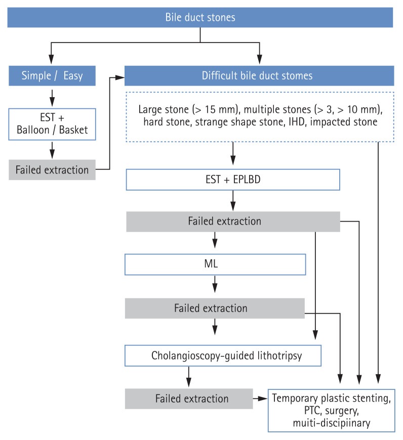
It is important that proven and effective techniques (e.g., ML/EPLBD) for the treatment of difficult BDSs are adequately considered before proceeding with SOC-guided lithotripsy. Future randomized studies comparing conventional ERCP and SOC-guided lithotripsy are needed to define the treatment algorithm for difficult BDSs considering cost-effectiveness (Fig. 4).
Figure 4.
Treatment algorithm for the management of difficult bile-duct stones. EST, endoscopic sphincterotomy; IHD, intrahepatic duct; EPLBD, endoscopic papillary large balloon dilatation; ML, mechanical lithotripsy; PTC, percutaneous transhepatic cholangioscopy.
The technical limitations of SOC-guided lithotripsy should also be considered. As mentioned earlier, treatment using SOC may be difficult if it is not possible to fill the bile duct with saline, if it is difficult to insert the EHL or LL probe through the channel of the cholangioscope (SpyScope), or if the location of the stones is difficult to access with a cholangioscope (such as the IHD). Therefore, the optimal management of difficult BDSs requires a multidisciplinary approach, including an endoscopist, a surgeon, and an interventional radiologist, before and/or after surgery. Furthermore, it is important to remember that what is challenging for one ERCP endoscopist may not be as challenging for another.
CONCLUSIONS
In clinical practice, clearance of BDSs is often not achievable with conventional ERCP. If the removal of these difficult BDSs is impossible with ML or EPLBD, cholangioscopy-guided lithotripsy represents an emerging alternative technique for stone fragmentation and extraction. Subsequent to the development and widespread use of SOC, SOC-guided lithotripsy has been established as an excellent treatment option for the management of difficult BDSs. In cases for which SOC-guided lithotripsy is needed, endoscopists should be aware of the indications, techniques, treatment results, and management of possible complications of the procedure.
Footnotes
Conflict of interest
No potential conflict of interest relevant to this article was reported.
REFERENCES
- 1.Collins C, Maguire D, Ireland A, Fitzgerald E, O’Sullivan GC. A prospective study of common bile duct calculi in patients undergoing laparoscopic cholecystectomy: natural history of choledocholithiasis revisited. Ann Surg. 2004;239:28–33. doi: 10.1097/01.sla.0000103069.00170.9c. [DOI] [PMC free article] [PubMed] [Google Scholar]
- 2.Freitas ML, Bell RL, Duffy AJ. Choledocholithiasis: evolving standards for diagnosis and management. World J Gastroenterol. 2006;12:3162–3167. doi: 10.3748/wjg.v12.i20.3162. [DOI] [PMC free article] [PubMed] [Google Scholar]
- 3.Yasuda I, Itoi T. Recent advances in endoscopic management of difficult bile duct stones. Dig Endosc. 2013;25:376–385. doi: 10.1111/den.12118. [DOI] [PubMed] [Google Scholar]
- 4.Manes G, Paspatis G, Aabakken L, et al. Endoscopic management of common bile duct stones: European Society of Gastrointestinal Endoscopy (ESGE) guideline. Endoscopy. 2019;51:472–491. doi: 10.1055/a-0862-0346. [DOI] [PubMed] [Google Scholar]
- 5.Ersoz G, Tekesin O, Ozutemiz AO, Gunsar F. Biliary sphincterotomy plus dilation with a large balloon for bile duct stones that are difficult to extract. Gastrointest Endosc. 2003;57:156–159. doi: 10.1067/mge.2003.52. [DOI] [PubMed] [Google Scholar]
- 6.Li G, Pang Q, Zhang X, et al. Dilation-assisted stone extraction: an alternative method for removal of common bile duct stones. Dig Dis Sci. 2014;59:857–864. doi: 10.1007/s10620-013-2914-4. [DOI] [PubMed] [Google Scholar]
- 7.Jun Bo Q, Li Hua X, Tian Min C, Liu Gen G, Yan Mei Y, Hua Sheng L. Small endoscopic sphincterotomy plus large-balloon dilation for removal of large common bile duct stones during ERCP. Pak J Med Sci. 2013;29:907–912. doi: 10.12669/pjms.294.3662. [DOI] [PMC free article] [PubMed] [Google Scholar]
- 8.Kim JH, Yang MJ, Hwang JC, Yoo BM. Endoscopic papillary large balloon dilation for the removal of bile duct stones. World J Gastroenterol. 2013;19:8580–8594. doi: 10.3748/wjg.v19.i46.8580. [DOI] [PMC free article] [PubMed] [Google Scholar]
- 9.Riemann JF, Seuberth K, Demling L. Mechanical lithotripsy of common bile duct stones. Gastrointest Endosc. 1985;31:207–210. doi: 10.1016/s0016-5107(85)72047-0. [DOI] [PubMed] [Google Scholar]
- 10.Garg PK, Tandon RK, Ahuja V, Makharia GK, Batra Y. Predictors of unsuccessful mechanical lithotripsy and endoscopic clearance of large bile duct stones. Gastrointest Endosc. 2004;59:601–605. doi: 10.1016/s0016-5107(04)00295-0. [DOI] [PubMed] [Google Scholar]
- 11.Cipolletta L, Costamagna G, Bianco MA, et al. Endoscopic mechanical lithotripsy of difficult common bile duct stones. Br J Surg. 1997;84:1407–1409. [PubMed] [Google Scholar]
- 12.Lee SH, Park JK, Yoon WJ, et al. How to predict the outcome of endoscopic mechanical lithotripsy in patients with difficult bile duct stones? Scand J Gastroenterol. 2007;42:1006–1010. doi: 10.1080/00365520701204253. [DOI] [PubMed] [Google Scholar]
- 13.Chang WH, Chu CH, Wang TE, Chen MJ, Lin CC. Outcome of simple use of mechanical lithotripsy of difficult common bile duct stones. World J Gastroenterol. 2005;11:593–596. doi: 10.3748/wjg.v11.i4.593. [DOI] [PMC free article] [PubMed] [Google Scholar]
- 14.Chung SC, Leung JW, Leong HT, Li AK. Mechanical lithotripsy of large common bile duct stones using a basket. Br J Surg. 1991;8:1448–1450. doi: 10.1002/bjs.1800781214. [DOI] [PubMed] [Google Scholar]
- 15.Thomas M, Howell DA, Carr-Locke D, et al. Mechanical lithotripsy of pancreatic and biliary stones: complications and available treatment options collected from expert centers. Am J Gastroenterol. 2007;102:1896–1902. doi: 10.1111/j.1572-0241.2007.01350.x. [DOI] [PubMed] [Google Scholar]
- 16.Ryozawa S, Yasuda I. Current strategies for the endoscopic management of difficult-to-treat bile duct stones in Japan. Dig Endosc. 2018;30(Suppl 1):54–58. doi: 10.1111/den.13020. [DOI] [PubMed] [Google Scholar]
- 17.Doshi B, Yasuda I, Ryozawa S, Lee GH. Current endoscopic strategies for managing large bile duct stones. Dig Endosc. 2018;30(Suppl 1):59–66. doi: 10.1111/den.13019. [DOI] [PubMed] [Google Scholar]
- 18.Franzini T, Moura RN, Bonifacio P, et al. Complex biliary stones management: cholangioscopy versus papillary large balloon dilation. A randomized controlled trial. Endosc Int Open. 2018;6:E131–E138. doi: 10.1055/s-0043-122493. [DOI] [PMC free article] [PubMed] [Google Scholar]
- 19.Bhandari S, Bathini R, Sharma A, Maydeo A. Usefulness of single-operator cholangioscopy-guided laser lithotripsy in patients with Mirizzi syndrome and cystic duct stones: experience at a tertiary care center. Gastrointest Endosc. 2016;84:56–61. doi: 10.1016/j.gie.2015.12.025. [DOI] [PubMed] [Google Scholar]
- 20.Ishida Y, Itoi T, Okabe Y. Types of peroral cholangioscopy: how to choose the most suitable type of cholangioscopy. Curr Treat Options Gastroenterol. 2016;14:210–219. doi: 10.1007/s11938-016-0090-2. [DOI] [PubMed] [Google Scholar]
- 21.Ishida Y, Itoi T, Okabe Y. Current status and future perspective in cholangiopancreatoscopy. Curr Treat Options Gastroenterol. 2019;17:327–341. doi: 10.1007/s11938-019-00238-1. [DOI] [PubMed] [Google Scholar]
- 22.Lux G, Ell C, Hochberger J, Muller D, Demling L. The first successful endoscopic retrograde laser lithotripsy of common bile duct stones in man using a pulsed neodymium-YAG laser. Endoscopy. 1986;18:144–145. doi: 10.1055/s-2007-1018356. [DOI] [PubMed] [Google Scholar]
- 23.Trikudanathan G, Navaneethan U, Parsi MA. Endoscopic management of difficult common bile duct stones. World J Gastroenterol. 2013;19:165–173. doi: 10.3748/wjg.v19.i2.165. [DOI] [PMC free article] [PubMed] [Google Scholar]
- 24.Patel SN, Rosenkranz L, Hooks B, et al. Holmium-yttrium aluminum garnet laser lithotripsy in the treatment of biliary calculi using single-operator cholangioscopy: a multicenter experience (with video) Gastrointest Endosc. 2014;79:344–348. doi: 10.1016/j.gie.2013.07.054. [DOI] [PubMed] [Google Scholar]
- 25.Maydeo A, Kwek BE, Bhandari S, Bapat M, Dhir V. Single-operator cholangioscopy-guided laser lithotripsy in patients with difficult biliary and pancreatic ductal stones (with videos) Gastrointest Endosc. 2011;74:1308–1314. doi: 10.1016/j.gie.2011.08.047. [DOI] [PubMed] [Google Scholar]
- 26.Wong JC, Tang RS, Teoh AY, Sung JJ, Lau JY. Efficacy and safety of novel digital single-operator peroral cholangioscopy-guided laser lithotripsy for complicated biliary stones. Endosc Int Open. 2017;5:E54–E58. doi: 10.1055/s-0042-118701. [DOI] [PMC free article] [PubMed] [Google Scholar]
- 27.Brewer Gutierrez OI, Bekkali NLH, Raijman I, et al. Efficacy and safety of digital single-operator cholangioscopy for difficult biliary stones. Clin Gastroenterol Hepatol. 2018;16:918–926. doi: 10.1016/j.cgh.2017.10.017. [DOI] [PubMed] [Google Scholar]
- 28.Korrapati P, Ciolino J, Wani S, et al. The efficacy of peroral cholangioscopy for difficult bile duct stones and indeterminate strictures: a systematic review and meta-analysis. Endosc Int Open. 2016;4:E263–E275. doi: 10.1055/s-0042-100194. [DOI] [PMC free article] [PubMed] [Google Scholar]
- 29.Bokemeyer A, Gerges C, Lang D, et al. Digital single-operator video cholangioscopy in treating refractory biliary stones: a multicenter observational study. Surg Endosc. 2020;34:1914–1922. doi: 10.1007/s00464-019-06962-0. [DOI] [PubMed] [Google Scholar]
- 30.Kurihara T, Yasuda I, Isayama H, et al. Diagnostic and therapeutic single-operator cholangiopancreatoscopy in biliopancreatic diseases: prospective multicenter study in Japan. World J Gastroenterol. 2016;22:1891–1901. doi: 10.3748/wjg.v22.i5.1891. [DOI] [PMC free article] [PubMed] [Google Scholar]
- 31.Navaneethan U, Hasan MK, Kommaraju K, et al. Digital, single-operator cholangiopancreatoscopy in the diagnosis and management of pancreatobiliary disorders: a multicenter clinical experience (with video) Gastrointest Endosc. 2016;84:649–655. doi: 10.1016/j.gie.2016.03.789. [DOI] [PubMed] [Google Scholar]
- 32.Turowski F, Hugle U, Dormann A, et al. Diagnostic and therapeutic single-operator cholangiopancreatoscopy with SpyGlassDS™: results of a multicenter retrospective cohort study. Surg Endosc. 2018;32:3981–3988. doi: 10.1007/s00464-018-6141-0. [DOI] [PubMed] [Google Scholar]
- 33.Angsuwatcharakon P, Kulpatcharapong S, Ridtitid W, et al. Digital cholangioscopy-guided laser versus mechanical lithotripsy for large bile duct stone removal after failed papillary large-balloon dilation: a randomized study. Endoscopy. 2019;51:1066–1073. doi: 10.1055/a-0848-8373. [DOI] [PubMed] [Google Scholar]
- 34.Chen YK, Parsi MA, Binmoeller KF, et al. Single-operator cholangioscopy in patients requiring evaluation of bile duct disease or therapy of biliary stones (with videos) Gastrointest Endosc. 2011;74:805–814. doi: 10.1016/j.gie.2011.04.016. [DOI] [PubMed] [Google Scholar]
- 35.Jin Z, Wei Y, Tang X, et al. Single-operator peroral cholangioscope in treating difficult biliary stones: a systematic review and meta-analysis. Dig Endosc. 2019;31:256–269. doi: 10.1111/den.13307. [DOI] [PubMed] [Google Scholar]
- 36.Veld JV, van Huijgevoort NCM, Boermeester MA, et al. A systematic review of advanced endoscopy-assisted lithotripsy for retained biliary tract stones: laser, electrohydraulic or extracorporeal shock wave. Endoscopy. 2018;50:896–909. doi: 10.1055/a-0637-8806. [DOI] [PubMed] [Google Scholar]
- 37.Buxbaum J, Sahakian A, Ko C, et al. Randomized trial of cholangioscopy-guided laser lithotripsy versus conventional therapy for large bile duct stones (with videos) Gastrointest Endosc. 2018;87:1050–1060. doi: 10.1016/j.gie.2017.08.021. [DOI] [PubMed] [Google Scholar]
- 38.Andriulli A, Loperfido S, Napolitano G, et al. Incidence rates of post-ERCP complications: a systematic survey of prospective studies. Am J Gastroenterol. 2007;102:1781–1788. doi: 10.1111/j.1572-0241.2007.01279.x. [DOI] [PubMed] [Google Scholar]
- 39.Lenze F, Bokemeyer A, Gross D, Nowacki T, Bettenworth D, Ullerich H. Safety, diagnostic accuracy and therapeutic efficacy of digital single-operator cholangioscopy. United European Gastroenterol J. 2018;6:902–909. doi: 10.1177/2050640618764943. [DOI] [PMC free article] [PubMed] [Google Scholar]
- 40.Imanishi M, Ogura T, Kurisu Y, et al. A feasibility study of digital single-operator cholangioscopy for diagnostic and therapeutic procedure (with videos) Medicine (Baltimore) 2017;96:e6619. doi: 10.1097/MD.0000000000006619. [DOI] [PMC free article] [PubMed] [Google Scholar]
- 41.Kamiyama R, Ogura T, Okuda A, et al. Electrohydraulic lithotripsy for difficult bile duct stones under endoscopic retrograde cholangiopancreatography and peroral transluminal cholangioscopy guidance. Gut Liver. 2018;12:457–462. doi: 10.5009/gnl17352. [DOI] [PMC free article] [PubMed] [Google Scholar]
- 42.Ogura T, Imanishi M, Kurisu Y, et al. Prospective evaluation of digital single-operator cholangioscope for diagnostic and therapeutic procedures (with videos) Dig Endosc. 2017;29:782–789. doi: 10.1111/den.12878. [DOI] [PubMed] [Google Scholar]
- 43.Sethi A, Chen YK, Austin GL, et al. ERCP with cholangiopancreatoscopy may be associated with higher rates of complications than ERCP alone: a single-center experience. Gastrointest Endosc. 2011;73:251–256. doi: 10.1016/j.gie.2010.08.058. [DOI] [PubMed] [Google Scholar]
- 44.Lubbe J, Arnelo U, Lundell L, et al. ERCP-guided cholangioscopy using a single-use system: nationwide register-based study of its use in clinical practice. Endoscopy. 2015;47:802–807. doi: 10.1055/s-0034-1391990. [DOI] [PubMed] [Google Scholar]
- 45.Deprez PH, Garces Duran R, Moreels T, et al. The economic impact of using single-operator cholangioscopy for the treatment of difficult bile duct stones and diagnosis of indeterminate bile duct strictures. Endoscopy. 2018;50:109–118. doi: 10.1055/s-0043-121268. [DOI] [PubMed] [Google Scholar]


