PLOS ONE logoLink to PLOS ONE
. 2021 Mar 18;16(3):e0248689. doi: 10.1371/journal.pone.0248689

Adenosine A2A receptor agonist polydeoxyribonucleotide ameliorates short-term memory impairment by suppressing cerebral ischemia-induced inflammation via MAPK pathway

Il-Gyu Ko 1, Jun-Jang Jin 1, Lakkyong Hwang 1, Sang-Hoon Kim 1, Chang-Ju Kim 1, Jung Won Jeon 2, Jun-Young Chung 3, Jin Hee Han 4,*
Editor: Giuseppe Pignataro5
PMCID: PMC7971468  PMID: 33735236

Abstract

Cerebral ischemia causes tissue death owing to occlusion of the cerebral blood vessels, and cerebral ischemia activates mitogen-activated protein kinase (MAPK) and induces secretion of pro-inflammatory cytokines. Adenosine A2A receptor agonist, polydeoxyribonucleotide (PDRN), suppresses the secretion of pro-inflammatory cytokines and exhibits anti-inflammatory effect. In the current study, the therapeutic effect of PDRN on cerebral ischemia was evaluated using gerbils. For the induction of cerebral ischemia, the common carotid arteries were exposed, and then aneurysm clips were used to occlude the common carotid arteries bilaterally for 7 minutes. In the PDRN-treated groups, the gerbils were injected intraperitoneally with 0.3 mL of saline containing 8 mg/kg PDRN, per a day for 7 days following cerebral ischemia induction. In order to confirm the participation of the adenosine A2A receptor in the effects mediated by PDRN, 8 mg/kg 7-dimethyl-1-propargylxanthine (DMPX), adenosine A2A receptor antagonist, was treated with PDRN. In the current study, induction of ischemia enhanced the levels of pro-inflammatory cytokines and increased phosphorylation of MAPK signaling factors in the hippocampus and basolateral amygdala. However, treatment with PDRN ameliorated short-term memory impairment by suppressing the production of pro-inflammatory cytokines and inactivation of MAPK signaling factors in cerebral ischemia. Furthermore, PDRN treatment enhanced the concentration of cyclic adenosine-3,5’-monophosphate (cAMP) as well as phosphorylation of cAMP response element-binding protein (p-CREB). Co-treatment of DMPX and PDRN attenuated the therapeutic effect of PDRN on cerebral ischemia. Based on these findings, PDRN may be developed as the primary treatment in cerebral ischemia.

1. Introduction

Cerebral ischemia, also known as stroke, is an acute disease induced by insufficient blood supply to the brain, which can result in severe complications and a high mortality rate [1, 2]. Currently, there is no effective drug therapy for acute ischemic stroke other than intravenous or intraarterial thrombolysis. However, as the time scope for treatment with a thrombolytic agent is narrow, the utility of thrombolytic agents is very limited [3, 4].

After an ischemic stroke, the development of lesions is generally associated with severity of inflammatory reaction [5]. During stroke, inflammation-inducing mediators are created, and inflammation serves to exacerbate the symptoms and progress ischemic stroke [6]. Inflammation exacerbates ischemic damage through the secretion of pro-inflammatory cytokines such as tumor necrosis factor-α (TNF-α), interleukin (IL)-1β and IL-6 [7].

Cerebral ischemia activates mitogen-activated protein kinase (MAPK), and MAPK activation plays a selective role in determining neuronal survival or death [8]. Activation of MAPK functions primarily as a mediator for cell survival or apoptosis through phosphorylation of intracellular enzymes, transcription factors and cytoplasmic proteins [9, 10]. Extracellular signal-regulated kinase (ERK), c-Jun NH2-terminal kinase (JNK) and p38 kinase are included in the MAPK system, and the MAPK signaling pathway regulates expressions of inflammatory cytokines and apoptosis factors during stroke. Therefore, this MAPK signaling pathway can be a therapeutic target when developing appropriate therapeutic agents [11, 12]. Thus, studies aimed at regulating inflammation through the MAPK system in ischemic brain tissue may provide a basis for discovering new therapeutic agents targeting stroke patients.

Adenosine and its receptors are essential neuromodulators playing important roles in various pathophysiological conditions. Adenosine A1, A2A, A2B and A3A are included in the adenosine receptors, and the adenosine receptors are expressed in inflammatory cells and immune cells. Among them, stimulation of A2A has been reported to reduce the secretion of pro-inflammatory cytokines in various neurological disease conditions [13, 14]. The physiological role of adenosine A2A receptor-induced BDNF production was demonstrated through synapse formation from immature and mature neurons, as well as protecting neurons from excitatory toxicity and increasing neurite expansion [15].

The adenosine A2A receptor agonist, polydeoxyribonucleotide (PDRN), exhibits an anti-inflammatory effect by inhibiting pro-inflammatory cytokine production. PDRN is also known to inhibit apoptosis in several disease states such as gastric ulcers, acute lung injury, and osteoarthritis [1618]. Despite the excellent pharmacological effects of PDRN, few studies have evaluated the efficacy of PDRN in cerebral ischemia.

In the current study, the effect of PDRN on short-term memory and inflammation in the hippocampus after induction of transient global ischemia was evaluated using gerbils. For the experiment, the step-down avoidance task was conducted for short-term memory, and the concentrations of TNF-α, IL-1β and cyclic adenosine-3’,5’-monophosphate (cAMP) were analyzed by enzyme-linked immunoassay (ELISA). In addition, the expression of neuronal nuclei (NeuN) was determined by immunohistochemical analysis, and the levels of adenosine A2A receptor, TNF-α, IL-1β, ERK, JNK, p38, cAMP response element-binding protein (CREB) and protein kinases A (PKA) were analyzed by western blotting.

2. Materials and methods

2.1. Animals and classification

The male adult Mongolian gerbils, weighing 50 ± 2 g (15 weeks old), were bred in the controlled temperature (23 ± 2°C) and lighting (08:00 to 20:00 h) conditions, and food and water were provided freely. The gerbils were randomly classified into the four groups such as sham-operation group, cerebral ischemia-induced group, cerebral ischemia-induced and PDRN-treated group, cerebral ischemia-induced and PDRN with 7-dimethyl-1-propargylxanthine (DMPX)-treated group (n = 10 in each group).

This experimental procedure was approved by the Institutional Animal Care and Use Committee of Kyung Hee University and received the following approval number (KHUASP[SE]-17-071). The experimental procedures were conducted in good faith in accordance with the guidelines for animal care from the National Institutes of Health and the Korean Institute of Medical Sciences. The gerbils underwent sufficient anesthesia with Zoletil 50® (Vibac Laboratories, Carros, France) to performed surgery or sacrifice. No gerbils died or were euthanized before the end of the experiment.

2.2. Transient global ischemia induction

Transient global ischemia was surgically made in the same manner as described above [19, 20]. A bilateral neck incision was made after anesthetizing the gerbils by Zoletil 50® (10 mg/kg; Vibac Laboratories). Then two common carotid arteries were exposed and closed using surgical clips for 7 minutes. By a Homeothermic Blanket Control Unit (Harvard Apparatus, Massachusetts, MA, USA) that wrapped around the head and body, the temperature of the head and body was maintained at 36 ± 0.5°C during the course of operation. After 7 minutes of closing, the surgical clips were removed and cerebral blood flow was allowed to reflow. Local brain blood flow on either side of the forebrain was determined using a BLF21D laser Doppler flowmeter (Transonic Systems Inc., New York, NY, USA). To prevent hypothermia, the gerbils were observed for an additional 4 hours after recovery. The animals from the sham-operation group were managed in a similar manner, but both common carotid arteries were not closed during the neck operation.

2.3. Treatment

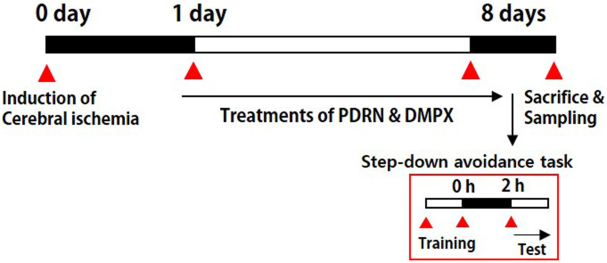
The gerbils from the PDRN-treated groups were injected intraperitoneally with 0.3 mL of normal saline (0.9%) containing 8 mg/kg of PDRN (Rejuvenex®, PharmaResearch Products, Pangyo, Korea), one time per a day continued for 7 days, started a day after operation. Based on preliminary data and previous studies, the PDRN concentration considered to be the most effective was used in this experiment [17, 21]. Additionally, in order to confirm that the adenosine A2A receptor is involved in the effect mediated by PDRN, 8 mg/kg DMPX (Sigma Chemical Co., St. Louis, MO, USA), an adenosine A2A receptor antagonist, was simultaneously administered with PDRN. In the sham-operation group and in the cerebral ischemia-induced group, the gerbils received 0.3 mL of normal saline without drugs according to the same timetable. The experiment schedule is shown in Fig 1.

Fig 1. Experimental schedule.

Fig 1

2.4. Step-down avoidance task

In the same manner as described above [22, 23], the step-down avoidance task was performed to determine the short-term memory. Eight days after induction of cerebral ischemia, the gerbils implemented a step-down avoidance task. Each gerbil was placed on a 7 × 25 cm platform at height of 2.5 cm, and the platform faced a grid of parallel steel bars (45 × 25 cm) with a diameter of 0.1 cm and a spacing of 1 cm. Going down the grid during training, the animal immediately took the out of the box after receiving a 0.3 mA foot shock for 2 seconds. After 2 hours of training period, the latency of each gerbil was determined. During the test time, the gerbils were placed back on the platform, and the latency time was defined as the time until the animal descended and placed all four feet on the grid. A delay of latency time more than 180 seconds was also calculated as 180 seconds.

2.5. Tissues preparation

On the 8th day after ischemia induction, immediately after measuring the latency of the step-down avoidance task, the animals were sacrificed. After anesthetizing the gerbils using the Zoletil 50® (10 mg/kg, ip; Vibac Laboratories), heart puncture was used to collect blood, left at room temperature for 1 hour and centrifugation was performed at 3,000 rpm for 20 minutes to obtain serum. After blood sampling, 50 mM phosphate-buffered saline (PBS) was transcardially perfused, and then fixed with a solution consisting of 4% paraformaldehyde in 100 mM phosphate buffer (PB, pH 7.4). After removing the brains, the brains were with the same fixative solution and then transferred to 30% sucrose solution to prevent freezing. Thereafter, 40-μm thick coronal sections were made using a freezing microtome (Leica, Nussloch, Germany), and an average of 10 sections was obtained in the CA1 region of each gerbil.

2.6. Pro-inflammatory cytokines and cAMP concentrations

Serum and hippocampus levels of pro-inflammatory cytokines (TNF-α and IL-1β) and cAMP concentrations were determined using enzyme-linked immunosorbent assay (ELISA). Enzyme immunoassay kits were used to detect the concentrations of TNF-α, IL-1β and cAMP in accordance with the manufacturer’s instructions (Abcam, Cambridge, UK), in the same manner as described above (n = 3 in each group) [17].

2.7. Western blot analysis

According to the same manner as described above [24, 25], analysis of western blotting was conducted (n = 4 in each group). Priority, approximately 30 mg of hippocampal tissues were extracted using 100 mg/mL of lysis buffer. The tissues were homogenized using a lysis buffer consisting of 1 mM PMSF, 1 mM EGTA, 1 mM Na2VO4, 1.5 mM MgCl2·6H2O, 50 mM Tris-HCl (pH 8.0), 100 mM NaF, 150 mM NaCl, 1% Triton X-100 and 10% glycerol, and then this homogeneous mixture was centrifuged at 14,000 rpm for 30 minutes. Concentration of protein was detected by a colorimetric protein analysis kit (Bio-Rad, Hercules, CA, USA). Protein 30 μg was separated from each sample on SDS-polyacrylamide gel and transferred to a nitrocellulose membrane. The antibodies for rabbit CREB antibody (1:1,000; Santa Cruz Biotechnology, Santa Cruz, CA, USA), phosphorylated (p)-CREB antibody (1:1,000; Santa Cruz Biotechnology), rabbit PKA antibody (1:1,000; Santa Cruz Biotechnology), p-PKA antibody (1:1,000; Santa Cruz Biotechnology), rabbit ERK antibody (1:2,000; Cell Signaling Technology, Danvers, USA), rabbit p-ERK antibody (1:2,000; Cell Signaling Technology), rabbit JNK antibody (1:2,000; Cell Signaling Technology), rabbit p-JNK antibody (1:2,000; Cell Signaling Technology), rabbit p38 antibody (1:2,000; Cell Signaling Technology), rabbit p-p38 antibody, (1:2,000; Cell Signaling Technology), mouse TNF-α antibody (1:1,000; Santa Cruz Biotechnology), mouse IL-1β antibody (1:1,000; Santa Cruz Biotechnology), mouse adenosine A2A receptor antibody (1:1,000; Santa Cruz Biotechnology), and β-actin antibody (1:1,000; Santa Cruz Biotechnology) used were as the primary antibodies. Horseradish peroxidase-conjugated anti-mouse antibodies (1:2,000; Vector Laboratories, Burlingame, CA, USA) for β-actin, TNF-α, IL-1β, adenosine A2A receptor and horseradish peroxidase-conjugated anti-rabbit antibodies (1:3,000; Vector Laboratories) for p-CREP, CREB, p-PKA, PKA, p-ERK, ERK, p-JNK, JNK, p-p38, p38 were used as the secondary antibodies.

The entire experimental step was carried out under normal laboratory conditions, except for membrane transfer performed at 4°C using pre-cooled buffer with cold pack. An enhanced chemiluminescence (ECL) detection kit (Santa Cruz Biotechnology) was used for band measurements. Each sample was loaded twice, and the number of samples was 4 per group.

2.8. Immunohistochemistry for NeuN

Immunohistochemistry for NeuN was conducted in the same manner as described above [26, 27]. The sections were treated with mouse anti-NeuN antibody (1:500; Abcam, Cambridge, MA, USA) overnight, and then treated with biotinylated mouse secondary antibody (1:200; Vector Laboratories, Burlingame, CA, USA) for an additional 1 hour. Secondary antibody was amplified using the Vector Elite ABC kit® (1:100; Vector Laboratories), and then 0.03% 3,3′-diaminobenzidine was used to show antibody-biotin-avidin-peroxidase complexes. The sections were then mounted on gelatin-coated slides, air-dried at room temperature overnight, and finally Permount® (Fisher Scientific, New Jersey, NJ, USA) was used to mount the coverslips.

2.9. Data analysis

The area of the hippocampal CA1 region on each slide was observed by optical microscope (Olympus, Tokyo, Japan), and the number of NeuN-positive cells was calculated using the Image-Pro® plus computer-assisted image analysis system (Media Cybernetics Inc., Silver Spring, MD, USA). The number of NeuN-positive cells was calculated using the following equation: N = Nv × Vref. N is the total number of NeuN-positive cells of the CA1 region, which is counted by multiplying the NeuN-positive cell numerical density Nv by the reference volume (mm3) Vref. Nv is the average numerical density of NeuN-positive cells and it is calculated based on the sum of counts within the CA1 region of each section and the volume of the CA1 region contained in each section. Vref is calculated according to Cavalieri’s method as follows [28, 29]: Vref = a × t × s. In this equation, a is the average area of the CA1 cell layer, t represents the average thickness (40 μm) of the microtome section, and s indicates the total number of sections through the reference volume.

In order to compare the relative levels of protein expressions, the bands were detected densitometrically by Molecular AnalystTM version 1.4.1 (Bio-Rad, Hercules). For relative quantification, a random value of 1.00 was given to the results of the sham-operation group (western blotting). One-way ANOVA with Duncan’s post-hoc test was used for data analysis, and the results are presented as mean ± standard error of the mean. For statistical analysis, the value of P < 0.05 was determined to be statistically significant.

3. Results

3.1. Changes of cerebral blood flow

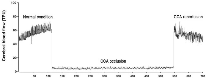
The flow of brain blood during carotid artery occlusion and reperfusion is shown in Fig 2. Occlusion of the carotid artery reduced brain blood flow and increased brain blood flow during reperfusion.

Fig 2. Brain blood flow during occlusion and reperfusion of both common carotid arteries.

Fig 2

3.2. Changes of short-term memory latency and NeuN-positive cell number in the CA 1 region

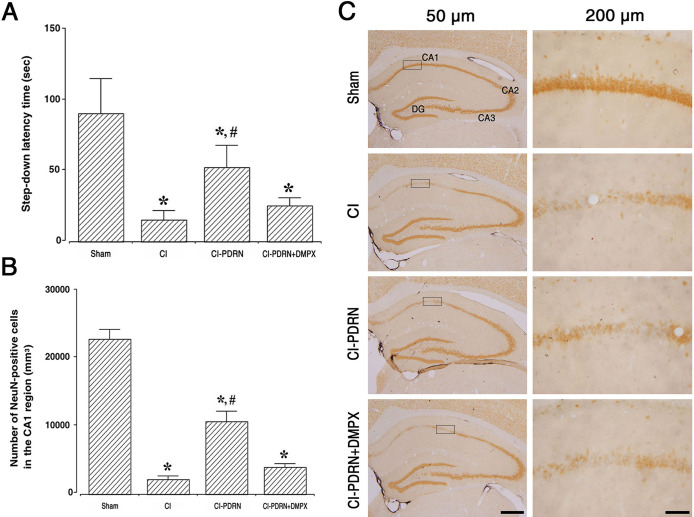
Fig 3A shows the results of the latency in the step-down avoidance task. Following the induction of cerebral ischemic damage, short-term memory was impaired (P < 0.05), and treatment with PDRN ameliorated this ischemia-induced memory impairment (P < 0.05). The co-administration of PDRN and DMPX failed to increase the latency observed with PDRN in cerebral ischemia.

Fig 3. Changes in short-term memory and neuronal survival in the CA1 region.

Fig 3

A. Latency of the step-down avoidance task in each group. B. Number of NeuN-positive cells in each group. C. Photomicrographs of NeuN-positive cells in the hippocampal CA1 region. The scale bar represents 50 μm (left) and 200 μm (right). (□) Area of magnification at 200 μm in CA1 region. Sham, sham-operation group; CI, cerebral ischemia-induced group; CI-PDRN, cerebral ischemia-induced and polydeoxyribonucleotide (PDRN)-treated group; CI-PDRN+DMPX, cerebral ischemia-induced and PDRN with 7-dimethyl-1-propargylxanthine (DMPX)-treated group. * indicates P < 0.05 compared with the sham-operation group. # indicates P < 0.05 compared with the cerebral ischemia-induced group.

Fig 3B and 3C are the photomicrographs of NeuN-positive cells in the hippocampal CA1 region. Cerebral ischemic damage decreased the number of NeuN-positive cells, indicating reduced neuronal survival in the hippocampal CA1 region (P < 0.05), and treatment with PDRN improved neuronal survival in cerebral ischemia (P < 0.05). The co-administration of PDRN and DMPX failed to enhance the neuronal survival observed with PDRN in cerebral ischemia.

3.3. Changes of pro-inflammatory cytokine expression

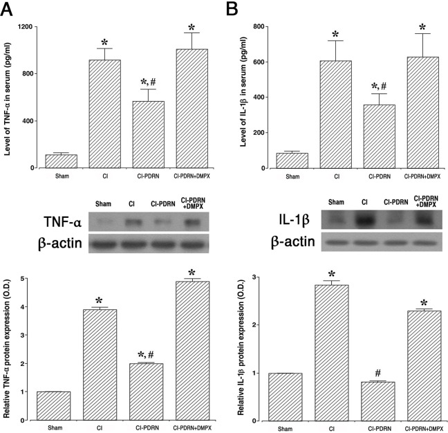
To determine whether PDRN improves cerebral ischemia, ELISA and western blot analysis were performed by examining the effect on PDRN on production of pro-inflammatory cytokines, TNF-α (Fig 4A) and IL-1β (Fig 4B). Following cerebral ischemic damage, TNF-α and IL-1β expressions were enhanced in the serum and hippocampus (P < 0.05), and treatment with PDRN suppressed the expressions of TNF-α and IL-1β (P < 0.05). The co-administration of PDRN and DMPX failed to decrease TNF-α and IL-1β expressions observed with PDRN in cerebral ischemia.

Fig 4. Altered expression of pro-inflammatory cytokines in the serum and hippocampus.

Fig 4

A-upper. Concentration of tumor necrosis factor-α (TNF-α) in the serum. A-lower. The relative level of TNF-α in the hippocampus. B-upper. Concentration of interleukin (IL)-1β in the serum. B-lower. The relative level of IL-1β in the hippocampus. Sham, sham-operation group; CI, cerebral ischemia-induced group; CI-PDRN, cerebral ischemia-induced and polydeoxyribonucleotide (PDRN)-treated group; CI-PDRN+DMPX, cerebral ischemia-induced and PDRN with 7-dimethyl-1-propargylxanthine (DMPX)-treated group. * indicates P < 0.05 compared with the sham-operation group. # indicates P < 0.05 compared with the cerebral ischemia-induced group.

3.4. Changes of phosphorylation of MAPK cascade

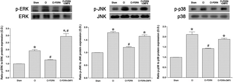
To determine whether cerebral ischemia is improved by PDRN, the effect of PDRN on MAPK phosphorylation was investigated using western blotting (Fig 5). Induction of cerebral ischemia promoted MAPK cascade phosphorylation, such as ERK, JNK and p38 (P < 0.05). Interestingly, treatment with PDRN more enhanced the phosphorylation of ERK, JNK and p38 (P < 0.05). The co-administration of PDRN and DMPX failed to further enhance ERK, JN and p38 phosphorylation observed with PDRN in cerebral ischemia.

Fig 5. Changes in the mitogen-activated protein kinase (MAPK) cascade in the hippocampus.

Fig 5

Left. Ratio of phosphorylated extracellular signal-regulated kinases (p-ERK) to ERK. Middle. Ratio of phosphorylated c-Jun NH2-terminal kinases (p-JNK) to JNK. Right. Ratio of phosphorylated p38 kinase (p-p38) to p38 in the hippocampus. Sham, sham-operation group; CI, cerebral ischemia-induced group; CI-PDRN, cerebral ischemia-induced and polydeoxyribonucleotide (PDRN)-treated group; CI-PDRN+DMPX, cerebral ischemia-induced and PDRN with 7-dimethyl-1-propargylxanthine (DMPX)-treated group. * indicates P < 0.05 compared with the sham-operation group. # indicates P < 0.05 compared with the cerebral ischemia-induced group.

3.5. Changes of cAMP concentration and adenosine A2A receptor expression

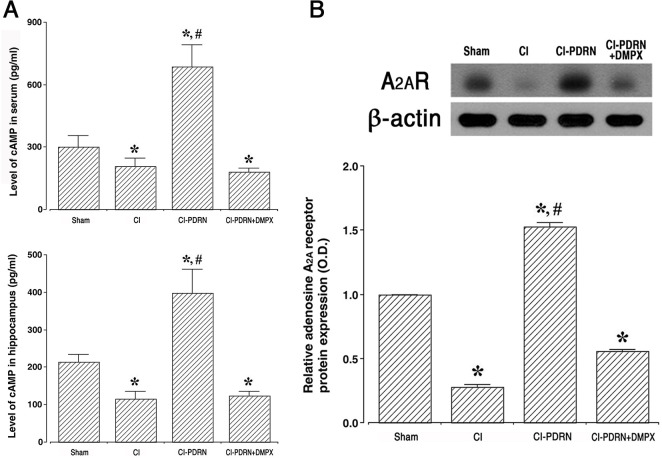
The cAMP concentration in the serum and hippocampus and adenosine A2A receptor expression in the hippocampus are shown in Fig 6. Induction of cerebral ischemia decreased the levels of cAMP concentration and adenosine A2A receptor expression (P < 0.05), and treatment with PDRN improved the levels of cAMP concentration and adenosine A2A receptor expression (P < 0.05). The co-administration of PDRN and DMPX failed to increase the levels of cAMP concentration and adenosine A2A receptor expression observed with PDRN in cerebral ischemia.

Fig 6. Changes in cAMP concentration and adenosine A2A receptor expression.

Fig 6

A-upper. Concentration of cAMP in serum. A-lower. Concentration of cAMP in the hippocampus. B. The relative expression of the adenosine A2A receptor in the hippocampus. Sham, sham-operation group; CI, cerebral ischemia-induced group; CI-PDRN, cerebral ischemia-induced and polydeoxyribonucleotide (PDRN)-treated group; CI-PDRN+DMPX, cerebral ischemia-induced and PDRN with 7-dimethyl-1-propargylxanthine (DMPX)-treated group. * indicates P < 0.05 compared with the sham-operation group. # indicates P < 0.05 compared with the cerebral ischemia-induced group.

3.6. Changes of ratio in p-CREB vs CREB and p-PKA vs PKA

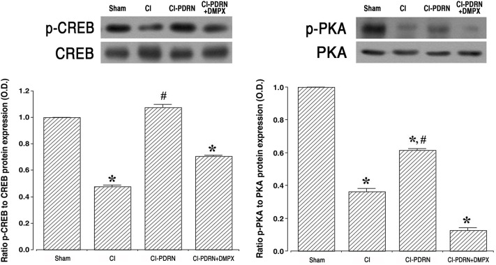
Western blot analysis was used to measure the relative expressions of p-CREB vs CREB and p-PKA vs PKA (Fig 7). Induction of cerebral ischemia decreased the ratio of p-CREB vs CREB and the ratio of p-PKA vs PKA when compared with the sham-operation group (P < 0.05). Treatment with PDRN increased the ratio of p-CREB vs CREB and ratio of p-PKA vs PKA in the hippocampus (P < 0.05). The co-administration of PDRN and DMPX failed to enhance the phosphorylation of CREB and PKA observed with PDRN in cerebral ischemia.

Fig 7. Changes in phosphorylated cAMP response element-binding protein (p-CREB) to CREB ratio and phosphorylated protein kinases A (p-PKA) to PKA ratio.

Fig 7

Left. Ratio of p-CREB to CREB in the hippocampus. Right. Ratio of p-PKA to PKA in the hippocampus. Sham, Sham-operation group; CI, cerebral ischemia-induced group; CI-PDRN, cerebral ischemia-induced and polydeoxyribonucleotide (PDRN)-treated group; CI-PDRN+DMPX, cerebral ischemia-induced and PDRN with 7-dimethyl-1-propargylxanthine (DMPX)-treated group. * indicates P < 0.05 compared with the sham-operation group. # indicates P < 0.05 compared with the cerebral ischemia-induced groups.

4. Discussion

Damage by ischemia particularly destroys pyramidal neurons in the hippocampal CA1 region, and ischemia induces apoptotic cell death in the hippocampal CA1 neurons [19, 30]. Pyramidal neurons are crucial for learning and memory, and appearance of passive avoidance memory impairment after ischemia is associated with damage in the neurons of the CA1 region [31, 32]. In the current study, the cerebral ischemic injury resulted in cell loss in the hippocampal CA1 neurons, and this neuronal cell loss reduced short-term memory when compared with the gerbils of the sham-operation group. The present findings are similar to previous studies showing that loss of neurons in the CA1 region caused short-term memory impairment [31, 33].

The ischemic brain exhibits inflammation characterized by the accumulation of inflammatory cells and mediators. Previous studies have suggested that induction of ischemic damage enhances neuronal cell loss owing to increased levels of inflammatory exudates and pro-inflammatory cytokines [6, 7, 34]. Furthermore, an increment of inflammatory cytokines in the brain acts as a major causes of neuronal cell loss and memory impairment [35, 36]. Based on current findings, enhanced secretion of TNF-α and IL-1β, pro-inflammatory cytokines, in the serum, hippocampus, and basolateral amygdala (Supplement 1 in S1 File) exacerbated the symptoms of ischemic injury. These results indicate that symptoms were worsened by excessive production of pro-inflammatory cytokines during cerebral ischemia. Inhibiting the secretion of pro-inflammatory cytokines is one of the important treatment strategies for the brain ischemic injury.

Most of cells involved in wound healing express the adenosine A2A receptor [17], and this adenosine A2A receptor is locates in several brain regions and modulates the pathophysiological response to ischemic stroke [14]. Agonists on adenosine A2A receptor have been reported to be useful for the treating of inflammatory diseases [14, 37]. In the previous studies, adenosine A2A receptor knockout mice exhibited symptoms of chronic cerebral ischemia such as working memory impairment, increased demyelination, glial proliferation, and increased pro-inflammatory cytokines [4, 38]. Reduced functional capacity of BDNF in adenosine A2A receptor knockout mice was associated with a decrease in hippocampal BDNF level, and pharmacological blockade of adenosine A2A receptors significantly reduced BDNF level in the hippocampus of normal mice [39]. These results indicated that tonic activation of adenosine A2A receptor is required for BDNF-induced potentiation of synaptic transmission and for sustaining a normal BDNF tone in the hippocampus [39]. The facilitating action of BDNF on hippocampal long-term potentiation is critically dependent on the presence of extracellular adenosine and activation of the A2A receptor through a cAMP/PKA-dependent mechanism [40]. Activation of the adenosine A2A receptor regulates BDNF production in rat cortical neurons, which provides neuroprotective action [15].

In the current study, PDRN treatment substantially suppressed the secretion of pro-inflammatory cytokines. Inflammation is a compensatory response to cellular and tissue damage caused by ischemia-reperfusion injury, and the inflammatory response is primarily regulated by the signaling pathway of the MAPK cascade [8, 12]. MAPK is essential for the regulation of various inflammatory mediators, and MAPK is a kind of kinases that regulate cellular response to external stress signals or inflammatory cytokines [2, 41]. It was demonstrated that the induction of ischemic damage activated phosphorylation of the MAPK cascade, and this activation of MAPK controls a wide range of cellular processes [2, 12]. Our current study found that phosphorylation of the MAPK cascade pathway was increased by ischemic damage and this phosphorylation of the MAPK cascade pathway was reduced by PDRN treatment.

The intracellular concentration of cAMP is increased by stimulation of the adenosine A2A receptor, and this increased cAMP concentration serves as a physiological inhibitor to function of inflammatory neutrophil [42]. Furthermore, adenosine A2A receptor activation promotes signal from the cAMP-PKA pathway and accelerates the level of CREB phosphorylation. Elevated cAMP concentration suppresses the level of phosphorylation of the MAPK cascade pathway in stimulated cells [42, 43]. Adenosine A2A receptor agonist, PDRN, has been proposed to have therapeutic potential in inflammatory diseases [16, 17]. In the current study, PDRN treatment increased the cAMP concentration in gerbils presenting cerebral ischemia, and this increased cAMP concentration inhibited phosphorylation of the MAPK cascade pathway, thereby inactivating the MAPK cascade pathway in the hippocampus and basolateral amygdala (Supplements 2–4 in S1 File).

The current study has revealed that PDRN treatment inhibits inflammation, improves neuronal cell survival, and prevents a decline in short-term memory in a brain ischemia animal model (S1 Graphic abstract). Co-administration of PDRN and adenosine A2A receptor antagonist DMPX attenuated the therapeutic effect of PDRN in cerebral ischemia. Based on these findings, PDRN may be developed as the primary treatment in cerebral ischemia.

Supporting information

S1 File

(DOCX)

S1 Graphic abstract

(TIF)

S1 Raw images

(PDF)

Data Availability

All relevant data are within the paper and its Supporting Information files.

Funding Statement

• Jin Hee Han • NRF-2017R1D1A1B03032827 • National Research Foundation of Korea • https://www.nrf.re.kr/ • Research fund support.

References

  • 1.Kwakkel G, Kollen B, Lindeman E. Understanding the pattern of functional recovery after stroke: facts and theories. Restor Neurol Neurosci. 2004; 22(3–5):281–99. [PubMed] [Google Scholar]
  • 2.Wang T, Wang F, Yu L, Li Z. Nobiletin alleviates cerebral ischemic-reperfusion injury via MAPK signaling pathway. Am J Transl Res. 2019; 11(9):5967–77. [PMC free article] [PubMed] [Google Scholar]
  • 3.Chen F, Qi Z, Luo Y, Hinchliffe T, Ding G, Xia Y, et al. Non-pharmaceutical therapies for stroke: mechanisms and clinical implications. Prog Neurobiol. 2014; 115(2):246–69. 10.1016/j.pneurobio.2013.12.007 [DOI] [PMC free article] [PubMed] [Google Scholar]
  • 4.Pedata F, Pugliese AM, Coppi E, Dettori I, Maraula G, Cellai L, et al. Adenosine A2A receptors modulate acute injury and neuroinflammation in brain ischemia. Mediators Inflamm. 2014; 2014:805198. 10.1155/2014/805198 [DOI] [PMC free article] [PubMed] [Google Scholar]
  • 5.Eltzschig HK, Eckle T. Ischemia and reperfusion-from mechanism to translation. Nat Med. 2011; 17(11):1391–401. 10.1038/nm.2507 [DOI] [PMC free article] [PubMed] [Google Scholar]
  • 6.Kong LL, Wang ZY, Han N, Zhuang XM, Wang ZZ, Li H, et al. Neutralization of chemokine-like factor 1, a novel C-C chemokine, protects against focal cerebral ischemia by inhibiting neutrophil infiltration via MAPK pathways in rats. J Neuroinflammation. 2014; 11:112. 10.1186/1742-2094-11-112 [DOI] [PMC free article] [PubMed] [Google Scholar]
  • 7.Ahmad M, Graham SH. Inflammation after stroke: mechanisms and therapeutic approaches. Transl Stroke Res. 2010; 1(2):74–84. 10.1007/s12975-010-0023-7 [DOI] [PMC free article] [PubMed] [Google Scholar]
  • 8.Kovalska M, Kovalska L, Pavlikova M, Janickova M, Mikuskova K, Adamkov M, et al. Intracellular signaling MAPK pathway after cerebral ischemia-reperfusion injury. Neurochem Res. 2012; 37(7):1568–77. 10.1007/s11064-012-0752-y [DOI] [PubMed] [Google Scholar]
  • 9.Kyriakis JM, Avruch J. Mammalian MAPK signal transduction pathways activated by stress and inflammation: a 10-year update. Physiol Rev. 2012; 92(2):689–737. 10.1152/physrev.00028.2011 [DOI] [PubMed] [Google Scholar]
  • 10.Jiang M, Li J, Peng Q, Liu Y, Liu W, Luo C, et al. Neuroprotective effects of bilobalide on cerebral ischemia and reperfusion injury are associated with inhibition of pro-inflammatory mediator production and down-regulation of JNK1/2 and p38 MAPK activation. J Neuroinflammation. 2014; 11:167. 10.1186/s12974-014-0167-6 [DOI] [PMC free article] [PubMed] [Google Scholar]
  • 11.Yang CH, Yen TL, Hsu CY, Thomas PA, Sheu JR, Jayakumar T. Multi-targeting andrographolide, a novel NF-κB inhibitor, as a potential therapeutic agent for stroke. Int J Mol Sci. 2017; 18(8):E1638. 10.3390/ijms18081638 [DOI] [PMC free article] [PubMed] [Google Scholar]
  • 12.Meng X, Xie W, Xu Q, Liang T, Xu X, Sun G, et al. Neuroprotective effects of radix scrophulariae on cerebral ischemia and reperfusion injury via MAPK pathways. Molecules. 2018; 23(9):E2401. 10.3390/molecules23092401 [DOI] [PMC free article] [PubMed] [Google Scholar]
  • 13.Melani A, Pugliese AM, Pedata F. Adenosine receptors in cerebral ischemia. Int Rev Neurobiol. 2014; 119:309–48. 10.1016/B978-0-12-801022-8.00013-1 [DOI] [PubMed] [Google Scholar]
  • 14.Zhou Y, Zeng X, Li G, Yang Q, Xu J, Zhang M, et al. Inactivation of endothelial adenosine A2A receptors protects mice from cerebral ischaemia-induced brain injury. Br J Pharmacol. 2019;176(13):2250–63. 10.1111/bph.14673 [DOI] [PMC free article] [PubMed] [Google Scholar]
  • 15.Jeon SJ, Rhee SY, Ryu JH, Cheong JH, Kwon K, Yang SI, et al. Activation of adenosine A2A receptor up-regulates BDNF expression in rat primary cortical neurons. Neurochem Res. 2011; 36(12):2259–69. 10.1007/s11064-011-0550-y [DOI] [PubMed] [Google Scholar]
  • 16.An J, Park SH, Ko IG, Jin JJ, Hwang L, Ji ES, et al. Polydeoxyribonucleotide ameliorates lipopolysaccharide-induced lung injury by inhibiting apoptotic cell death in rats. Int J Mol Sci. 2017; 18(9):E1847. 10.3390/ijms18091847 [DOI] [PMC free article] [PubMed] [Google Scholar]
  • 17.Ko IG, Kim SE, Jin JJ, Hwang L, Ji ES, Kim CJ, et al. Combination therapy with polydeoxyribonucleotide and proton pump inhibitor enhances therapeutic effectiveness for gastric ulcer in rats. Life Sci. 2018; 203:12–9. 10.1016/j.lfs.2018.04.009 [DOI] [PubMed] [Google Scholar]
  • 18.Yoon S, Kang JJ, Kim J, Park S, Kim JM. Efficacy and safety of intra-articular injections of hyaluronic acid combined with polydeoxyribonucleotide in the treatment of knee osteoarthritis. Ann Rehabil Med. 2019; 43(2):204–14. 10.5535/arm.2019.43.2.204 [DOI] [PMC free article] [PubMed] [Google Scholar]
  • 19.Ko IG, Shin MS, Kim BK, Kim SE, Sung YH, Kim TS, et al. Tadalafil improves short-term memory by suppressing ischemia-induced apoptosis of hippocampal neuronal cells in gerbils. Pharmacol Biochem Behav. 2009;91(4):629–35. 10.1016/j.pbb.2008.10.009. [DOI] [PubMed] [Google Scholar]
  • 20.Lee SH, Ko IG, Kim SE, Hwang L, Jin JJ, Choi HH, et al. Aqueous extract of Cordyceps alleviates cerebral ischemia-induced short-term memory impairment in gerbils. J Exerc Rehabil. 2016; 12(2):69–78. 10.12965/jer.1632586.293 [DOI] [PMC free article] [PubMed] [Google Scholar]
  • 21.Jeon JW, Lee JI, Shin HP, Cha JM, Joo KR, Kim SH, et al. Adenosine A2A-receptor agonist polydeoxyribonucleotide promotes gastric ulcer healing in Mongolian gerbils. Animal Cells Syst. 2014; 18(6): 399–406. 10.1080/19768354.2014.983968. [DOI] [Google Scholar]
  • 22.Park JH, Kim SE, Jin JJ, Choi HS, Kim CJ, Ko IG. Pentoxifylline alleviates perinatal hypoxic-ischemia-induced short-term memory impairment by suppressing apoptosis in the hippocampus of rat pups. Int Neurourol J. 2016; 20(2):107–13. 10.5213/inj.1632532.266 [DOI] [PMC free article] [PubMed] [Google Scholar]
  • 23.Ko IG, Kim CJ, Kim H. Treadmill exercise improves memory by up-regulating dopamine and down-regulating D2 dopamine receptor in traumatic brain injury rats. J Exerc Rehabil. 2019; 15(4):504–11. 10.12965/jer.1938316.158 [DOI] [PMC free article] [PubMed] [Google Scholar]
  • 24.Ko IG, Kim SE, Hwang L, Jin JJ, Kim CJ, Kim BK, et al. Late starting treadmill exercise improves spatial leaning ability through suppressing CREP/BDNF/TrkB signaling pathway following traumatic brain injury in rats. J Exerc Rehabil. 2018; 14(3):327–34. 10.12965/jer.1836248.124 [DOI] [PMC free article] [PubMed] [Google Scholar]
  • 25.Shin MS, Kim TW, Park SS, Ko IG, Kim CJ, Kim M, et al. Long-term surgical and chemical castration deteriorates memory function through downregulation of PKA/CREB/BDNF and c-Raf/MEK/ERK pathways in hippocampus. Int Neurourol J. 2019; 23(2):116–24. 10.5213/inj.1938103.052 [DOI] [PMC free article] [PubMed] [Google Scholar]
  • 26.Cho JW, Jung SY, Kim DY, Chung YR, Choi HH, Jeon JW, et al. PI3K-Akt-Wnt pathway is implicated in exercise-induced improvement of short-term memory in cerebral palsy rats. Int Neurourol J. 2018; 22(3):S156–64. 10.5213/inj.1836224.112 [DOI] [PMC free article] [PubMed] [Google Scholar]
  • 27.Lee JM, Ji ES, Kim TW, Kim CJ, Shin MS, Lim BV, et al. Treadmill exercise improves memory function by inhibiting hippocampal apoptosis in pilocarpine-induced epileptic rats. J Exerc Rehabil. 2018; 14(5):713–23. 10.12965/jer.36394.197 [DOI] [PMC free article] [PubMed] [Google Scholar]
  • 28.Kempermann G, Kuhn HG, Gage FH. More hippocampal neurons in adult mice living in an enriched environment. Nature. 1997; 386(6624):493–5. 10.1038/386493a0 [DOI] [PubMed] [Google Scholar]
  • 29.Hwang L, Ko IG, Jin JJ, Kim SH, Kim CJ, Chang B, et al. Dexmedetomidine ameliorates memory impairment in sleep-deprived mice. Anim Cells Syst (Seoul). 2019; 23(6):371–9. 10.1080/19768354.2019.1688185 [DOI] [PMC free article] [PubMed] [Google Scholar]
  • 30.Schmidt-Kastner R, Freund TF. Selective vulnerability of the hippocampus in brain ischemia. Neuroscience. 1991; 40(3):599‐636. 10.1016/0306-4522(91)90001-5 [DOI] [PubMed] [Google Scholar]
  • 31.Erfani S, Khaksari M, Oryan S, Shamsaei N, Aboutaleb N, Nikbakht F, et al. Visfatin reduces hippocampal CA1 cells death and improves learning and memory deficits after transient global ischemia/reperfusion. Neuropeptides. 2015; 49:63–8. 10.1016/j.npep.2014.12.004 [DOI] [PubMed] [Google Scholar]
  • 32.Cho CH, Byun HR, Jover-Mengual T, Pontarelli F, Dejesus C, Cho AR, et al. Gadd45b acts as neuroprotective effector in global ischemia-induced neuronal death. Int Neurourol J. 2019; 23(1):S11–21. 10.5213/inj.1938040.020 [DOI] [PMC free article] [PubMed] [Google Scholar]
  • 33.Zamani M, Hassanshahi J, Soleimani M, Zamani F. Neuroprotective effect of olive oil in the hippocampus CA1 neurons following ischemia: Reperfusion in mice. J Neurosci Rural Pract. 2013; 4(2):164‐70. 10.4103/0976-3147.112753 [DOI] [PMC free article] [PubMed] [Google Scholar]
  • 34.Lambertsen KL, Biber K, Finsen B. Inflammatory cytokines in experimental and human stroke. J Cereb Blood Flow Metab. 2012; 32(9):1677–98. 10.1038/jcbfm.2012.88 [DOI] [PMC free article] [PubMed] [Google Scholar]
  • 35.Brea D, Sobrino T, Ramos-Cabrer P, Castillo J. Inflammatory and neuroimmunomodulatory changes in acute cerebral ischemia. Cerebrovasc Dis. 2009; 27:S48–S64. 10.1159/000200441 [DOI] [PubMed] [Google Scholar]
  • 36.Naderi Y, Sabetkasaei M, Parvardeh S, Moini Zanjani T. Neuroprotective effects of pretreatment with minocycline on memory impairment following cerebral ischemia in rats. Behav Pharmacol. 2017; 28:214–22. 10.1097/FBP.0000000000000297 [DOI] [PubMed] [Google Scholar]
  • 37.Milne GR, Palmer TM. Anti-inflammatory and immunosuppressive effects of the A2A adenosine receptor. ScientificWorldJournal. 2011; 11:320–39. 10.1100/tsw.2011.22 [DOI] [PMC free article] [PubMed] [Google Scholar]
  • 38.Adén U, Halldner L, Lagercrantz H, Dalmau I, Ledent C, Fredholm BB. Aggravated brain damage after hypoxic ischemia in immature adenosine A2A knockout mice. Stroke. 2003; 34(3):739–44. 10.1161/01.STR.0000060204.67672.8B [DOI] [PubMed] [Google Scholar]
  • 39.Tebano MT, Martire A, Potenza RL, Grò C, Pepponi R, Armida M, et al. Adenosine A2A receptors are required for normal BDNF levels and BDNF-induced potentiation of synaptic transmission in the mouse hippocampus. J Neurochem. 2008; 104(1):279–86. 10.1111/j.1471-4159.2007.05046.x [DOI] [PubMed] [Google Scholar]
  • 40.Fontinha BM, Diógenes MJ, Ribeiro JA, Sebastião AM. Enhancement of long-term potentiation by brain-derived neurotrophic factor requires adenosine A2A receptor activation by endogenous adenosine. Neuropharmacology. 2008; 54(6):924–33. 10.1016/j.neuropharm.2008.01.011 [DOI] [PubMed] [Google Scholar]
  • 41.Fann DY, Lim YA, Cheng YL, Lok KZ, Chunduri P, Baik SH, et al. Evidence that NF-κB and MAPK signaling promotes NLRP inflammasome activation in neurons following ischemic stroke. Mol Neurobiol. 2018; 55(2):1082–96. 10.1007/s12035-017-0394-9 [DOI] [PubMed] [Google Scholar]
  • 42.Giambelluca MS, Pouliot M. Early tyrosine phosphorylation events following adenosine A2A receptor in human neutrophils: identification of regulated pathways. J Leukoc Biol. 2017; 102(3):829–36. 10.1189/jlb.2VMA1216-517R [DOI] [PMC free article] [PubMed] [Google Scholar]
  • 43.Chera S, Ghila L, Wenger Y, Galliot B. Injury-induced activation of the MAPK/CREB pathway triggers apoptosis-induced compensatory proliferation in hydra head regeneration. Dev Growth Differ. 2011; 53(2):186–201. 10.1111/j.1440-169X.2011.01250.x [DOI] [PubMed] [Google Scholar]

Decision Letter 0

Giuseppe Pignataro

18 Dec 2020

PONE-D-20-25445

Adenosine A2A receptor agonist polydeoxyribonucleotide ameliorates short-term memory impairment by suppressing cerebral ischemia-induced inflammation via MAPK pathway

PLOS ONE

Dear Dr. Han,

Thank you for submitting your manuscript to PLOS ONE. After careful consideration, we feel that it has merit but does not fully meet PLOS ONE’s publication criteria as it currently stands. Therefore, we invite you to submit a revised version of the manuscript that addresses the points raised during the review process.

Please submit your revised manuscript by Jan 29 2021 11:59PM. If you will need more time than this to complete your revisions, please reply to this message or contact the journal office at plosone@plos.org. When you're ready to submit your revision, log on to https://www.editorialmanager.com/pone/ and select the 'Submissions Needing Revision' folder to locate your manuscript file.

Please include the following items when submitting your revised manuscript:

  • A rebuttal letter that responds to each point raised by the academic editor and reviewer(s). You should upload this letter as a separate file labeled 'Response to Reviewers'.

  • A marked-up copy of your manuscript that highlights changes made to the original version. You should upload this as a separate file labeled 'Revised Manuscript with Track Changes'.

  • An unmarked version of your revised paper without tracked changes. You should upload this as a separate file labeled 'Manuscript'.

If you would like to make changes to your financial disclosure, please include your updated statement in your cover letter. Guidelines for resubmitting your figure files are available below the reviewer comments at the end of this letter.

If applicable, we recommend that you deposit your laboratory protocols in protocols.io to enhance the reproducibility of your results. Protocols.io assigns your protocol its own identifier (DOI) so that it can be cited independently in the future. For instructions see: http://journals.plos.org/plosone/s/submission-guidelines#loc-laboratory-protocols

We look forward to receiving your revised manuscript.

Kind regards,

Giuseppe Pignataro, MD, PhD

Academic Editor

PLOS ONE

Journal Requirements:

When submitting your revision, we need you to address these additional requirements.

1. Please ensure that your manuscript meets PLOS ONE's style requirements, including those for file naming. The PLOS ONE style templates can be found at

https://journals.plos.org/plosone/s/file?id=wjVg/PLOSOne_formatting_sample_main_body.pdf and

https://journals.plos.org/plosone/s/file?id=ba62/PLOSOne_formatting_sample_title_authors_affiliations.pdf

2. As part of your revisions, we kindly request additional details pertaining to animal care, use and research procedures. Please address the following items in your Methods section: (1) all methods undertaken to minimize potential pain and distress; (2) environmental enrichments that the gerbils were provided (if applicable); (3) the number of animals that died prior to the experimental endpoints and the cause of death for any that were found dead; (4) the monitoring parameters and the humane endpoints/criteria for early euthanasia for animals who became severely ill. We thank you for your attention to this matter.

3. PLOS ONE now requires that authors provide the original uncropped and unadjusted images underlying all blot or gel results reported in a submission’s figures or Supporting Information files. This policy and the journal’s other requirements for blot/gel reporting and figure preparation are described in detail at https://journals.plos.org/plosone/s/figures#loc-blot-and-gel-reporting-requirements and https://journals.plos.org/plosone/s/figures#loc-preparing-figures-from-image-files. When you submit your revised manuscript, please ensure that your figures adhere fully to these guidelines and provide the original underlying images for all blot or gel data reported in your submission. See the following link for instructions on providing the original image data: https://journals.plos.org/plosone/s/figures#loc-original-images-for-blots-and-gels.

In your cover letter, please note whether your blot/gel image data are in Supporting Information or posted at a public data repository, provide the repository URL if relevant, and provide specific details as to which raw blot/gel images, if any, are not available. Email us at plosone@plos.org if you have any questions.

[Note: HTML markup is below. Please do not edit.]

Reviewers' comments:

Reviewer's Responses to Questions

Comments to the Author

1. Is the manuscript technically sound, and do the data support the conclusions?

The manuscript must describe a technically sound piece of scientific research with data that supports the conclusions. Experiments must have been conducted rigorously, with appropriate controls, replication, and sample sizes. The conclusions must be drawn appropriately based on the data presented.

Reviewer #1: Yes

Reviewer #2: Partly

**********

2. Has the statistical analysis been performed appropriately and rigorously?

Reviewer #1: Yes

Reviewer #2: I Don't Know

**********

3. Have the authors made all data underlying the findings in their manuscript fully available?

The PLOS Data policy requires authors to make all data underlying the findings described in their manuscript fully available without restriction, with rare exception (please refer to the Data Availability Statement in the manuscript PDF file). The data should be provided as part of the manuscript or its supporting information, or deposited to a public repository. For example, in addition to summary statistics, the data points behind means, medians and variance measures should be available. If there are restrictions on publicly sharing data—e.g. participant privacy or use of data from a third party—those must be specified.

Reviewer #1: Yes

Reviewer #2: Yes

**********

4. Is the manuscript presented in an intelligible fashion and written in standard English?

PLOS ONE does not copyedit accepted manuscripts, so the language in submitted articles must be clear, correct, and unambiguous. Any typographical or grammatical errors should be corrected at revision, so please note any specific errors here.

Reviewer #1: Yes

Reviewer #2: Yes

**********

5. Review Comments to the Author

Please use the space provided to explain your answers to the questions above. You may also include additional comments for the author, including concerns about dual publication, research ethics, or publication ethics. (Please upload your review as an attachment if it exceeds 20,000 characters)

Reviewer #1: Results

In the present work Il-Gyu Ko and colleagues demonstrated that the administration of an Adenosine A2a receptor agonist, polydeoxyribonucleotide (PDRN), following the induction of transient global ischemia in gerbils results in an improvement in short-term memory tested 8 days following induction of ischemia and in the survival of NeuN-positive cells in the CA1 region of Hippocampus. PDRN induced a decrease in proinflammatory cytokine production and in the activation of MAPK signalling which were increased following ischemia. Moreover an increase in the concentration of C-AMP in serum and hippocampus and of A2a receptor expression, accompanied by an increase of CREB and PKA phosphorylation and a decrease of the phosphorylation of ERK, p38 and JNK were observed. The co-treatment with the A2a receptor antagonist DMPX attenuated the therapeutic effect of PDRN.

The manuscript provides a demonstration of the efficacy of an adenosine A2a receptor agonist on cognitive decline following cerebral ischemia by inhibiting the activation of endogenous inflammation mechanisms.

However, the neuroprotective effect of adenosine neurotransmission in ischemia is not emphasized in the introduction and a greater emphasis could underline the involvement of the pathway involved in neuronal plasticity in the discussion.

English should be checked in all the text and in some places there is no correspondence between text and figures.

Regarding the treatment, the injection volume is unclear as different values ​​are reported in the abstract and in the text.

In Figure 2C the spatial reference of the magnification at 200 μm is missing.

Regarding the cognitive task it is not clear when the training phase was carried out with respect to induction of ischemia.

Moreover, only the hippocampus was considered in the measurements. Other brain areas, for example the basolateral amygdala, are also important in the acquisition and consolidation of the memory trace relating to the association between the shock and the position of the animal.

Reviewer #2: Paper by Jin-Hee Han and collegues describes the effects of adenosine A2A receptor agonist, polydeoxyribonucleotide (PDRN), on short-term memory in gerbils subjected to global ischemia. The authors speculate that treatment with PDRN ameliorated short-term memory impairment by suppressing the production of pro-inflammatory cytokines and inactivation of MAPK signaling factors in cerebral ischemia. The work is discreet, well written and is the first to analyze the effect of polydeoxyribonucleotide (PDRN) in an ischemia model. A couple of issues need to be resolved:

1. There is no indication of the calculation of the sample size for in vivo experiments together with the number of experimental groups for western blotting evaluations etc.

2. What evidence is available regarding the ability of the experimental compound PNDR to cross the blood brain barrier when administered intraperitoneally?

**********

6. PLOS authors have the option to publish the peer review history of their article (what does this mean?). If published, this will include your full peer review and any attached files.

If you choose “no”, your identity will remain anonymous but your review may still be made public.

Do you want your identity to be public for this peer review? For information about this choice, including consent withdrawal, please see our Privacy Policy.

Reviewer #1: No

Reviewer #2: Yes: Antonio Vinciguerra

[NOTE: If reviewer comments were submitted as an attachment file, they will be attached to this email and accessible via the submission site. Please log into your account, locate the manuscript record, and check for the action link "View Attachments". If this link does not appear, there are no attachment files.]

While revising your submission, please upload your figure files to the Preflight Analysis and Conversion Engine (PACE) digital diagnostic tool, https://pacev2.apexcovantage.com/. PACE helps ensure that figures meet PLOS requirements. To use PACE, you must first register as a user. Registration is free. Then, login and navigate to the UPLOAD tab, where you will find detailed instructions on how to use the tool. If you encounter any issues or have any questions when using PACE, please email PLOS at figures@plos.org. Please note that Supporting Information files do not need this step.

PLoS One. 2021 Mar 18;16(3):e0248689. doi: 10.1371/journal.pone.0248689.r002

Author response to Decision Letter 0


5 Feb 2021

Answer to Reviewers’ Comments

Manuscript ID: PONE-D-20-25445

Title: Adenosine A2A receptor agonist polydeoxyribonucleotide ameliorates short-term memory impairment by suppressing cerebral ischemia-induced inflammation via MAPK pathway

Authors: Il-Gyu Ko, Jun-Jang Jin, Lakkyong Hwang, Sang-Hoon Kim, Chang-Ju Kim, Jung Won Jeon, Jun-Young Chung, Jin Hee Han

We sincerely appreciate for your kind advice and comments to our manuscript. We revised the manuscript according to the reviewer’s comments. We added new experimental data, and modifications were expressed in red (Please check the attached file).

Reviewer #1: Results

In the present work Il-Gyu Ko and colleagues demonstrated that the administration of an Adenosine A2a receptor agonist, polydeoxyribonucleotide (PDRN), following the induction of transient global ischemia in gerbils results in an improvement in short-term memory tested 8 days following induction of ischemia and in the survival of NeuN-positive cells in the CA1 region of Hippocampus. PDRN induced a decrease in proinflammatory cytokine production and in the activation of MAPK signalling which were increased following ischemia. Moreover an increase in the concentration of C-AMP in serum and hippocampus and of A2a receptor expression, accompanied by an increase of CREB and PKA phosphorylation and a decrease of the phosphorylation of ERK, p38 and JNK were observed. The co-treatment with the A2a receptor antagonist DMPX attenuated the therapeutic effect of PDRN.

Q1. The manuscript provides a demonstration of the efficacy of an adenosine A2a receptor agonist on cognitive decline following cerebral ischemia by inhibiting the activation of endogenous inflammation mechanisms. However, the neuroprotective effect of adenosine neurotransmission in ischemia is not emphasized in the introduction and a greater emphasis could underline the involvement of the pathway involved in neuronal plasticity in the discussion.

Answer 1. According to reviewer comment, we have added sentences to emphasize the neuroprotective effect of adenosine A2A receptor in the manuscript.

• Following sentence was added into the Introduction.

The physiological role of adenosine A2A receptor-induced BDNF production was demonstrated through synapse formation from immature and mature neurons, as well as protecting neurons from excitatory toxicity and increasing neurite expansion [15].

• Following sentences were added into the Discussion.

Reduced functional capacity of BDNF in adenosine A2A receptor knockout mice was associated with a decrease in hippocampal BDNF level, and pharmacological blockade of adenosine A2A receptors significantly reduced BDNF level in the hippocampus of normal mice [39]. These results indicated that tonic activation of adenosine A2A receptor is required for BDNF-induced potentiation of synaptic transmission and for sustaining a normal BDNF tone in the hippocampus [39]. The facilitating action of BDNF on hippocampal long-term potentiation is critically dependent on the presence of extracellular adenosine and activation of the A2A receptor through a cAMP/PKA-dependent mechanism [40]. Activation of the adenosine A2A receptor regulates BDNF production in rat cortical neurons, which provides neuroprotective action [15].

• Following references were added into the Reference list.

15. Jeon SJ, Rhee SY, Ryu JH, Cheong JH, Kwon K, Yang SI, et al. Activation of adenosine A2A receptor up-regulates BDNF expression in rat primary cortical neurons. Neurochem Res. 2011; 36(12):2259-69. http://doi.org/10.1007/s11064-011-0550-y.

39. Tebano MT, Martire A, Potenza RL, Grò C, Pepponi R, Armida M, et al. Adenosine A2A receptors are required for normal BDNF levels and BDNF-induced potentiation of synaptic transmission in the mouse hippocampus. J Neurochem. 2008; 104(1):279-86. http://doi.org/10.1111/j.1471-4159.2007.05046.x.

40. Fontinha BM, Diógenes MJ, Ribeiro JA, Sebastião AM. Enhancement of long-term potentiation by brain-derived neurotrophic factor requires adenosine A2A receptor activation by endogenous adenosine. Neuropharmacology. 2008; 54(6):924-33. http://doi.org/10.1016/j.neuropharm.2008.01.011.

Q2. English should be checked in all the text and in some places there is no correspondence between text and figures.

Answer 2. According to reviewer comment, we carefully checked spelling and grammar throughout text. Also, the text and figure have been modified to match.

• (Fig. 1) was deleted from the “Transient global ischemia induction” of the Materials and Methods.

Local brain blood flow on either side of the forebrain was determined using a BLF21D laser Doppler flowmeter (Transonic Systems Inc., New York, NY, USA) (Fig. 1).

• Fig. 1 was inserted in the “Treatment” of the Materials and Methods.

The experiment schedule is shown in Fig 1.

• Following words were modified in the Results.

To determine whether PDRN improves cerebral ischemia, ELISA and western blot analysis were performed by examining the effect on PDRN on production of pro-inflammatory cytokines, TNF-α (Fig. 4A) and IL-1β (Fig. 4B).

• Following hyphens were added in the Fig. 4 and Fig. 6.

Fig 4. Altered expression of pro-inflammatory cytokines in the serum and hippocampus. A-upper. Concentration of tumor necrosis factor-α (TNF-α) in the serum. A-lower. The relative level of TNF-α in the hippocampus. B-upper. Concentration of interleukin (IL)-1β in the serum. B-lower. The relative level of IL-1β in the hippocampus. Sham, sham-operation group; CI, cerebral ischemia-induced group; CI-PDRN, cerebral ischemia-induced and polydeoxyribonucleotide (PDRN)-treated group; CI-PDRN+DMPX, cerebral ischemia-induced and PDRN with 7-dimethyl-1-propargylxanthine (DMPX)-treated group. * indicates P < 0.05 compared with the sham-operation group. # indicates P < 0.05 compared with the cerebral ischemia-induced group.

Fig 6. Changes in cAMP concentration and adenosine A2A receptor expression. A-upper. Concentration of cAMP in serum. A-lower. Concentration of cAMP in the hippocampus. B. The relative expression of the adenosine A2A receptor in the hippocampus. Sham, sham-operation group; CI, cerebral ischemia-induced group; CI-PDRN, cerebral ischemia-induced and polydeoxyribonucleotide (PDRN)-treated group; CI-PDRN+DMPX, cerebral ischemia-induced and PDRN with 7-dimethyl-1-propargylxanthine (DMPX)-treated group. * indicates P < 0.05 compared with the sham-operation group. # indicates P < 0.05 compared with the cerebral ischemia-induced group.

Q3. Regarding the treatment, the injection volume is unclear as different values are reported in the abstract and in the text.

Answer 3. According to reviewer comment, we corrected injection volume in the Abstract.

• Following word was modified in the Abstract.

In the PDRN-treated groups, the gerbils were injected intraperitoneally with 0.3 mL of saline containing 8 mg/kg PDRN, per a day for 7 days following cerebral ischemia induction.

Q4. In Figure 2C the spatial reference of the magnification at 200 μm is missing.

Answer 4. According to reviewer comment, we have marked the reference of the enlarged figure in the form of a box.

• Following figure was modified in the Results.

Q5. Regarding the cognitive task it is not clear when the training phase was carried out with respect to induction of ischemia.

Answer 5. The step-down avoidance task evaluates short-term memory ability. In this study, training was performed 8 days after the induction of cerebral ischemia, and testing was performed 2 hours after training. This was explained in the “Step-down avoidance task” of the

Materials and Methods.

According to reviewer comment, the experiment schedule has been newly inserted to make it easier to check the experiment schedule.

• Following sentence was added in the Materials and Methods.

The experiment schedule is shown in Fig 1.

• Following figure was added in the Results.

Q6. Moreover, only the hippocampus was considered in the measurements. Other brain areas, for example the basolateral amygdala, are also important in the acquisition and consolidation of the memory trace relating to the association between the shock and the position of the animal.

Answer 6. According to reviewer comment, we further evaluated the factors identified in the hippocampal region in the basolateral amygdala region. In the isolated amygdala, the concentration of inflammatory cytokines (TNF-α and IL-1β) was evaluated using ELISA. The expression of MAPK cascade (ERK, JNK, and p38), adenosine A2A receptor, CREB, and PKA was confirmed using western blotting. The results of additional research are same as in this study, and the results are shown in supplement 1-4.

In the results of additional study, concentration of pro-inflammatory cytokines and expression of MAPK cascade were increased in the basolateral amygdala following cerebral ischemic insult. Expression of adenosine A2A receptor, p-CREB, and p-PKA was decreased in the basolateral amygdala by cerebral ischemic damage. However, PDRN treatment suppressed concentration of pro-inflammatory cytokines and expression of MAPK cascade, whereas enhanced adenosine A2A receptor, p-CREB, and p-PKA expression in the basolateral amygdala. The co-treatment of PDRN and DMPX did not show any improvement effect in basolateral amygdala, as shown in the hippocampus. Additional study results indicate that PDRN treatment is effective in improving cerebral ischemia by acting effectively on the basolateral amygdala.

• Following sentences were added in the Discussion.

Based on current findings, enhanced secretion of TNF-α and IL-1β, pro-inflammatory cytokines, in the serum, hippocampus, and basolateral amygdala (supplement 1) exacerbated the symptoms of ischemic injury.

In the current study, PDRN treatment increased the cAMP concentration in gerbils presenting cerebral ischemia, and this increased cAMP concentration inhibited phosphorylation of the MAPK cascade pathway, thereby inactivating the MAPK cascade pathway in the hippocampus and basolateral amygdala (supplement 2-4).

• Following figures and figure legends were added in the supplement results.

Reviewer #2

Paper by Jin-Hee Han and collegues describes the effects of adenosine A2A receptor agonist, polydeoxyribonucleotide (PDRN), on short-term memory in gerbils subjected to global ischemia. The authors speculate that treatment with PDRN ameliorated short-term memory impairment by suppressing the production of pro-inflammatory cytokines and inactivation of MAPK signaling factors in cerebral ischemia. The work is discreet, well written and is the first to analyze the effect of polydeoxyribonucleotide (PDRN) in an ischemia model. A couple of issues need to be resolved:

Q1. There is no indication of the calculation of the sample size for in vivo experiments together with the number of experimental groups for western blotting evaluations etc.

Answer 1. According to reviewer comment, we have inserted the sentences about the sample size for in vivo experiments together with the number of experimental groups for western blotting evaluations in the manuscript.

• Following sentences were added in the “Western blot analysis” of the Materials and Methods.

According to the same manner as described above [24,25], analysis of western blotting was conducted (n = 4 in each group). Priority, approximately 30 mg of hippocampal tissues were extracted using 100 mg/mL of lysis buffer.

Each sample was loaded twice, and the number of samples was 4 per group.

Q2. What evidence is available regarding the ability of the experimental compound PNDR to cross the blood brain barrier when administered intraperitoneally?

Answer 2. When considering the pharmacological action of PDRN, there is reason to infer that it acts on the brain through the BBB. The rationale for the action of PDRN can be classified into two categories: 1. Adenosine receptor signaling and BBB permeability regulation and intracellular action. 2. Adenosine receptor signaling increases immune cell entry by increasing BBB permeability. On this basis, it is likely that an active carried-mediated transport enables PDRN to traverse the blood-brain barrier as already reported for nucleosides.

1. Adenosine receptor signaling and BBB permeability regulation and intracellular action.

Studies have shown that adenosine-based drugs may play a substantial modulatory role in CNS barrier permeability. Koszalka et al. (2004) reported that adenosine produced extracellularly during disease in an experimental autoimmune encephalomyelitis (EAE) animal model positively regulates lymphocyte entry into the brain and spinal cord. Mills et al. (2008) suggested that experimental recruitment of adenosine receptors either by the broad-spectrum agonist NECA or the engagement of both A1 and A2A receptors by selective agonists (CCPA and CGS21680) cumulatively and transiently augmented BBB permeability facilitating the entry of intravenously infused macromolecules into the CNS. Furthermore, the analysis of engineered mice lacking adenosine receptors reveals a limited entry of macromolecules into the brain upon exposure to adenosine receptor agonists.

CNS entry of intravenously delivered macromolecules was also induced by the FDA-approved, adenosine A2A receptor agonist Lexiscan: 10 kDa dextran was detectable within the CNS of mice as soon as 5 min after drug injection (Carman et al., 2011). Actually, adenosine receptor activation by agonists was indeed associated with augmented actinomyosin stress fiber formation indicating that adenosine receptors signaling initiates changes in cytoskeletal organization and cell shape. These processes are reversed as the half-life of the adenosine receptor agonist decreases. In agreement with these findings and with the observation that human brain endothelial cells do respond to adenosine in vitro, agonist-induced A2A receptor signaling transiently permeabilized a primary human brain endothelial cell monolayer to the passage of both drugs and human T cells in vitro (Kim and Bynoe, 2014).

Hence, by regulating the expression level of factors crucially involved in tight junction integrity/function, signaling induced through receptors for adenosine acts as a potent, endogenous modulator of BBB permeability in mouse models or human brain endothelial cells.

2. Adenosine receptor signaling increases immune cell entry by increasing BBB permeability.

Adenosine contributes to restraining leukocyte recruitment and platelet aggregation and might be important to control vascular inflammation. This is notable as most studies show that leukocyte migration into the CNS or in vitro BBB models occur by both paracellular and transcellular pathways (Zimmermann, 1992; Yegutkin, 2008). Thus, in vivo paracellular T-cell transendothelial migration under physiological conditions may be mediated by adenosine receptor signaling. Previous study observed that CD73-generated adenosine promotes the entry of inflammatory lymphocytes into the CNS during EAE development (Mills et al., 2008). For adenosine to exert biological effect, CD73 and adenosine receptors must be present on the same cell or on adjacent cells, because adenosine acts locally due to its short half-life. CD73, adenosine A1 and adenosine A2A receptor are indeed expressed on BBB endothelial cells in mice and humans. While CD73 is highly and constitutively expressed on choroid plexus epithelial cells that form the blood to CSF barrier, its expression on brain endothelial barrier cells is low under steady state conditions.

In addition, selective adenosine A2A receptor agonist CGS21680 caused an increase in CX3CL1 level in the brain of treated mice. Conversely, the A2A adenosine receptor antagonist SCH58261 protected mice from CNS lymphocyte infiltration and EAE induction recapitulating the phenotype of CD73 null mutant mice (Imai et al.,1997; Mills et al., 2012). Thus, the augmented CX3CL1 expression level seen in the brain of EAE developing mice can be regulated by adenosine A2A receptor signaling. This suggests that CD73/A2A receptor signaling may preferentially regulate inflammatory immune cells entry into the CNS but confers less stringency on these suppressor T cells.

Moreover, pharmacological activation or inhibition of the adenosine A2A receptor expressed on BBB cells opens and tightens the BBB, respectively, to entry of macromolecules or cells in the mice. The observation that adenosine can modulate BBB permeability upon A2A receptor activation suggest that this pathway might represent a valuable strategy for modulating BBB permeability and promote drug delivery within the CNS (Kim and Bynoe, 2014). Especially, FDA-approved, adenosine A2A receptor agonist, Lexiscan, or a broad-spectrum agonist, NECA, increased BBB permeability and supported macromolecule delivery to the CNS (Carman et al., 2011).

In other words, inhibiting adenosine receptor signaling on BBB cells restricts the entry of macromolecules and inflammatory immune cells into the CNS with limited impact on anti-inflammatory, T regulatory cells. Conversely, activation of adenosine receptors on BBB cells promotes entry of small molecules and macromolecules in the CNS in a time-dependent manner. The duration of BBB permeabilization depends on the half-life of the adenosine receptor activating agent or agonist, suggesting that adenosine receptor modulation of the BBB is a tunable system.

These actions can eventually be seen as a predictable mechanism for the in vivo actions of PDRN in relation to BBB permeability through adenosine A2A receptor stimulation. PDRN acting on adenosine A2 subtype receptors in the cerebrovasculature (increase in E and I prostaglandin levels with consequent regional vasodilatation, inhibition of platelet aggregation, reduction of leukotriene B4 levels) may increase oxygen and substrate supply using a range of doses between 30 and 200 mg/kg in different species (Palmer and Goa, 1993). Furthermore, PDRN has been shown to directly inhibit lipid peroxidation activated during ischemic events in the rats. In addition, PDRN has been shown to inhibit the activation of neutrophils through adenosine A2 subtype receptors, so indirectly reducing the release of free oxygen radicals and other cytotoxic substances (Di Perri et al., 1991).

It is likely that an active carried-mediated transport enables PDRN to traverse the blood-brain barrier as already reported for nucleosides (Pardridge, 1983; Paschen et al., 1988). Finally, it can be hypothesized that PDRN could interact with polyamines whose synthesis can be greatly stimulated in response to ischemia.

<References>

Carman AJ, Mills JH, Krenz A, Kim DG, Bynoe MS. Adenosine receptor signaling modulates permeability of the blood-brain barrier. J Neurosci. 2011;31(37):13272-80.

Di Perri T, Pasini FL, Ceccatelli L, Pasqui AL, Capecchi PL. Defibrotide inhibits Ca2+ dependent neutrophil activation: implications for its pharmacological activity in vascular disorders. Angiology. 1991;42(12):971-8.

Imai T, Hieshima K, Haskell C, Baba M, Nagira M, Nishimura M, Kakizaki M, Takagi S, Nomiyama H, Schall TJ, Yoshie O. Identification and molecular characterization of fractalkine receptor CX3CR1, which mediates both leukocyte migration and adhesion. Cell. 1997;91(4):521-30.

Kim DG, Bynoe MS. A2A Adenosine Receptor Regulates the Human Blood-Brain Barrier Permeability. Mol Neurobiol. 2015;52(1):664-78.

Koszalka P, Ozüyaman B, Huo Y, Zernecke A, Flögel U, Braun N, Buchheiser A, Decking UK, Smith ML, Sévigny J, Gear A, Weber AA, Molojavyi A, Ding Z, Weber C, Ley K, Zimmermann H, Gödecke A, Schrader J. Targeted disruption of cd73/ecto-5'-nucleotidase alters thromboregulation and augments vascular inflammatory response. Circ Res. 2004;95(8):814-21.

Mills JH, Alabanza LM, Mahamed DA, Bynoe MS. Extracellular adenosine signaling induces CX3CL1 expression in the brain to promote experimental autoimmune encephalomyelitis. J Neuroinflamm. 2012;9:193.

Mills JH, Thompson LF, Mueller C, Waickman AT, Jalkanen S, Niemela J, Airas L, Bynoe MS. CD73 is required for efficient entry of lymphocytes into the central nervous system during experimental autoimmune encephalomyelitis. Proc Natl Acad Sci USA. 2008;105(27):9325-30.

Palmer KJ, Goa KL. Defibrotide. A review of its pharmacodynamic and pharmacokinetic properties, and therapeutic use in vascular disorders. Drugs. 1993;45(2):259-94.

Pardridge WM. Neuropeptides and the blood brain barrier. Annu Rev Physiol. 1983;45:73-82.

Paschen W, Schmidt-Kastner R, Hallmayer J, Djuricic B. Polyamines in cerebral ischemia. Neurochem Pathol. 1988;9:1-20.

Yegutkin GG. Nucleotide- and nucleoside-converting ectoenzymes:important modulators of purinergic signalling cascade. Biochim Biophys Acta. 2008;1783(5):673-94.

Zimmermann H. 5′-Nucleotidase: molecular structure and functional aspects. Biochem J. 1992;285(2):345-65.

Attachment

Submitted filename: Response to Reviewers.docx

Decision Letter 1

Giuseppe Pignataro

12 Feb 2021

PONE-D-20-25445R1

Adenosine A2A receptor agonist polydeoxyribonucleotide ameliorates short-term memory impairment by suppressing cerebral ischemia-induced inflammation via MAPK pathway

PLOS ONE

Dear Dr. Han,

Thank you for submitting your manuscript to PLOS ONE. After careful consideration, we feel that it has merit but does not fully meet PLOS ONE’s publication criteria as it currently stands. Therefore, we invite you to submit a revised version of the manuscript that addresses the points raised during the review process.

Please submit your revised manuscript by Mar 29 2021 11:59PM. If you will need more time than this to complete your revisions, please reply to this message or contact the journal office at plosone@plos.org. When you're ready to submit your revision, log on to https://www.editorialmanager.com/pone/ and select the 'Submissions Needing Revision' folder to locate your manuscript file.

Please include the following items when submitting your revised manuscript:

  • A rebuttal letter that responds to each point raised by the academic editor and reviewer(s). You should upload this letter as a separate file labeled 'Response to Reviewers'.

  • A marked-up copy of your manuscript that highlights changes made to the original version. You should upload this as a separate file labeled 'Revised Manuscript with Track Changes'.

  • An unmarked version of your revised paper without tracked changes. You should upload this as a separate file labeled 'Manuscript'.

If you would like to make changes to your financial disclosure, please include your updated statement in your cover letter. Guidelines for resubmitting your figure files are available below the reviewer comments at the end of this letter.

If applicable, we recommend that you deposit your laboratory protocols in protocols.io to enhance the reproducibility of your results. Protocols.io assigns your protocol its own identifier (DOI) so that it can be cited independently in the future. For instructions see: http://journals.plos.org/plosone/s/submission-guidelines#loc-laboratory-protocols

We look forward to receiving your revised manuscript.

Kind regards,

Giuseppe Pignataro, MD, PhD

Academic Editor

PLOS ONE

[Note: HTML markup is below. Please do not edit.]

Reviewers' comments:

Reviewer's Responses to Questions

Comments to the Author

1. If the authors have adequately addressed your comments raised in a previous round of review and you feel that this manuscript is now acceptable for publication, you may indicate that here to bypass the “Comments to the Author” section, enter your conflict of interest statement in the “Confidential to Editor” section, and submit your "Accept" recommendation.

Reviewer #1: All comments have been addressed

Reviewer #2: (No Response)

**********

2. Is the manuscript technically sound, and do the data support the conclusions?

The manuscript must describe a technically sound piece of scientific research with data that supports the conclusions. Experiments must have been conducted rigorously, with appropriate controls, replication, and sample sizes. The conclusions must be drawn appropriately based on the data presented.

Reviewer #1: Yes

Reviewer #2: Yes

**********

3. Has the statistical analysis been performed appropriately and rigorously?

Reviewer #1: Yes

Reviewer #2: Yes

**********

4. Have the authors made all data underlying the findings in their manuscript fully available?

The PLOS Data policy requires authors to make all data underlying the findings described in their manuscript fully available without restriction, with rare exception (please refer to the Data Availability Statement in the manuscript PDF file). The data should be provided as part of the manuscript or its supporting information, or deposited to a public repository. For example, in addition to summary statistics, the data points behind means, medians and variance measures should be available. If there are restrictions on publicly sharing data—e.g. participant privacy or use of data from a third party—those must be specified.

Reviewer #1: Yes

Reviewer #2: Yes

**********

5. Is the manuscript presented in an intelligible fashion and written in standard English?

PLOS ONE does not copyedit accepted manuscripts, so the language in submitted articles must be clear, correct, and unambiguous. Any typographical or grammatical errors should be corrected at revision, so please note any specific errors here.

Reviewer #1: Yes

Reviewer #2: Yes

**********

6. Review Comments to the Author

Please use the space provided to explain your answers to the questions above. You may also include additional comments for the author, including concerns about dual publication, research ethics, or publication ethics. (Please upload your review as an attachment if it exceeds 20,000 characters)

Reviewer #1: The manuscript is in line with the previous comments.

The abstract lacks a reference to the brain areas where the measurements were made.

Reviewer #2: The present paper by Il-Gyo Ko and collegues aims to demonstrate the therapeutic effect of PDRN (polydeoxyribonucleotide), an adenosine A2A receptor agonist, on cerebral ischemia in gerbils by suppressing the secretion of pro-inflammatory cytokines with an anti-inflammatory effect. This action is accompanied by increased phosphorylation of MAPK in hippocampus and an amelioration in short-term memory in gerbils. The model of global cerebral ischemia performed is of 2-common carotid arteries occlusion for 7 minutes.

The manuscript is not original, since there is a lot of evidence about the neuroprotective role for PDRN in cerebral ischemia in rodents. At the same time, it has been widely demonstrated the involvement of A2A adenosine receptors in cerebral ischemia reperfusion injury, in the signaling to phosphorylated extracellular signal-regulated protein kinase (pERK1/2). The survival pathway evoked is clearly linked to an amelioration in short-term memory performances in rodent models of global cerebral ischemia, where CA1 ippocampal populations are mainly impaired. Therefore the paper is not innovative, not suitable of publication on PLOS journal.

**********

7. PLOS authors have the option to publish the peer review history of their article (what does this mean?). If published, this will include your full peer review and any attached files.

If you choose “no”, your identity will remain anonymous but your review may still be made public.

Do you want your identity to be public for this peer review? For information about this choice, including consent withdrawal, please see our Privacy Policy.

Reviewer #1: Yes: Mario Campanile

Reviewer #2: No

[NOTE: If reviewer comments were submitted as an attachment file, they will be attached to this email and accessible via the submission site. Please log into your account, locate the manuscript record, and check for the action link "View Attachments". If this link does not appear, there are no attachment files.]

While revising your submission, please upload your figure files to the Preflight Analysis and Conversion Engine (PACE) digital diagnostic tool, https://pacev2.apexcovantage.com/. PACE helps ensure that figures meet PLOS requirements. To use PACE, you must first register as a user. Registration is free. Then, login and navigate to the UPLOAD tab, where you will find detailed instructions on how to use the tool. If you encounter any issues or have any questions when using PACE, please email PLOS at figures@plos.org. Please note that Supporting Information files do not need this step.

PLoS One. 2021 Mar 18;16(3):e0248689. doi: 10.1371/journal.pone.0248689.r004

Author response to Decision Letter 1


1 Mar 2021

Answers to Reviewers’ Comments

Manuscript ID: PONE-D-20-25445

Title: Adenosine A2A receptor agonist polydeoxyribonucleotide ameliorates short-term memory impairment by suppressing cerebral ischemia-induced inflammation via MAPK pathway

Authors: Il-Gyu Ko, Jun-Jang Jin, Lakkyong Hwang, Sang-Hoon Kim, Chang-Ju Kim, Jung Won Jeon, Jun-Young Chung, Jin Hee Han

We sincerely appreciate for your kind advice and comments to our manuscript. We revised the manuscript according to the reviewer’s comments. We added new experimental data, and modifications were expressed in red.

Reviewer #1

The manuscript is in line with the previous comments. The abstract lacks a reference to the brain areas where the measurements were made.

Answer: According to reviewer comment, we have added word in the abstract.

• Following word was added into the Abstract.

In the current study, induction of ischemia enhanced the levels of pro-inflammatory cytokines and increased phosphorylation of MAPK signaling factors in the hippocampus and basolateral amygdala.

Reviewer #2

The present paper by Il-Gyo Ko and collegues aims to demonstrate the therapeutic effect of PDRN (polydeoxyribonucleotide), an adenosine A2A receptor agonist, on cerebral ischemia in gerbils by suppressing the secretion of pro-inflammatory cytokines with an anti-inflammatory effect. This action is accompanied by increased phosphorylation of MAPK in hippocampus and an amelioration in short-term memory in gerbils. The model of global cerebral ischemia performed is of 2-common carotid arteries occlusion for 7 minutes.

The manuscript is not original, since there is a lot of evidence about the neuroprotective role for PDRN in cerebral ischemia in rodents. At the same time, it has been widely demonstrated the involvement of A2A adenosine receptors in cerebral ischemia reperfusion injury, in the signaling to phosphorylated extracellular signal-regulated protein kinase (pERK1/2). The survival pathway evoked is clearly linked to an amelioration in short-term memory performances in rodent models of global cerebral ischemia, where CA1 hippocampal populations are mainly impaired. Therefore, the paper is not innovative, not suitable of publication on PLOS journal.

Answer: A search for "PDRN" and "ischemia" in PubMed only shows 6 results: renal ischemia, skin ischemia, testicular ischemia, and peripheral arterial occlusion. There have been no studies on the effect of PDRN on the central nervous system, particularly the brain. Therefore, this study is considered to be the first to evaluate the action and mechanism of PDRN on brain ischemia. In this study, we first identified the pharmacological action of PDRN in brain ischemia, and proved that this paper has sufficient originality.

We have demonstrated that PDRN treatment promoted the signaling of the cAMP-PKA pathway by activating the adenosine A2A receptor and enhanced phosphorylation of CREB. It was also found that increased cAMP concentration reduced the expression of pro-inflammatory cytokines by inhibiting the phosphorylation level of the MAPK cascade pathway. This mechanism of action of PDRN has been proven to be effective in ameliorating short-term memory loss. In this paper, we identified the mechanism of action of PDRN during brain ischemia, and contained very innovative contents.

In addition, the PDRN used in this study is one of many adenosine A2A receptor agonists, whose origin differs from conventional drugs. PDRN (source: DNA extracted from salmon sperm cells) used in this study has very high stability and pharmacological action unlike compounds extracted from mammalian organs or PDRN (eg Defibrotide). Thus, this study evaluated the therapeutic efficacy of cerebral ischemia using PDRN, a highly stable drug. In this paper, we deal with PDRN, a very stable drug, and contain novel discoveries.

Therefore, we disagree with the reviewers' opinion that our work is not innovative. We think this paper has originality containing innovative contents and novel discoveries.

Attachment

Submitted filename: Response to Reviewers.docx

Decision Letter 2

Giuseppe Pignataro

4 Mar 2021

Adenosine A2A receptor agonist polydeoxyribonucleotide ameliorates short-term memory impairment by suppressing cerebral ischemia-induced inflammation via MAPK pathway

PONE-D-20-25445R2

Dear Dr. Han,

We’re pleased to inform you that your manuscript has been judged scientifically suitable for publication and will be formally accepted for publication once it meets all outstanding technical requirements.

Within one week, you’ll receive an e-mail detailing the required amendments. When these have been addressed, you’ll receive a formal acceptance letter and your manuscript will be scheduled for publication.

An invoice for payment will follow shortly after the formal acceptance. To ensure an efficient process, please log into Editorial Manager at http://www.editorialmanager.com/pone/, click the 'Update My Information' link at the top of the page, and double check that your user information is up-to-date. If you have any billing related questions, please contact our Author Billing department directly at authorbilling@plos.org.

If your institution or institutions have a press office, please notify them about your upcoming paper to help maximize its impact. If they’ll be preparing press materials, please inform our press team as soon as possible -- no later than 48 hours after receiving the formal acceptance. Your manuscript will remain under strict press embargo until 2 pm Eastern Time on the date of publication. For more information, please contact onepress@plos.org.

Kind regards,

Giuseppe Pignataro, MD, PhD

Academic Editor

PLOS ONE

Additional Editor Comments (optional):

Reviewers' comments:

Acceptance letter

Giuseppe Pignataro

9 Mar 2021

PONE-D-20-25445R2

Adenosine A2A receptor agonist polydeoxyribonucleotide ameliorates short-term memory impairment by suppressing cerebral ischemia-induced inflammation via MAPK pathway

Dear Dr. Han:

I'm pleased to inform you that your manuscript has been deemed suitable for publication in PLOS ONE. Congratulations! Your manuscript is now with our production department.

If your institution or institutions have a press office, please let them know about your upcoming paper now to help maximize its impact. If they'll be preparing press materials, please inform our press team within the next 48 hours. Your manuscript will remain under strict press embargo until 2 pm Eastern Time on the date of publication. For more information please contact onepress@plos.org.

If we can help with anything else, please email us at plosone@plos.org.

Thank you for submitting your work to PLOS ONE and supporting open access.

Kind regards,

PLOS ONE Editorial Office Staff

on behalf of

Prof. Giuseppe Pignataro

Academic Editor

PLOS ONE

Associated Data

    This section collects any data citations, data availability statements, or supplementary materials included in this article.

    Supplementary Materials

    S1 File

    (DOCX)

    S1 Graphic abstract

    (TIF)

    S1 Raw images

    (PDF)

    Attachment

    Submitted filename: Response to Reviewers.docx

    Attachment

    Submitted filename: Response to Reviewers.docx

    Data Availability Statement

    All relevant data are within the paper and its Supporting Information files.


    Articles from PLoS ONE are provided here courtesy of PLOS

    RESOURCES