The purpose of this pictorial essay is to provide a visual guide to the frontal sinus drainage pathway (FSDP), associated anatomic structures, and normal variations in sinus anatomy (Figs 1–9). Suggested readings provide more detailed descriptions of paranasal sinus anatomy and terminology for further study (1–9).
Fig 1.
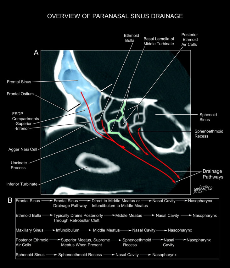
Overview of the drainage pathways of the paranasal sinuses.
A, Composite image displaying the drainage pathways of the frontal, ethmoid, and sphenoid sinuses in relation to a sagittal CT section through the skull illustrated in Figs 3 and 4. The frontal ostium forms the upper border of the superior compartment of the FSDP. The superior compartment of the FSDP drains posteroinferiorly into a narrow inferior compartment situated always anterior to the ethmoid bulla. The inferior compartment of the FSDP is formed by the ethmoid infundibulum when the superior portion of the uncinate process attaches to the skull base but is formed instead by the middle meatus when the uncinate process attaches to the lamina papyracea. The basal lamella of the middle turbinate (colored green) divides the ethmoid air cells into anterior and posterior groups. The agger nasi air cell is classified as an extramural ethmoid air cell, because it projects anterior to the ethmoid bone. This cell frequently extends to the lacrimal bone and the frontal process of the maxilla.
B, Summary of the typical drainage pathways of the paranasal sinuses, including the drainage pathway of the maxillary sinus. Ultimately, all sinus drainage is directed toward the nasopharynx.
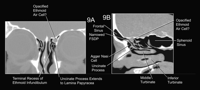
Fig 9.
Opacified anterior ethmoid air cell or suprabullar recess encroaches on the posterior aspect of the FSDP. Differentiation of an osseous air cell from an osseous recess probably would have to be determined surgically. A, Coronal CT. B, Reformatted sagittal CT. The narrowing of the FSDP is shown best in the reformatted sagittal CT (B). The coronal CT (A) shows lateral extension of the uncinate process to join the lamina papyracea, forming a terminal recess of the ethmoid infundibulum (see also Fig 5A).
Sinus nomenclature may be both redundant and confusing (10, 11). Originally, the terms sinus, antrum, and recess all referred to a cavity, then a cavity within a bone. Now an osseous recess is an air space with more than one drainage ostium (as distinct from an osseous air cell, which has only one drainage ostium), whereas the terms maxillary sinus and maxillary antrum may be used interchangeably. Both cribriform and ethmoid mean “sievelike,” and both bulla and agger signify a “projecting anatomic structure,” but convention has limited usage of each term to specific structures. For these reasons, and because anatomic variations are frequent, the exact terminology employed to designate paranasal air spaces may be subjective. Herein one coherent set of names for the structures of the FSDP is used that specifically excludes the term “frontal recess” because that term is difficult to define anatomically (6).
Figure 1 provides an overview of the drainage pathways of the paranasal sinuses that are based upon the skull illustrated in Figures 3 and 4.
Fig 3.
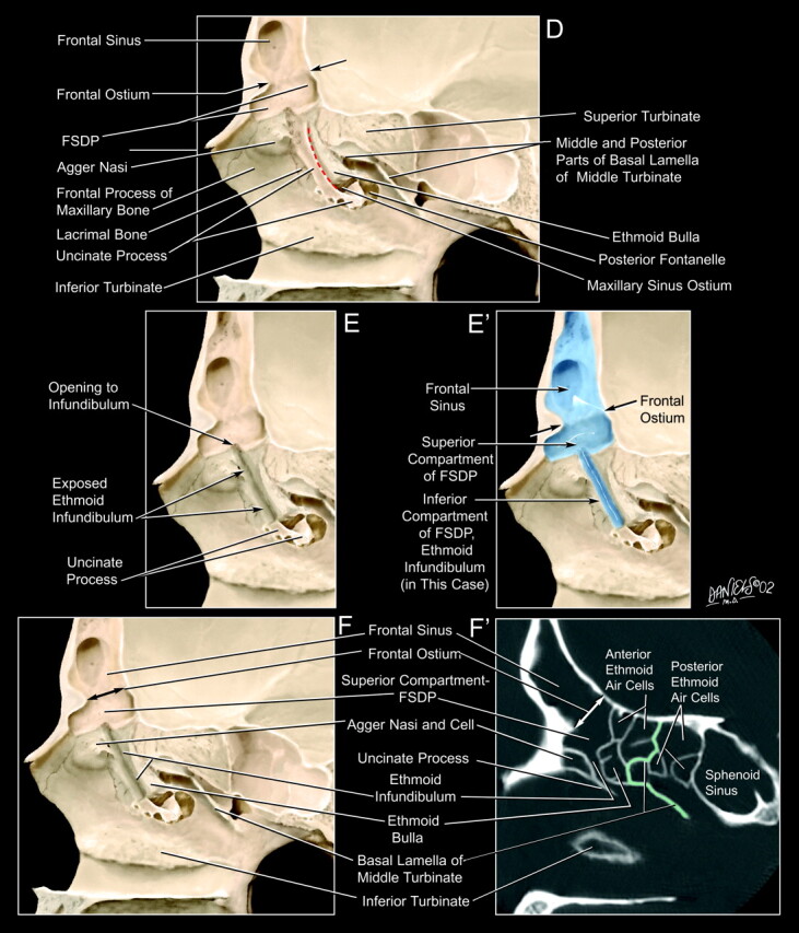
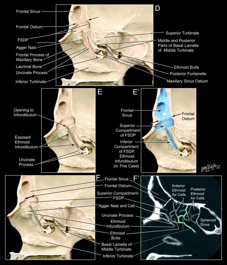
Sequential, more lateral dissections of the skull and the FSDP (A–F and E′) matched to sagittal CT sections of the skull (A′–C′ and F′).
A, A′. Sagittal structures. The nasal septum is a composite structure, formed by the perpendicular plate of the ethmoid bone anterosuperiorly and by the vomer posteroinferiorly. The inferior margin of the vomer rests upon the nasal crest of the maxillary and palatine bones. The superior margin of the vomer supports the perpendicular plate of the ethmoid bone.
B, B′. Paramedian structures. Removal of the nasal septum reveals the inferior, middle, and superior turbinates. The superior turbinate attaches to the skull base. The middle turbinate shown in B is superimposed (green) onto the sagittal CT (B′) to illustrate how the vertical portion of the basal lamella extends to the skull base at the cribriform plate. Its superior border has variable shape.
C, C′. Image C displays a simplified posteromedial oblique view of the middle turbinate and its basal lamella. The basal lamella has three portions. The vertical portion of the basal lamella attaches to the cribriform plate, the middle and posterior portions attach to the lamella papyracea, and the posterior margin of the basal lamella attaches to the perpendicular plate of the palatine bone. Typically, the vertical, middle, and posterior portions of the basal lamella are oriented in near-sagittal, coronal, and horizontal planes, respectively. Usually, the middle and posterior portions appear irregular in shape because of encroachment by adjacent ethmoid air cells. In C, the inferior edge of the middle turbinate and the medial edge of the middle and posterior portions of the basal lamella together have an angular configuration in the sagittal CT (C′) that highlights the middle and posterior portions of the basal lamella (green). The basal lamella in C′ can be conceptualized by removing the most medial portion of the middle turbinate and the vertical portion of the basal lamella from B′.
Continued
D, Removing the middle turbinate and the vertical portion of the basal lamella exposes, from anterior to posterior, the agger nasi extending to the frontal process of the maxillary bone, the lacrimal bone, the uncinate process of the ethmoid bone extending upward toward the skull base, the hiatus semilunaris (dashed red line), the ethmoid bulla, and the middle and posterior portions of the basal lamella extending laterally toward the lamina papyracea. The hiatus semilunaris is the narrow, slitlike passage between the uncinate process and the ethmoid bulla, through which the ethmoid infundibulum communicates with the middle meatus (6). The ethmoid infundibulum is the space bounded by the uncinate process anteromedially, the ethmoid bulla posterolaterally, and the lamina papyracea anterolaterally (6). Simultaneous removal of the wall of the frontal sinus shows the relationship of the frontal ostium to the superior compartment of the FSDP and how the sinus cavity tapers inferiorly toward the frontal ostium.
E, E′. Removal of the anterior portion of the uncinate process exposes the ethmoid infundibulum. In this specimen, the superior compartment of the FSDP drains directly into the ethmoid infundibulum, which then drains through the hiatus semilunaris to the middle meatus. E′ displays a three-dimensional view of the frontal sinus and the superior and inferior compartments of the FSDP (blue).
F, F′. The frontal ostium extends posteriorly from the anterior frontal bone ridge to the posterior wall of the frontal sinus and is oriented nearly perpendicular to the posterior wall of the sinus. The superior compartment of the FSDP lies inferior to the frontal ostium. In this specimen, the inferior compartment of the FSDP is the ethmoid infundibulum. In F′, the ethmoid infundibulum is bounded anteriorly by the agger nasi cell and the uncinate process and posteriorly by the ethmoid bulla. The middle and posterior portions of the basal lamella of the middle turbinate (green) separate the anterior and posterior ethmoid air cells (see also Fig 4E, the coronal CT of the same specimen).
Fig 4.
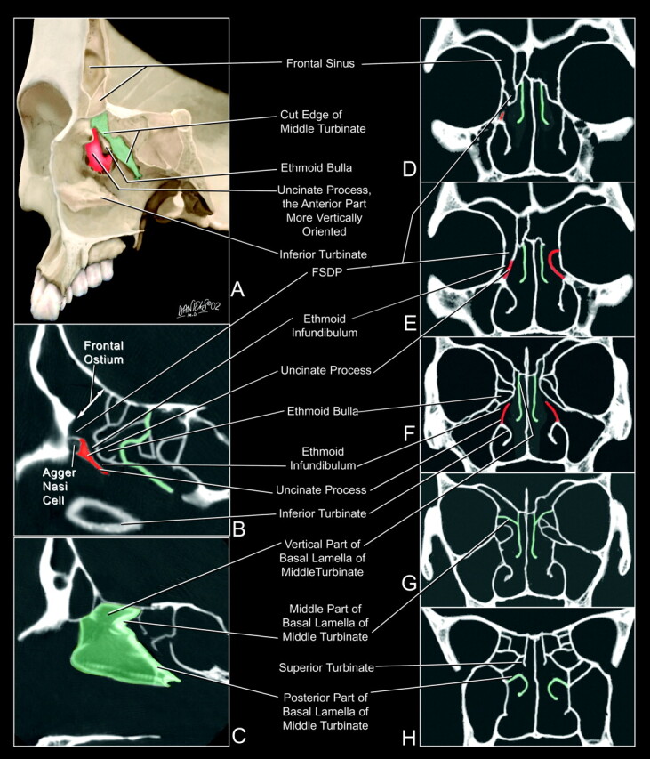
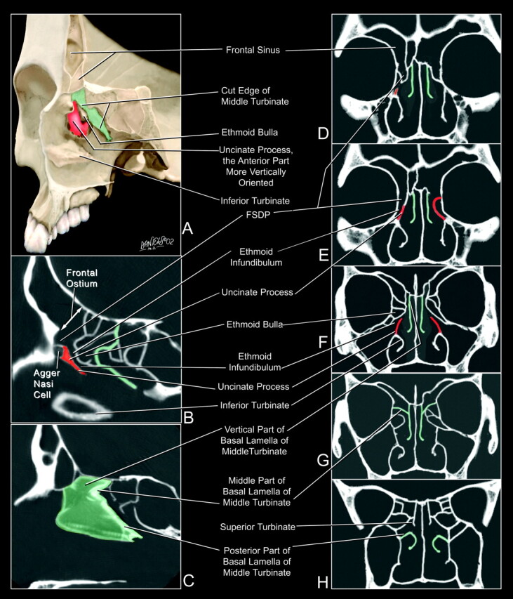
Anatomic relationships in the coronal plane.
A, Frontal oblique view of the same skull specimen illustrated in Fig 3. Resection of the anteroinferior portion of the middle turbinate (green) exposes the curved uncinate process (red) just superior to the inferior turbinate. In this specimen, the anterior portion of the uncinate process extends to the skull base.
B and C, Sagittal CT sections color coded to display the uncinate process (red) and the middle turbinate including the basal lamella (green). Note that the different portions of the basal lamella are oriented more nearly vertical, or horizontal, facilitating their identification on sagittal and coronal CT sections.
D–H, Sequential coronal CT sections, displayed from anterior to posterior to illustrate the changing relationships and attachments of the color-coded structures.
D, The superior compartment of the FSDP lies superior to the ethmoid infundibulum. The frontal ostium is more easily identified in the sagittal plane (B) than in the anterior coronal plane (D), where the border between frontal sinus and FSDP is difficult to define.
E, The anterior portion of the uncinate process (red) extends upward to the skull base in the right side of this specimen, so the FSDP includes the ethmoid infundibulum. The frontal ostium remains poorly localized.
Continued
F, The posterior portion of the uncinate process (red) lies just inferior to the ethmoid bulla. The inferior margin of the uncinate process lies just superior to the point of attachment of the inferior turbinate. The vertical portion of the basal lamella (green) of the middle turbinate extends to the skull base at the cribriform plate.
G, The midportion of the basal lamella of the middle turbinate (green) extends laterally to join the lamina papyracea. It is continuous with the vertical portion that joins the cribriform plate.
H, The posterior portion of the basal lamella (green) of the middle turbinate lies inferior to the superior turbinate and also extends laterally to join the perpendicular plate of the palatine bone in this CT section.
The frontal sinus has the most complex and variable drainage of any paranasal sinus. Each frontal sinus narrows down to an inferior margin designated the frontal ostium (Figs 1 and 3D-F′). The frontal ostium extends between the anterior and posterior walls of the frontal sinus, is demarcated by a variably shaped ridge of bone on the anterior wall of the sinus, and is oriented nearly perpendicular to the posterior wall of the sinus. It may be difficult to define when air cells marginate the ostium (Fig 8).
Fig 8.
Prominent anterior air cell encroaches on the frontal sinus and the FSDP.
A, Coronal CT.
B, Reformatted sagittal CT.
The FSDP has superior and inferior compartments. The superior compartment of the FSDP is formed by the union of adjacent air spaces at the anteroinferior portion of the frontal bone and the anterosuperior portion of the ethmoid bone (Fig 2). Its upper border is the frontal ostium. Its size and shape vary with the variable anatomy of the frontoethmoid air cells (8; Figs 6–9). The superior compartment communicates directly with the inferior compartment. The inferior compartment of the FSDP is a narrow passageway formed by either the ethmoid infundibulum or the middle meatus (6, 7). When the anterior portion of the uncinate process extends superiorly to attach to the skull base, the inferior compartment of the FSDP is the ethmoid infundibulum (Figs 3D–F′, 4E, 5). This then communicates with the middle meatus via the hiatus semilunaris; however, when the anterior portion of the uncinate process attaches to the lamina papyracea instead of the skull base, the inferior compartment of the FSDP is then the middle meatus itself (Fig 5).
Fig 2.
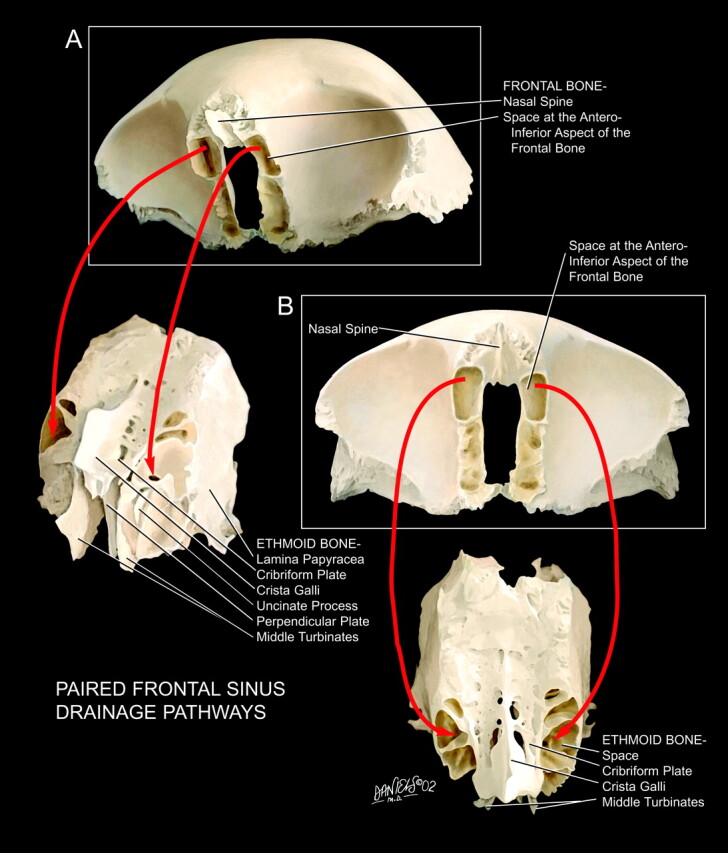
The superior compartment of the FSDP. Shown are oblique (A) and straight (B) views of the superior and inferior surfaces of the frontal and ethmoid bones of two different specimens. The paired air spaces situated beneath the frontal sinuses and the frontal ostia at the anteroinferior aspect of the frontal bone drain (red arrows) into the paired labyrinthine air spaces at the anterosuperior aspect of the ethmoid bone. In the intact skull of a single specimen, these spaces would join smoothly to form the paired superior compartments of the FSDPs. Common anatomic variations affecting the superior compartment of the FSDP include osseous septations (Fig 6) and marginating air cells (Fig 7). These anatomic variations cause substantial variation in the size and shape of the superior compartments. Nonetheless, the superior compartments ultimately drain into the middle meatus, either directly or via the ethmoid infundibulum (Fig 5).
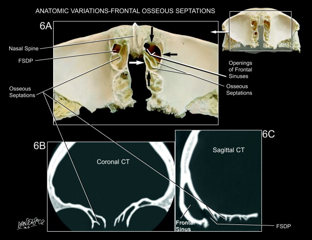
Fig 6.
Anatomic variations in the frontal osseous septations. A, Inferior surface of the frontal bone. B, Coronal CT section. C, Sagittal CT section. Variations in the union of the osseous septations of the frontal bone with the subjacent air cells lead to great diversity in the arrangements of air spaces bordering the FSDP. A well-corticated osseous rim (arrows) marginates the uppermost portion of the superior compartment of the FSDP at the anteroinferior aspect of the frontal bone. The left side shows multiple openings of the frontal sinuses into the FSDP.
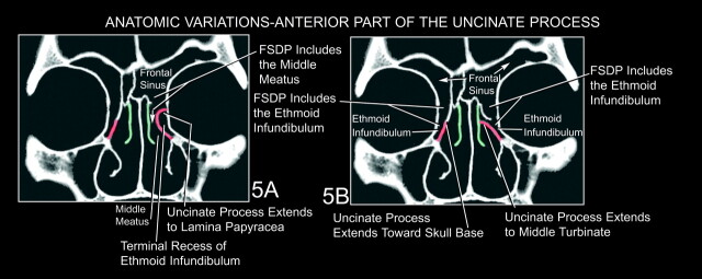
Fig 5.
Anatomic variations in the anterior uncinate process.
A and B, Coronal CT sections modified from the skull illustrated in Figs 3 and 4. Coronal CT sections display the three major variations in the superior attachment of the uncinate process (7): extension laterally to join the lamina papyracea (A, left), extension superiorly to join the skull base (A and B, right), and extension medially to join the lateral surface of the middle turbinate (B, left). Because of these variations in the uncinate process, the frontal sinus drainage on the left side of panel A passes through the superior compartment directly into the middle meatus, not the ethmoid infundibulum. In contrast, the frontal sinus drainage on the right side of panel A and both sides of panel B passes through the superior compartment into the ethmoid infundibulum before entering the middle meatus. The ethmoid infundibulum forms the inferior compartment of the FSDP in these cases. In A, the attachment of the left uncinate process with the lamina papyracea forms a terminal recess of the ethmoid infundibulum.
Together, the frontal sinus and the superior compartment of the FSDP resemble an Erlenmeyer flask, with the neck of the flask representing the frontal ostium. The inferior compartment of the FSDP could then be conceptualized as directly communicating with a small opening in the base of the flask.
The basal lamella of the middle turbinate is a thin bony plate that forms part of the ethmoid infrastructure. It has three portions (6, 7). The vertical portion of the basal lamella attaches to the cribriform plate. The middle and posterior portions extend laterally to join the lamina papyracea, thereby dividing the anterior ethmoid air cells from the posterior ethmoid air cells and the posterior margin of the basal lamella attaches to the perpendicular plate of the palatine bone (Figs 3C and 3F′). The basal lamella is best displayed in three-dimensional models and sagittal and coronal CT images. Its vertical portion is seen best on coronal CT images, whereas the middle and posterior portions are seen best on sagittal CT images (Figs 3C′, 3F′, and 4F–H).
Figure 2 separates the frontal bone from the ethmoid bone to illustrate how they join to form the superior compartments of the FSDPs. In Figure 2B, the paired air spaces at the anteroinferior aspect of the frontal bone are not the frontal sinuses, but portions of the superior compartments of the FSDPs, inferior to the frontal ostia and the frontal sinuses.
Figures 3 and 4 correlate the lateral and frontal oblique views of the FSDP with sagittal and coronal CT scans of a skull to show the FSDP as it would be displayed in clinical CT scans. In the specimen illustrated, the uncinate process extends superiorly to reach the skull base, so the inferior compartment of the FSDP is formed by the ethmoid infundibulum (Figs 3D–F′ and 4E).
Figures 5–9 illustrate variations in the anatomy of the air cells and the uncinate process, which affect the FSDP.
The complex anatomy and frequent anatomic variations of the FSDP may challenge the skills of the radiologist; however, use of helical CT studies and multiplanar CT reformations now provide the radiologist with a clearer depiction of the anatomy of each patient and a clearer understanding of the anatomic diversity of the FSDP within the population. Sagittal specimen and reformatted clinical CT scans display better the size and margins of the frontal ostium, the FSDP, and the adjacent air cells (Figs 1, 3F′, 8, and 9). Coronal CT scans demonstrate the superior attachment of the uncinate process, which determines whether the inferior compartment of the FSDP is formed by the ethmoid infundibulum or the middle meatus (Figs 4E and 5). Coronal CT scans also document patency of the FSDP.
Display of the FSDP is useful when evaluating the cause and potential surgical therapy for obstruction of the frontal sinus (4). Air cells may encroach upon the anterior, posterior, lateral, and/or superior margins of the FSDP (5, 8; Figs 7, 8, and 9). The nomenclature used to describe these cells varies with their position. Ethmoid air cells that encroach upon the anterior aspect of the superior compartment of the FSDP or upon the frontal sinus itself are designated frontal air cells. A classification of frontal cells is given by Bent et al (8); however, further work is needed to understand the complex anatomy and variations of the frontal sinuses and FSDPs.
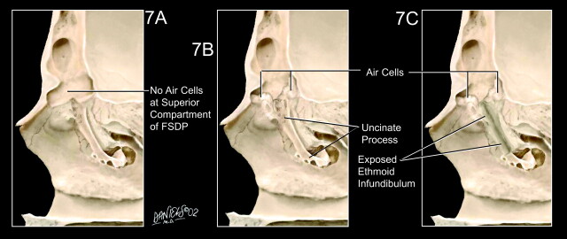
Fig 7.
Variations in the shape of the superior compartment of the FSDP due to variations in the marginating air cells. A, No marginating air cells. B and C, Marginating air cells anteriorly and posteriorly encroach upon the lower portion of the superior compartment and its junction with the inferior compartment. In B, the uncinate process obscures the ethmoid infundibulum. Removing the uncinate process (C) exposes the course and configuration of the ethmoid infundibulum.
Acknowledgments
We thank Lindell R. Gentry, Leo F. Czervionke, Hugh D. Curtin, William P. Dillon, Katherine A. Shaffer, Lowell A. Sether, Mehmet Kocak, Scott A. Koss, and John R. Grogan, for their help in preparing this article.
References
- 1.Mafee MF, Chow JM, Meyers R. Functional endoscopic sinus surgery: anatomy, CT screening, indications, and complications. AJR Am J Radiol 1993;160:735–744 [DOI] [PubMed] [Google Scholar]
- 2.Mafee MF. Preoperative imaging anatomy of nasal-ethmoid complex for functional endoscopic sinus surgery. Radiol Clin North Am 1993;31:1:1–20 [PubMed] [Google Scholar]
- 3.Harnsberger HR. Handbook of head and neck imaging. 2nd ed. St Louis: Mosby;1995. :339–395
- 4.Smith MM, Smith TL. Frontal Sinus DrainageProcedures: Postoperative Imaging Appearance. Chicago: Radiological Society of North America;2000
- 5.Bolger WE, Mawn CB. Analysis of the suprabullar and retrobullar recesses for endoscopic sinus surgery. Ann Otol Rhinol Laryngol (suppl)2001;186:3–14 [DOI] [PubMed] [Google Scholar]
- 6.Stammberger HR, Kennedy DW, Bolger WE, et al. Paranasal sinuses: anatomic terminology and nomenclature. Ann Otol Rhinol Laryngol (suppl)1995;167:7–16 (especially helpful source) [PubMed] [Google Scholar]
- 7.Stammberger H. Functional endoscopic sinus surgery. Philadelphia: BC Decker;1991
- 8.Bent JP, Cuilty-Siller C, Kuhn FA. The frontal cell as a cause of frontal sinus obstruction. Am J Rhinol 1994;8:185–191 [Google Scholar]
- 9.Masala W, Perugini S, Salvolini U, Teatin GP. Multiplanar reconstructions in the study of ethmoid anatomy. Neuroradiology 1989;31:151–155 [DOI] [PubMed] [Google Scholar]
- 10.Skinner HA. The Origin of Medical Terms. New York: Hafner;1970
- 11.Dorland’s Illustrated Medical Dictionary. 29th ed. Philadelphia: Saunders;2002


