Abstract
Summary: Vertebroplasty is a procedure in which polymethylmethacrylate (PMMA) is injected into a vertebral body compression fracture. It has been suggested that fractures greater than 65% to 70% of the original vertebral body height are too compressed to be treated successfully with this procedure. We describe six patients with severe compression fractures that were successfully treated with vertebroplasty in which the trocar was inserted in the far lateral aspect of the vertebral body, because of the typical morphology of severe compression fractures.
Vertebroplasty has been reported to alleviate pain and to prevent further collapse in patients with vertebral compression fractures (1–6). Some clinicians, however, have declined to use the procedure in patients in whom the compression fracture is greater than 70% of the original height of the vertebral body (1, 2). We describe six patients with severe vertebral body fractures in whom vertebroplasty was technically successful and was associated with significant pain relief in four patients.
Technique
Five patients with fractures greater than 70% of the original vertebral body height and one patient with a 65% fracture underwent vertebroplasty as a pain relief measure. The patients included five women and one man, with an average age of 75 years. The compression fractures that were treated occurred in the thoracic and lumbar spine. A scale system was used to assess the level of pain, mobility, and medication use in each patient both before and after surgery. The scale for each is provided in Table 1. The average follow-up time was 3 months. The degree of compression was established by comparing the height of the compressed vertebral body with the height of the closest normal vertebral body superior or inferior to it.
TABLE 1:
Scale used for patient assessment

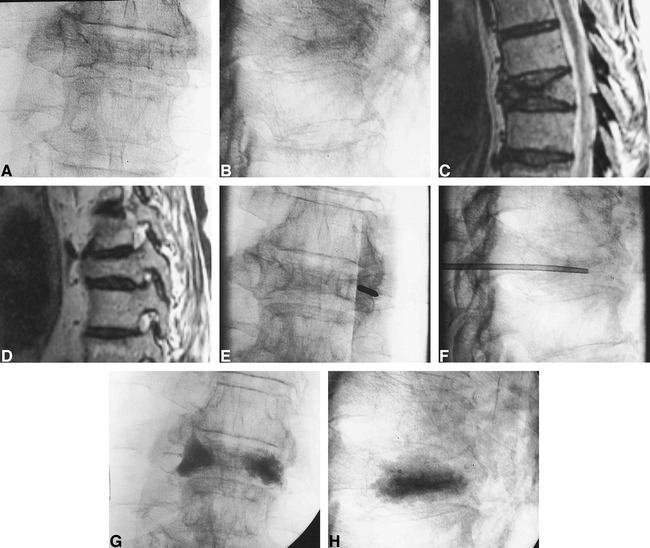
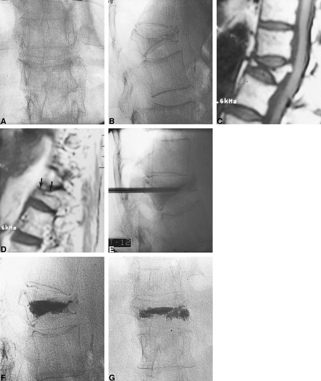
The imaging studies from patients 4 and 5 are used as representative examples to illustrate the procedure in Figures 1 and 2, respectively. In patients with severe compression deformities, the greatest loss of height usually occurs at the center of the vertebral body. Viewed in the coronal plane, the resulting deformity resembles a bow tie (Figs 1A and 2A). The far lateral aspect of the vertebral body generally is significantly less compressed (Figs 1B and 2B). MR imaging reveals the sparing of height of the lateral aspect of the vertebral body relative to the more severe compression at the mid-vertebral body (Figs 1C, 1D, 2C, and 2D). Consequently, it may be difficult to place a bone trocar into or near the center of the vertebral body, but it is usually possible to place the trocar far laterally and still remain in the bone marrow. Also, attempts to apply polymethylmethacrylate (PMMA) mixed with 10 to 15 mL of sterile barium sulfate in the center of a severely compressed vertebral body without entering the disk space frequently result in rapid extravasation of the PMMA into the disk space, whereas the bone marrow present at the far lateral location generally allows for better filling of the vertebral body.
fig 1.

Case 4.
A, Anteroposterior radiograph of the T8 vertebral body.
B, Lateral radiograph of the same vertebral body shows a severe compression fracture.
C, Sagittal T2-weighted MR image of the T8 vertebral body again shows a severe compression fracture. This mid-sagittal image shows the most severe portion of the compression fracture at the center of the vertebral body.
D, Sagittal T2-weighted MR image of the T8 vertebral body shows relative preservation of vertebral body height far laterally.
E, Anteroposterior radiograph shows the 13-gauge trocar in the vertebral body via a parapedicular approach. Note the far lateral approach in the area of the vertebral body with relative preservation of bone marrow.
F, Lateral radiograph shows placement of a 13-gauge bone trocar into the vertebral body.
G, Anteroposterior radiograph after vertebroplasty with PMMA in the far lateral aspect of the vertebral body bone marrow. The typical bow-tie appearance of vertebral bodies with severe compression fractures is shown.
H, Lateral radiograph after vertebroplasty with PMMA filling the residual vertebral body bone marrow.
Vertebroplasty Technique
The transpedicular approach (4) was used, but the trocar was placed as far laterally as possible (Figs 1F, 1E, and 2E). Given the small space being entered, a 13-gauge trocar proved useful, especially in the thoracic spine, where the pedicles are relatively small. With a 13-gauge trocar, a less viscous mixture of PMMA was used. Figures 1H, 1G, 2G, and 2F show the appearance of severely compressed vertebral bodies after vertebroplasty performed with this technique.
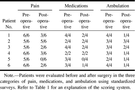
Data for the six patients studied are presented in Table 2. The average compression was 73%, with a range of 83% to 65%. The average volume of PMMA used per vertebroplasty was 5.2 mL. A bipedicular approach was necessary in all cases. In four of the six patients, the procedure provided significant pain relief as well as increased mobility and decreased analgesic use. Results for each patient are provided in Table 3. A “good” technical result was obtained in all six patients, which was defined as adequate filling of the remaining vertebral body.
TABLE 2:
Characteristics of vertebroplasties for six patients

TABLE 3:
Pre- and postoperative clinical results for six patients

In patient 1, a small amount of PMMA extravasated into the disk space below, as well as into the superior endplate fracture. In patient 2, there was a small amount of extravasation into the right epidural venous plexus. Neither complication produced any clinical sequelae.
Discussion
Some authors have recommended that vertebroplasty not be attempted in patients with compression fractures greater than 70% of previous vertebral body height. From Mathis et al: “Percutaneous vertebroplasty may be precluded technically if vertebral collapse is extreme. No exact percentage of collapse has been determined as an absolute limit, since compressions are often asymmetric; however, compressions of >65–70% become technically challenging” (1). In our six patients with severe compression fractures, vertebroplasty was technically feasible. We believe that the far lateral approach we used allowed us to deliver more PMMA into a severely compressed vertebral body than would have been possible with the more standard approach. The procedure afforded significant pain relief, improved mobility, and decreased need for pain medications for four of the six patients. Not every compression fracture in every patient needs to be treated. We determined which levels needed treatment on the basis of a combination of imaging findings and clinical examination. For example, patient 4 (Fig 1) had more than one compression fracture on imaging studies; however, clinical examination suggested that he was most tender at the T8 level, so this was the level treated, and he gained significant pain relief from treatment of this single level. The procedure did not relieve the pain of two patients, but it did not increase their pain or cause any complications. The rate of effectiveness of pain relief for this series of patients was lower than that reported by other investigators (1–6), which is not surprising considering the severe degree of the compression fractures in our patients. Also, our study group included only six patients, and the true frequency of pain relief for patients with severe compression fractures cannot be determined from such a small sample. Future studies may reveal the rate of pain relief from vertebroplasty in this subgroup of patients.
Conclusion
We propose that, contrary to statements in the literature, some patients can be effectively treated with vertebroplasty using the far lateral approach we describe.
fig 2.

Case 5
A, Anteroposterior radiograph of the T12 vertebral body.
B, Lateral radiograph of the same vertebral body shows a severe compression fracture.
C, Sagittal T1-weighted MR image shows a severe compression deformity at the center of the vertebral body with some retropulsion of the posterior vertebral body.
D, Sagittal T1-weighted MR image shows relative preservation of vertebral body bone marrow far laterally (arrows).
E, Lateral radiograph shows placement of an 11-gauge trocar into the T12 vertebral body.
F, Lateral radiograph of the T12 vertebral body after vertebroplasty.
G, Anteroposterior radiograph after vertebroplasty with PMMA in the far lateral position, as well as a small amount of PMMA underneath the superior endplate fracture in the midline.
Footnotes
Address reprint requests to Avery J. Evans, MD, 511 W Bay St, Suite 301, Tampa, FL 33606
References
- 1.Mathis JM, Petri M, Naff N. Percutaneous vertebroplasty treatment of steroid-induced osteoporotic compression fractures. Arthritis Rheum 1998;41:171-175 [DOI] [PubMed] [Google Scholar]
- 2.Weill A, Chiras J, Simon JM, et al. Spinal metastases: indications for and results of percutaneous injection of acrylic surgical cement. Radiology 1996;199:241-247 [DOI] [PubMed] [Google Scholar]
- 3.Cotten A, Dewatre F, Cortet B, et al. Percutaneous vertebroplasty for osteolytic metastases and myeloma: effects of the percentage of lesion filling and the leakage of methyl methacrylate at clinical follow-up. Radiology 1996;200:525-530 [DOI] [PubMed] [Google Scholar]
- 4.Jensen ME, Evans AJ, Mathis JM, Kallmes DF, Cloft HJ, Dion JE. Percutaneous polymethylmethacrylate vertebroplasty in the treatment of osteoporotic vertebral body compression fractures: technical aspects. AJNR Am J Neuroradiol 1997;18:1897-1904 [PMC free article] [PubMed] [Google Scholar]
- 5.Deramond H, Depriester C, Toussaint P. Percutaneous vertebroplasty with polymethylmethacrylate: technique, indications, and results. Radiol Clin North Am 1998;36:533-546 [DOI] [PubMed] [Google Scholar]
- 6.Martin JB, Jean B, Sugui K, et al. Vertebroplasty: clinical experience and follow-up results. Bone 1999;25:11S-15S [DOI] [PubMed] [Google Scholar]


