Abstract
BACKGROUND AND PURPOSE: The termination of the superficial middle cerebral vein is classically assimilated to the sphenoid portion of the sphenoparietal sinus. This notion has, however, been challenged in a sometimes confusing literature. The purpose of the present study was to evaluate the actual anatomic relationship existing between the sphenoparietal sinus and the superficial middle cerebral vein.
METHODS: The cranial venous system of 15 nonfixed human specimens was evaluated by the corrosion cast technique (12 cases) and by classic anatomic dissection (three cases). Angiographic correlation was provided by use of the digital subtraction technique.
RESULTS: The parietal portion of the sphenoparietal sinus was found to correspond to the parietal portion of the anterior branch of the middle meningeal veins. The sphenoid portion of the sphenoparietal sinus was found to be an independent venous sinus coursing under the lesser sphenoid wing, the sinus of the lesser sphenoid wing, which was connected medially to the cavernous sinus and laterally to the anterior middle meningeal veins. The superficial middle cerebral vein drained into a paracavernous sinus, a laterocavernous sinus, or a cavernous sinus but was never connected to the sphenoparietal sinus. All these venous structures were demonstrated angiographically.
CONCLUSION: The sphenoparietal sinus corresponds to the artificial combination of two venous structures, the parietal portion of the anterior branch of the middle meningeal veins and a dural channel located under the lesser sphenoid wing, the sinus of the lesser sphenoid wing. The classic notion that the superficial middle cerebral vein drains into or is partially equivalent to the sphenoparietal sinus is erroneous. Our study showed these structures to be independent of each other; we found no instance in which the superficial middle cerebral vein was connected to the anterior branch of the middle meningeal veins or the sinus of the lesser sphenoid wing. The clinical implications of these anatomic findings are discussed in relation to dural arteriovenous fistulas in the region of the lesser sphenoid wing.
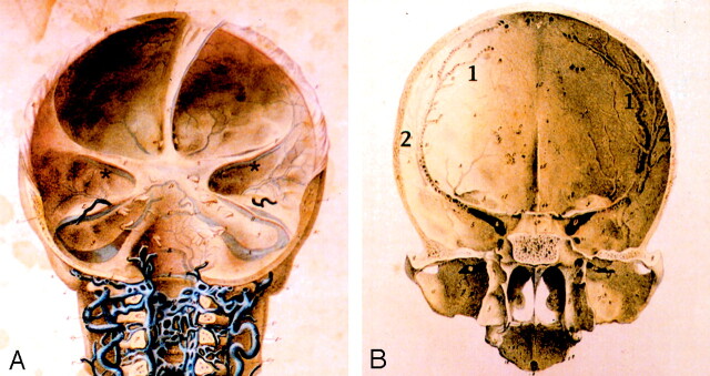
The term sphenoparietal sinus was introduced in the early 19th century by Gilbert Breschet (1) in an atlas of the venous system. If the sphenoparietal sinus was clearly depicted and labeled in the anatomic drawings illustrating the atlas, the manuscript itself was incompletely published, and no reference to the sphenoparietal sinus can be found in the accompanying text. Cruveilhier (2) later quoted Breschet’s description of the sphenoparietal sinus as a “sinus found within the limits of the anterior and medial portions of the base of the skull, which occupies a transversally oriented gutter that runs inward into the cavernous sinus. This sinus receives several branches from the skull bones, the dura mater, and the diploic vein of the temporal bone [author’s translation].” (The original quotation reads, “Un sinus situé sur la limite de la portion antérieure et de la portion moyenne de la base du crâne, sinus qui occupe une gouttière transversalement dirigée de dehors en dedans et s’abouche dans le sinus caverneux. Ce sinus reçoit plusieurs branches veineuses des os crâniens, de la dure-mère, et la veine diploïque du temporal.” Cruveilhier observed that book II, plate 3 could not be found or confirmed and does not correspond to Breschet’s atlas [1]). This account, consistent with Breschet’s illustrations, corresponds to the sphenoid portion of the sphenoparietal sinus, found under the lesser sphenoid wing and involved in diploic and meningeal venous drainage. This is the classic definition of the sphenoparietal sinus, which then came to be known as the sinus of Breschet (3).
The anatomic description became more complex and somewhat confusing when the notion was introduced that the superficial middle cerebral vein ended in the sphenoparietal sinus under the lesser sphenoid wing (4) or that the superficial middle cerebral vein was at least partially equivalent to the sphenoparietal sinus. This view, which can be traced back to Hédon’s monograph on the cerebral venous system published in 1888 (5), has been commonly reproduced in the radiologic, surgical, and anatomic literature. Oka et al (6) reported that the superficial middle cerebral vein could either join the sphenoparietal sinus or drain directly into the cavernous sinus. These authors also affirmed that the sphenoparietal sinus could drain into the sphenobasal or sphenopetrosal sinuses, which are better described conjointly as the paracavernous sinus, one of the well-documented drainage pathways of the superficial middle cerebral vein (7, 8). Bisaria (9) reported that the superficial middle cerebral vein terminated into the sphenoparietal sinus in 68% of cases reviewed. Wolf et al (10) reported that “in the region of the pterion, the vein [ie, superficial middle cerebral vein] enters the dura and runs along the lesser wing of the sphenoid to enter the anterior end of the cavernous sinus. The dural portion of this channel along the lesser wing is frequently referred to as the sphenoparietal sinus.” Wolf et al add, however, that “there is no significant dilution of the opaque material when the sinus [ie, sphenoparietal sinus] fills via the superficial sylvian vein [ie, superficial middle cerebral vein]”. They suggest that the sphenoparietal sinus drains exclusively into the superficial middle cerebral vein and that the term sinus of the lesser wing of the sphenoid should be preferred.
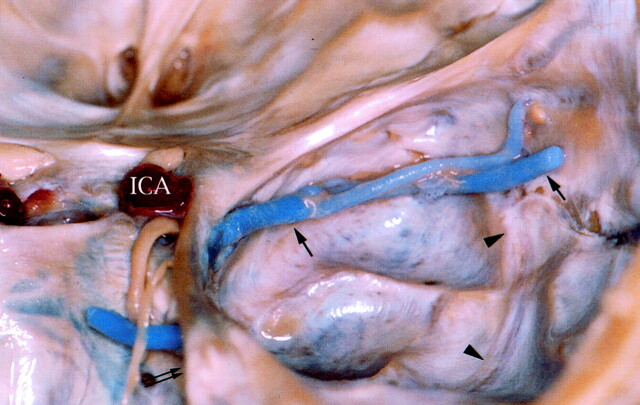
In a study on superficial middle cerebral vein termination, however, San Millán Ruïz et al (11) observed that injecting the superficial middle cerebral vein with colored gelatin was not followed by a concomitant filling of the middle meningeal veins, as would be expected following the classic description mentioned above. Their study also showed that when the superficial middle cerebral vein drained into the laterosellar region, it did not first constitute a dural venous sinus under the lesser sphenoid wing, but instead directly pierced the dura mater in the superior-anterior aspect of the lateral wall of the cavernous sinus (Fig 1). In rare cases in which the superficial middle cerebral vein coursed under the lesser sphenoid wing, it was attached to the dura mater overlying the sphenoid ridge yet maintained the macroscopic appearance of an arachnoid vein; that is, it did not become a venous sinus. These findings suggest that the superficial middle cerebral vein and the middle meningeal veins are not connected and that, if a dural venous sinus actually exists under the lesser sphenoid wing, it is not related to the superficial middle cerebral vein.
Fig 1.
Superior view of right middle cranial fossa. The superficial middle cerebral vein (arrows) drains into the laterosellar region into the cavernous sinus. The superficial middle cerebral vein is not related with the lesser sphenoid wing during its course and it pierces the dura mater of the lateral wall of the cavernous sinus directly. Transition from arachnoid vein to dural venous sinus is abrupt. ICA signifies internal carotid artery; double arrow, tentorial edge; arrowheads, middle meningeal veins.
The purpose of the present investigation was to better characterize the relationship between the sphenoparietal sinus, the sinus of the lesser sphenoid wing, and the distal superficial middle cerebral vein and analyze their respective participation in the cerebral venous drainage. The existence of the sphenoparietal sinus as a distinct anatomic entity is discussed. The clinical implications of these anatomic findings are discussed in relation to dural arteriovenous fistulas in the region of the lesser sphenoid wing and illustrated with a case.
Methods
Corrosion Cast Study
Corrosion casts of the cranial and cerebral venous system were prepared from 12 nonfixed human specimens (eight female and four male specimens; average age at time of death, 85 years). In each case, the internal jugular veins were carefully dissected in the neck and canulated with 6-mm metallic probes. The venous system was thoroughly rinsed with a saline solution. Leakage sites were identified and ligatured. A mixture of blue methylmethacrylate (Beracryl; Troller, Fulenbach, Switzerland) and barium sulfate powder (HD 200 Plus; Lafayette, Anaheim, CA) was injected through both internal jugular veins until the angular veins became engorged. The specimens were then placed in a potassium hydroxide bath (40% solution, 40°C) until all surrounding soft and osseous tissues were dissolved.
Of the 24 injected specimen sides, five showed complete filling of the cerebral, diploic, and meningeal veins. In these five sides, the injection of the meningeal and diploic veins of the middle cranial fossa, the lesser sphenoid wing region and the pterion was optimal; that is, the venous channels could be followed from their origin to their termination. These five sides were used for precise evaluation of the skull base venous anatomy. On the other hand, the superficial middle cerebral vein was adequately injected in 21 of the 24 specimen sides. These 21 sides were used for the analysis of connections between the superficial middle cerebral vein and the sphenoparietal sinus.
Standard Anatomic Dissection
Standard anatomic dissection was performed on three nonfixed specimens (two female and one male; average age at time of death, 82 years) after vascular injection. Blue methylmethacrylate was injected in the venous system as described above. Red methylmethacrylate was injected in the internal and external carotid territories via bilateral common carotid cannulations. The brain was removed before complete polymerization of the methylmethacrylate, with care taken to identify all the venous bridges attaching the brain to the base of the skull. The dura mater of the anterior and middle cranial fossa was dissected layer by layer to reveal any intradural vascular channel. The periosteal dural layer was then removed except for the dura surrounding the meningeal vessels coursing along the anterior and middle cranial fossae. The inner table of the sphenoid, frontal, and parietal bones was progressively removed so as to expose diploic vessels. Finally, the floor of the middle cranial fossa was resected to expose the pterygoid plexus. The three specimens (six sides) used for standard anatomic dissections showed complete filling of the cerebral, meningeal, and diploic venous systems.
Angiographic Illustrations
Typical angiographic appearance of the venous structures discussed in the anatomic study are presented to provide radioanatomic correlations. The venous phase of routine cerebral angiograms obtained on a biplane digital subtraction angiography equipment (BN3000; Philips, Best, the Netherlands) were used.
Clinical Case
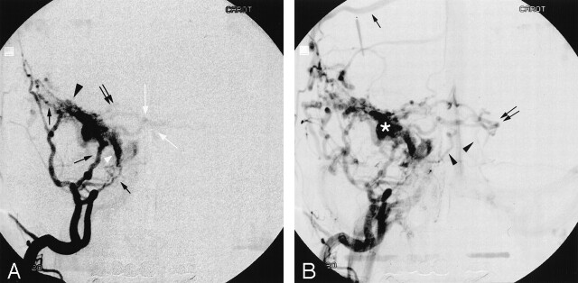
A 47-year-old man presented with a long history of headache when bending. There was no prior record of head trauma. The only clinical finding on examination was a pulsatile bruit upon auscultation of the right orbital region that was not subjectively perceived by the patient. MR imaging and MR angiography demonstrated a possible dural arteriovenous fistula of the lesser sphenoid wing. Digital subtraction angiography revealed a type IV right dural arteriovenous fistula under the lesser sphenoid wing, nourished by branches from the right middle meningeal artery and from the recurrent meningeal branch of the right ophthalmic artery (Fig 2A and B). The fistula was directly on the right superficial middle cerebral vein. The portion of the superficial middle cerebral vein that coursed under the sphenoid lesser wing showed a focal saccular dilatation. Superficial and deep cerebral veins were filled in a retrograde fashion via the superficial middle cerebral vein and the deep middle cerebral vein. Embolization into the recurrent meningeal branch of the ophthalmic artery followed by surgical clip placement and resection of the saccular dilatation of the superficial middle cerebral vein (performed by Prof. Nicolas de Tribolet, Division of Neurosurgery, Geneva University Hospital, Geneva, Switzerland) permitted closure of the fistula with a good clinical outcome.
Fig 2.
Dural arteriovenous fistula of the lesser sphenoid wing.
A, Early filling of a selective right external carotid artery injection in the anteroposterior projection. Nourishing branches from the right middle meningeal artery (black arrows) feeds a dilated superficial middle cerebral vein (black arrowhead). The superficial middle cerebral vein drains into a laterocavernous sinus (white arrowhead), which is also directly involved in the dural arteriovenous fistula and is fed by branches of the right middle meningeal artery. Early anterograde filling of the cavernous sinus is observed. There is retrograde filling into the right basal vein (white arrows) and into the right peduncular vein, through the right deep middle cerebral vein.
B, Selective right external carotid injection, later in the venous phase. Retrograde filling of the right superficial cortical veins (black arrow) via the right superficial middle cerebral vein is now observed. The left basal vein (black double arrow) is now also opacified via the peduncular veins, and the straight sinus is clearly delineated. There is infratentorial drainage of the fistula into the anterior and lateral pontomesencephalic veins (black arrowheads). The saccular dilatation of the superficial middle cerebral vein is clearly visible (asterisk).
Results
Anatomic Findings
The following anatomic description is based on observations made of the 11 specimen sides with adequate vascular filling: five sides were prepared as corrosion casts and six sides were used for standard dissection.
The major venous channels observed in the middle cranial fossa, in the lesser sphenoid wing and laterosellar regions, and along the cranial vault in the frontoparietal region were the anterior branch of the middle meningeal veins, a dural venous sinus coursing under the lesser sphenoid wing (thereafter called sinus of the lesser sphenoid wing), and the termination of the superficial middle cerebral vein.
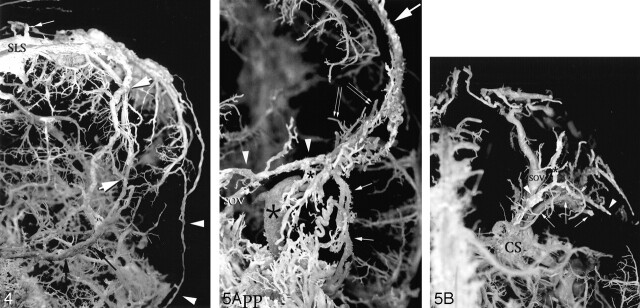
The anterior branch of the middle meningeal veins was composed of a sphenoid and a parietal portion. The sphenoid portion of the anterior branch of the middle meningeal veins (Figs 3A–C) crossed the floor of the middle cranial fossa from the lateral and anterior aspect of the greater sphenoid wing to the foramen ovale or spinosum, through which it joined the pterygoid plexus. This sphenoid portion was composed of two small parallel venous channels separated by the anterior branch of the middle meningeal artery. At the anterior margin of the greater sphenoid wing, the sphenoid portion entered a 3- to 10-mm-long osseous canal identified by Trolard (12) as the sphenoparietal canal. The parietal portion of the anterior branch of the middle meningeal veins (Figs 3B and 4) extended from the lateral opening of the sphenoparietal canal up to the venous lakes of the superior sagittal sinus. As it emerged from the sphenoparietal canal, the parietal portion of the anterior branch of the middle meningeal veins appeared as a single wide channel, which was larger than its two tributaries and met the definition of an anterior parietal diploic vein. Two thin individual veins corresponding to the classic anterior branch of the middle meningeal vein appearance were seen again for short distances wherever the anterior parietal diploic vein deviated from its more or less straight caudocranial course toward the superior sagittal sinus. The distinct anterior branch of the middle meningeal veins fused with the anterior parietal diploic vein once their respective courses met again.
Fig 3.
Serial dissection of a fresh specimen.
A, Superior topographic view of the right. The brain has been removed and the bridging veins of the temporal pole, in this case a single superficial middle cerebral vein (black arrowhead), have been sectioned close to the brain surface. The superficial middle cerebral vein is attached to the dura mater overlying the lesser sphenoid wing but keeps the appearance of an arachnoid vein. Note the different appearance between the superficial middle cerebral vein and the middle meningeal vessels (white arrowheads), the latter being embedded in the dura mater. The superficial middle cerebral vein terminates in a laterocavernous sinus (black arrows). Note that in this case the laterocavernous sinus shares both arachnoid and dural characteristics being as translucent as the superficial middle cerebral vein but more greatly embedded in the dura matter. The lateral wall of the cavernous sinus becomes a definite dural venous sinus more posteriorly (white arrows) as it drains into the superior petrosal sinus (asterisk). ICA signifies internal carotid artery.
B, The dura mater of the middle cranial fossa and the ridge of the lesser sphenoid wing have been removed to reveal the sphenoid (small black arrows) and parietal (large black arrow) portions of the anterior branch of the middle meningeal veins and the sinus of the lesser sphenoid wing (black arrowheads). The white arrows demonstrate the location of the sphenoparietal canal (of Trolard), whose roof has been removed. The superficial middle cerebral vein (black double arrow) has been kept in situ and no connections are demonstrated with the sinus of the lesser sphenoid wing. The anterior branch of the middle meningeal artery is seen between the two veins that constitute the anterior branch of the middle meningeal veins.
C, The inner bony plate of the frontal and sphenoid bones has been removed to expose diploic vessels. Part of the roof of the orbit has been removed to expose the superior ophthalmic vein (SOV). The diploic vein of the orbital roof (black arrowheads) is followed anteriorly and exits the skull through the supraorbital foramen (not shown here). The diploic vein of the orbital roof connects with a frontal diploic vein (white arrows) that drains into the superior longitudinal sinus. The diploic vein of the greater sphenoid wing (black arrows) drains into the pterygoid plexus extracranially.
Fig 4.
Anterior and lateral view of the left convexity of a corrosion cast showing the parietal portion of the anterior branch of the middle meningeal veins. The dual meningeal and diploic nature of the parietal portion of the anterior branch of the middle meningeal veins is demonstrated. The anterior parietal diploic vein (black arrows) and the parietal portion of the anterior branch of the middle meningeal veins may be individualized wherever their course does not overlap. Note how diploic veins enter the venous lacunae of the superior longitudinal sinus at a right angle (white arrow). A parietotemporal diploic vein is demonstrated (white arrowheads); it drained into the middle third of the left transverse sinus.
Fig 5. Corrosion cast specimens illustrating the venous structures in the region of the lesser sphenoid wing.
A, Anteroposterior view of left side of a corrosion cast showing the sinus of the lesser sphenoid wing (arrowheads), the parietal portion of the anterior branch of the middle meningeal veins (large arrow), and the sphenoid portion of the anterior branch of the middle meningeal veins (small arrows). Different branches of the superficial middle cerebral vein (double arrows) are seen behind the sinus of the lesser sphenoid wing. The superficial middle cerebral vein drains into a paracavernous sinus (large asterisk). The sinus of the lesser sphenoid wing is seen to cross over the superior ophthalmic vein (SOV). Only the dorsal aspect of the superior ophthalmic vein was filled in this side. Note the different aspects of the sphenoid and parietal portions of the anterior branch of the middle meningeal veins, in which the former offers a typical aspect of parallel meningeal channels, whereas the latter resembles a diploic vein. A diploic vein of the greater sphenoid wing (small asterisks) is seen to drain into the pterygoid plexus.
B, Superior view of the right side of a corrosion cast in the region of the right lesser sphenoid wing, demonstrating the sinus of the lesser sphenoid wing (white arrowheads), the diploic vein of the orbital roof (asterisk; the anterior portion of this vein is not filled), and the superficial middle cerebral vein (arrows) draining into a lateral wall of a cavernous sinus (not seen). Note how the superficial middle cerebral vein and the sinus of the lesser sphenoid wing are not connected and course on different anatomic planes. The sinus of the lesser sphenoid wing typically crosses over the dorsal portion of the superior ophthalmic vein. The anterior branch of the middle meningeal veins was not filled in this side. SOV signifies superior ophthalmic vein; CS, cavernous sinus; SLS, superior longitudinal sinus.
A venous sinus of the lesser sphenoid wing was constantly observed, both in the dissected specimens and in the corrosion casts (Figs 3B and C, 5A and B). In the dissected specimens, this venous channel was found within the dura mater under the lesser sphenoid wing. It was connected laterally with the sphenoid portion of the anterior branch of the middle meningeal veins proximal to its entry into the sphenoparietal canal. Medially, the sinus of the lesser sphenoid wing crossed over the superior ophthalmic vein before entering the most anterior and superior aspect of the cavernous sinus. The middle and lateral third of the sinus of the lesser sphenoid wing received several branches:
1) a diploic vein that coursed within the orbital roof toward the orbital process of the frontal bone, to exit the skull through the supraorbital foramen into the supraorbital veins (five of 11 sides; Figs 3C and 5B);
2) a diploic vein of the greater sphenoid wing coursing craniocaudally into the pterygoid plexus (seven of 11 sides; Fig 3C);
3) an orbital vein observed once in a dissected specimen. This vein was only injected over a few millimeters, and its orbital termination could not be confirmed. Its anterolateral orientation suggested, however, that it corresponded to an ophthalmomeningeal vein (of Hyrtl) (10).
The relation between the superficial middle cerebral vein and the dura mater in the lesser sphenoid wing region could be assessed only in the dissected specimens, the corrosion casts being devoid of cranial soft and osseous tissues. Of the six dissected sides, the superficial middle cerebral vein continued as a paracavernous sinus once (Fig 5A), as a laterocavernous sinus on three occasions (Fig 3A), or joined the cavernous sinus on two occasions (Fig 1). In three of the five sides where it drained toward the laterosellar region, either into a laterocavernous sinus, or into the cavernous sinus, the superficial middle cerebral vein pierced the dura mater of the cavernous sinus directly (Fig 1). In the two other cases, the superficial middle cerebral vein was superficially attached to the dura mater underlying the middle and medial third of the lesser sphenoid wing but kept the characteristics of an arachnoid vein. The transition between the arachnoid and dural components of the superficial middle cerebral vein occurred abruptly in all sides but one, in which the superficial middle cerebral vein drained into a laterocavernous sinus. The superficial middle cerebral vein never assumed the characteristics of a dural sinus. Connections between the superficial middle cerebral vein and the anterior branch of the middle meningeal veins or the sinus of the lesser sphenoid wing were not observed in these six dissected specimen sides.
The drainage of the superficial middle cerebral vein and its connections with other skull base venous channels were analyzed in the 21 corrosion cast sides that showed complete filling of the superficial middle cerebral vein. The superficial middle cerebral vein drained into the cavernous sinus in seven instances. It continued as a laterocavernous sinus in seven sides, and as a paracavernous sinus in five sides. No connections between the superficial middle cerebral vein and the anterior branch of the middle meningeal veins or the sinus of the lesser sphenoid wing were documented in these 21 corrosion cast sides.
Angiographic Correlation
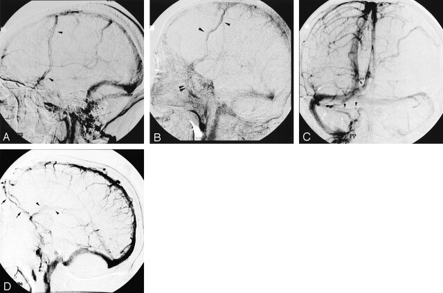
The superficial middle cerebral vein and its various drainage patterns, the sinus of the lesser sphenoid wing, and the sphenoid and parietal portions of the anterior branch of the middle meningeal veins were identified during the various venous phases of cerebral angiograms. Some examples are shown in Figure 6A–D.
Fig 6.
Various venous phases of digital subtraction angiography using selective internal carotid artery injections in three patients with no confirmed cerebrovascular disease.
A, Lateral projection of a late venous phase shows the characteristic venous sinus appearance of the parietal portion of the anterior branch of the middle meningeal veins, seen as two distinct thin channels coursing in parallel (arrowheads).
B, Lateral projection of a late venous phase depicts both the sphenoid (double arrowhead) and parietal (single arrowheads) portions of the anterior branch of the middle meningeal veins. The parietal portion is seen as a wide sinuous channel typical of a diploic channel.
C, Anteroposterior projection demonstrating simultaneously the sinus of the lesser sphenoid wing and the superficial middle cerebral vein. The sinus of the lesser sphenoid wing (black arrowheads) drains medially into the cavernous sinus and is connected laterally to the diploic vein of the orbital roof (black arrow). The superficial middle cerebral artery (white arrows) is seen to drain through emissary veins that drain into the pterygoid plexus (PP).
D, As in 5C, lateral view shows the diploic vein of the orbital roof (black arrows) connecting extracranially with the supraorbital veins (black double arrows). The superficial middle cerebral vein (black arrowheads) is well delineated and drains into the emissary veins of the middle cranial fossa.
Discussion
Anatomy
The anterior branch of the middle meningeal veins (sphenoid portion) crosses the floor of the middle cranial fossa, from the foramen ovale or spinosum to the pterion, taking the form of two parallel venous sinuses centered by the middle meningeal artery. It then progresses cranially along the anterior margin of the parietal squama to terminate into the venous lakes of the superior sagittal sinus (parietal portion). As they course under the most lateral aspect of the lesser sphenoid wing, the anterior branch of the middle meningeal veins and its arterial counterpart are contained for a short distance within an osseous canal, the sphenoparietal canal (of Trolard), before exiting into a sulcus of the parietal squama. This has been previously reported (7, 12, 13). Before entering this osseous canal, the anterior branch of the middle meningeal veins establishes a connection with a dural venous sinus located under the lesser sphenoid wing, the sinus of the lesser sphenoid wing. The sinus of the lesser sphenoid wing is connected laterally with the sphenoid portion of the anterior branch of the middle meningeal veins and medially with the anterior and superior aspect of the cavernous sinus. It crosses over the superior ophthalmic vein before reaching the cavernous sinus. A similar description of the sinus of the lesser sphenoid wing was given by Trolard (12). Three tributaries of the sinus of the lesser sphenoid wing were found, usually draining into its lateral portion:
1) a diploic vein of the orbital roof. It connected anteriorly and extracranially with the supraorbital veins and sometimes laterally with frontal diploic veins that drained into the superior sagittal sinus. We found no mention of this diploic vein in the literature.
2) a diploic vein located within the greater sphenoid wing and draining into the pterygoid plexus.
3) an orbital vein corresponding to the ophthalmomeningeal vein (of Hyrtl) (10).
Connections between the superficial middle cerebral vein and the sinus of the lesser sphenoid wing or the anterior branch of the middle meningeal veins were never demonstrated in this study. All the other possible terminations of the superficial cerebral vein in the middle cranial fossa; that is, into a paracavernous sinus, a laterocavernous sinus, or a cavernous sinus were encountered (11, 14). Our data also confirm previous reports showing that, whenever the superficial middle cerebral vein drains in the laterosellar region, it generally directly pierces the dura of the lateral wall of the cavernous sinus. Only rarely is the superficial middle cerebral vein attached to the dura mater underlying the inferior aspect of the lesser sphenoid wing. In such cases it maintains the characteristics of an arachnoid vein (9, 11). In our material, the superficial middle cerebral vein never assumed the characteristics of a dural sinus.
On the basis of their angiographic observation that opacified blood from the superficial middle cerebral vein was not diluted under the lesser sphenoid wing, Wolf et al (10) suggested that the sinus of the lesser sphenoid wing exclusively drained the superficial middle cerebral vein. With this assumption, they clearly—and, we think, erroneously—assimilated the distal portion of the superficial middle cerebral vein to the sinus of the lesser sphenoid wing. As early as 1890, however, Trolard (12) had challenged the already widespread impression that the superficial middle cerebral vein and the sinus of the lesser sphenoid wing were connected under the lesser sphenoid wing. This general misconception can probably be ascribed to the close topographic relationship between the sinus of the lesser sphenoid wing and a superficial middle cerebral vein attached to the dura mater of the lesser sphenoid wing. Furthermore, most of the anatomic studies evaluating the termination of the superficial middle cerebral vein have been performed with noninjected specimens, a fact that may easily explain why the sinus of the lesser sphenoid wing was overlooked.
That meningeal and diploic veins of the middle cranial fossa are not anatomically and functionally related to the superficial middle cerebral vein is also supported by their different developmental origins. According to Padget (7), the middle meningeal veins arise from the primitive middle meningeal sinuses, which are lateral tributaries of the prootic sinus. The prootic sinus is also the precursor of the cavernous sinus and of the inferior petrosal sinus, both of which occupy an extradural position in the adult (15). The primitive middle meningeal sinuses develop as extrachondrocranial vessels (around the 40-mm stage) and are involved in the vascularization of membranous bones that will form the skull vault (7). On the other hand, the superficial middle cerebral vein drains, along with the deep middle cerebral veins and the primitive tributaries of the basal vein of Rosenthal, into the primitive tentorial sinus of Padget (7). By the 60-mm ands 80-mm stages, with the development of the cerebral hemispheres and the subsequent caudal swing of the transverse sinus, the caudal end of the tentorial sinus migrates cranially and ventrally toward the junction between the sigmoid and transverse sinuses. As already noted by Padget, the final location of the tentorial sinus in the middle cranial fossa can vary from a medial to a lateral position. The adult remnant of the primitive tentorial sinus is, therefore, either incorporated to the cavernous sinus (medial position), courses within the lateral wall of the cavernous sinus (intermediate position), or remains on the floor of the middle cranial fossa as a paracavernous sinus (lateral position) (11, 14).
An interesting finding in our material was the observation that the anterior middle meningeal veins became assimilated to the anterior parietal diploic vein as they came out from the sphenoparietal canal (of Trolard) only to emerge as two distinct channels wherever their respective course did not overlap. The dual diploic and meningeal nature of the anterior branch of the middle meningeal veins has been reported by Trolard and Padget (7, 12). This phenomenon is probably related to aging. All the observations in our study are based on the cadavers of people who lived at least 80 years, on average. Diploic vessels appear postnatally concomitantly with the development of the cranial diploe and become prominent with old age (16). With age, the parietal impressions that correspond to the anterior branch of the middle meningeal veins (13) become deeper and more prominent (17, 18). Furthermore, there exist connections by way of small foramina between the parietal portion of the anterior branch of the middle meningeal veins and the parietal diploic vein (1, 12, 18). Because, with age, the anterior branch of the middle meningeal veins becomes more deeply positioned in the inner table of the skull, it seems reasonable to assume that a secondary assimilation of the parietal portion of the anterior branch of the middle meningeal veins to the underlying parietal diploic vein may ensue.
Angiographic Correlation
The parietal portion of the anterior branch of the middle meningeal veins can be identified during the late venous phase of cerebral angiography. When detectable, it is usually seen as two parallel, regular, and thin venous channels characteristic of meningeal veins, coursing from the pterion to the superior sagittal sinus. Alternatively, the parietal portion of the anterior branch of the middle meningeal veins may appear as a wide sinuous channel typical of a diploic vein.
The sphenoid portion of the anterior branch of the middle meningeal veins is uncommonly documented at routine cerebral angiography, either concomitantly to the superficial middle cerebral vein opacification, or slightly later in the venous phase. Distinction between the sphenoid portion the anterior branch of the middle meningeal veins and a paracavernous sinus or a lateral wall of a cavernous sinus on the lateral projection is based on the absence of cortical afferences from the superficial or deep middle cerebral veins.
The sinus of the lesser sphenoid wing is rarely observed angiographically. It appears concomitant to the superficial middle cerebral vein and can be identified in the anteroposterior projection as a thin channel coursing parallel to the inferiorly located superficial middle cerebral vein. The diploic vein of the orbital roof may be identified angiographically and must be distinguished from an ophthalmic vein, especially on lateral projections. Nonsubtracted images in the lateral projection demonstrate the position of both the superior ophthalmic vein and the diploic vein of the orbital roof in relation to the orbital roof. In the anteroposterior projection, distinction between the diploic vein of the orbital roof and the superior ophthalmic vein is made by identifying the characteristic inverted-S course of the superior ophthalmic vein and its dorsal connection with the cavernous sinus.
The Sphenoparietal Sinus of Breschet
As mentioned earlier, Breschet’s atlas of the venous system remained unfinished, and we found no written description of the sphenoparietal sinus by Breschet himself (1). Trolard (13) reached the same conclusion in 1890. Cruveilhier quoted Breschet’s definition of the sphenoparietal sinus as a dural venous channel located under the lesser sphenoid wing and draining meningeal and diploic blood, but the origin of this quotation could not be confirmed (2). In several of Breschet’s original illustrations (1), a venous channel, located under the lesser sphenoid wing and apparently dural in nature, is clearly identified and labeled as a sphenoparietal sinus (Figs 7A and B). The middle meningeal veins are also illustrated, and the legend mentions their connection with the sphenoparietal sinus. Nowhere in the 42 plates of Breschet’s atlas does the superficial middle cerebral vein appear to be connected with the sphenoparietal sinus.
Fig 7.
Plates from Breschet’s atlas (courtesy of Mme. B. Molitor, Section Histoire de la Médecine, Bibliotèque Inter Universitaire de Médecine, Paris, France).
A, Posterior view of the three cranial fossae. The sphenoid portion of the sphenoparietal sinus, corresponding to the sinus of the lesser sphenoid wing, is shown under the lesser sphenoid wing (brown asterisk). Connections between the sphenoparietal sinus and the anterior branch of the middle meningeal veins are illustrated and were described in the legends of the original text. Note that the superficial middle cerebral artery is not depicted.
B, Posterior view of the anterior and middle cranial fossae, in which the dura mater has been removed. The parietal portion of the sphenoparietal sinus (1) is depicted and artificially distinguished from the anterior branch of the middle meningeal veins (2). The floor of the sulcus of the sphenoparietal sinus is pierced by many small foramina. The legend describes these foramina, which are connections between the sphenoparietal sinus and underlying diploic veins.
According to his illustrations, Breschet seems to have combined the parietal portion of the anterior branch of the middle meningeal veins with a dural sinus located under the lesser sphenoid wing, to create a single continuous venous entity that he named the sphenoparietal sinus. We think, along with Trolard (12), that the parietal portion of Breschet’s sphenoparietal sinus is in fact the parietal prolongation of the anterior branch of the middle meningeal veins. The sphenoid portion of the sphenoparietal sinus, on the other hand, appears to be a distinct dural venous sinus coursing under the lesser sphenoid wing, with its own set of tributaries and secondary anastomoses. This venous structure is, in our opinion, adequately described by the term sinus of the lesser sphenoid wing proposed by Wolf et al (10).
It thus appears that the name sphenoparietal sinus does not describe an actual anatomic entity but rather corresponds to the arbitrary assimilation of two independent meningeal vessels. Furthermore, the frequent and incorrect assimilation made in the anatomic, radiologic, and surgical literature of the so-called sphenoparietal sinus with the termination of the superficial middle cerebral vein, generates further confusion. For these reasons, it appears that the term sphenoparietal sinus should be abandoned.
Clinical Relevance
Dural arteriovenous fistulas in the region of the sphenoid lesser wing are rare findings; there is usually a history of previous head trauma or prior surgery (19, 20). We present a case of type IV dural arteriovenous fistula under the lesser sphenoid wing. This fistula involved the superficial middle cerebral vein and not a sinus of the lesser sphenoid wing. Distinction between the two was possible by recognizing a retrograde filling into the deep and superficial cerebral venous system, the presence of a saccular dilatation more typical of a vein than a venous sinus, and the demonstration of a laterocavernous sinus, which, when present, drains the superficial middle cerebral vein or the deep middle cerebral vein or both (11, 14).
Development of a dural arteriovenous fistula in a superficial middle cerebral vein is rendered possible by its occasional attachment to the dura mater overlying the lesser sphenoid wing. This distinction is clinically relevant, because cerebral venous hypertension and related complications are only likely to result from a high-grade fistula involving a superficial middle cerebral vein. Bitoh et al (19) reported a case of dural arteriovenous fistula of the sinus of the lesser sphenoid wing in a patient with a 3-month history of exophthalmia and chemosis due to venous hypertension in the superior ophthalmic vein, which necessitated surgical correction. In general, however, asymptomatic dural arteriovenous fistulas of the sinus of the lesser sphenoid wing probably do not require any correction. This is supported by a case reported by Tsutsumi et al (20) of the spontaneous resolution of a dural arteriovenous fistula of the sinus of the lesser sphenoid wing that had appeared postoperatively.
Conclusion
The sphenoparietal sinus corresponds to the artificial combination of two venous structures, the parietal portion of the anterior branch of the middle meningeal veins and a dural channel located under the lesser sphenoid wing, the sinus of the lesser sphenoid wing. The classic notion that the superficial middle cerebral vein drains into or is partially equivalent to the sphenoparietal sinus is erroneous. These structures are independent, and, in our study, the superficial middle cerebral vein was never connected to the anterior branch of the middle meningeal veins or the sinus of the lesser sphenoid wing. Our findings indicate that the term sphenoparietal sinus would better be abandoned for the sake of anatomic precision and consistency. Knowledge of the venous anatomy in the region of the lesser sphenoid wing is of clinical importance in the diagnosis, classification, and therapeutic considerations of dural arteriovenous fistulas in this region.
Acknowledgments
We wish to thank Drs. P. and M. Francis for revision of the manuscript.
Footnotes
Presented at the 39th annual meeting of the American Society of Neuroradiology, Boston, MA, April 23–27, 2001.
References
- 1.Breschet G. Recherches Anatomiques,Physiologiques et Pathologiques sur le Système Veineux et Spécialement sur les Canaux Veineux des Os. Paris: Villeret et Rouen;1829. :1–42
- 2.Cruveilhier J. Traité d’anatomie Descriptive: Angéologie. 3rd ed. Paris: Labé;1852. :43
- 3.Rouvière H, Delmas A. Anatomie humaine. Tome I: Tête et Cou. 14th ed Paris: Masson;1997;233
- 4.Galligioni F, Bernardi R, Pellone M, et al. The superficial sylvian vein in normal and pathologic cerebral angiography. Am J Roentgenol Radium Ther Nucl Med 1969;107:565–578 [DOI] [PubMed] [Google Scholar]
- 5.Hédon CE. Etude anatomique sur la circulation veineuse de l’encéphale. Thèse de la Faculté de Médecine de Bordeaux;1888. :1–96
- 6.Oka K, Rhoton AL, Barry M, et al. Microsurgical anatomy of the superficial veins of the cerebrum. Neurosurgery 1985;17:711–748 [DOI] [PubMed] [Google Scholar]
- 7.Padget DH. Development of the cranial venous system in man, from viewpoint of comparative anatomy. Contrib Embryol 1957;36:79–140 [Google Scholar]
- 8.Hacker H. Superficial supratentorial veins and dural sinuses. In: Newton TH, Potts DG, eds. Radiology of the Skull and Brain. Vol 2. Book 3. St Louis: Mosby;1974. :1851–1877
- 9.Bisaria KK. The superficial sylvian vein in humans: with special reference to its termination. Anat Rec 1985;212:319–325 [DOI] [PubMed] [Google Scholar]
- 10.Wolf BS, Huang YP, Newman CM. The superficial sylvian venous drainage system. Am J Roentgenol Radium Ther Nucl Med 1963;89:389–410 [PubMed] [Google Scholar]
- 11.San Millán Ruïz D, Gailloud P, de Miquel Miquel MA, et al. Laterocavernous sinus. Anat Rec 1999;254:7–12 [DOI] [PubMed] [Google Scholar]
- 12.Trolard P. Les veines méningées moyennes. Rev Sci Biol 1890;485–499
- 13.Jones FW. On the grooves upon the ossa parietalia commonly said to be caused by the arteria meningea media. J Anat Physiol 1912;46:228–238 [PMC free article] [PubMed] [Google Scholar]
- 14.Gailloud P, San Millán Ruïz D, Muster M, et al. Angiographic anatomy of the laterocavernous sinus. AJNR Am J Neuroradiol 2000;21:1923–1929 [PMC free article] [PubMed] [Google Scholar]
- 15.Parkinson D. Extradural neural axis compartment. J Neurosurg 2000;92:585–588 [DOI] [PubMed] [Google Scholar]
- 16.Boismoreau MEP. Contribution à l’étude de la vascularisaton du diploé. Thèse de la Faculté de Médecine de Bordeaux;1904. :60 pp, 3 plates
- 17.Augier M. Crâne et cerveau chez le vieillard. L’Anthropologie 1932;42:315–321 [Google Scholar]
- 18.Saban R. Anatomie et Évolution des veines méningées chez les hommes fossiles. 1st ed. Paris: Comité des Travaux Historiques et Scientifiques. 1984. :15–132
- 19.Bitoh S, Arita N, Fujiwara M, et al. Dural arteriovenous malformation near the left sphenoparietal sinus. Surg Neurol 1980;13:345–349 [PubMed] [Google Scholar]
- 20.Tsutsumi K, Shiokawa Y, Kubota M, et al. Postoperative arteriovenous fistula between the middle meningeal artery and the sphenoparietal sinus. Neurosurgery 1990;26:869–870 [DOI] [PubMed] [Google Scholar]


