Abstract
BACKGROUND AND PURPOSE: Amygdalar and hippocampal volume measurements indicate a right-greater-than-left asymmetry for right-handed normal participants in most studies. The purpose of this study was to compare amygdalar and hippocampal volume ratios between right- and left-handed participants.
METHODS: Amygdalar and hippocampal volume measurements were performed in 34 (20 right-handed and 14 left-handed) normal participants. All participants completed a 10-item handedness questionnaire. The MR imaging sequence was a 3D T1-weighted gradient-echo acquisition of the whole brain (24/6 [TR/TE]; flip angle, 25 degrees). MR images were spatially normalized, and volumes were painted with a 1.0-mm3 resolution cursor on an SGI workstation.
RESULTS: In right-handed participants, the amygdala and hippocampus (P < .001 for both) were significantly larger on the right side than on the left. The left-handed group did not show a significant difference between right- and left-sided structures. Right-to-left volume ratios differed significantly between right- and left-handed participants for both amygdalar (P < .02) and hippocampal (P < .01) structures. Gender did not affect right-to-left amygdalar and hippocampal volume ratios in right- or left-handed participants.
CONCLUSION: Handedness, but not gender, seems to affect right-to-left amygdalar and hippocampal volume ratios.
Hippocampal volumetry has become a clinical tool in identifying unilateral or bilateral hippocampal volume changes in cases of temporal lobe epilepsy (1, 2). Amygdalar volumetry has been implemented more recently to help in the lateralization of a suspected temporal lobe seizure focus in the absence of hippocampal atrophy (3). Because of the differences in acquisition techniques, technical capabilities, processing, and techniques for delineation of the anatomic structures, absolute volumes vary among centers (1–6). Volume ratios have been more consistent among epilepsy surgery centers than have been the measurements of absolute volumes. Still, some centers show right-greater-than-left volume ratios in normal participants whereas others do not (4–6).
The effect of handedness has not been well documented in the past. Only one study included a small number of left-handed and ambidextrous participants, but the participants were not selected on the basis of a questionnaire (4). Although only approximately 10% of the general population is developmentally left-handed, left-handedness is found in ≤30% of patients with medically refractory epilepsy, especially in the setting of left hemispheric foci that become symptomatic during infancy or early childhood (7–9).
Hence, it is important to determine whether handedness affects amygdalar and hippocampal volume ratios. If volume ratios differ between right- and left-handed persons, normative data for developmentally left-handed participants may improve the sensitivity and specificity of amygdalar and hippocampal volumetry in the evaluation of patients with epilepsy. Furthermore, a difference between left- and right-handed epilepsy would identify the need to reevaluate asymmetry ratios in pathologically left-handed persons.
Methods
Participants
MR images of 20 right-handed participants (11 women and nine men) were randomly selected from a database of 170 normal participants (10). A total of 17 left-handed participants were also identified. MR images with the appropriate sequences and resolution were available for only 14 left-handed (eight women and six men) participants. All the participants completed a handedness questionnaire at the time of MR imaging. The questionnaire inquired about hand preference for 10 activities: writing, drawing, throwing, using scissors, toothbrushing, using a screwdriver, using a spoon, using a key, using a hammer, and striking a match. The questions were drawn from the Edinburgh Laterality Inventory and Reitan Handedness Scale. The handedness index for right-handed participants ranged between 8 and 10 (mode 10) and for left-handed participants between 0 and 4.9 (mode 1). Ten participants indicated complete right-handedness. Family history of left-handedness in at least one parent was identified for two (10%) of 21 right-handed participants and five (36%) of 14 left-handed participants.
Image Acquisition and Processing
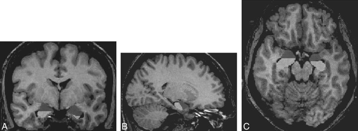
The MR imaging sequence used for volumetric analysis was a 3D T1-weighted gradient-echo acquisition of the whole brain (24/6; flip angle, 25 degrees; field of view, 256 × 256 mm; matrix, 256 × 192). The right side was marked by a 1-cm-diameter plastic tube filled with water, placed caudally from the participant's ear and additionally defined in the header file. Pixels were 1.0 × 1.0 mm, and section width was 1 mm without an interleave gap. Spatial normalization was performed on all images, using validated methods to remove brain size effects (10). All volumes were painted with a 1.0-mm3 resolution cursor on an SGI workstation using Display (MNI, Canada), which allowed simultaneous visualization of the structures in three planes (Fig 1). The use of Display for volumetric measurements of brain structures has been recently reported (11–13).
fig 1.
Amygdala and hippocampus in coronal (A), sagittal (B), and axial (C) planes
All volumetric measurements were performed by the principle investigator (C.A.S.), who was blinded to handedness in all the left-handed participants and more than half the right-handed participants. Five images of right-handed participants were chosen randomly for repeated measurements to assess intrarater intertrial reliability within 48 hr of the first measurement and in five left-handed participants 6 months after the first measurement.
Amygdalar Volumetry
The anterior border was defined in the axial and sagittal planes (Fig 1). The anterior border was delimited by the gyrus ambiens and the endorhinal sulcus at the level of the optic chiasm. Anteromedially, the amygdala included the cortical surface of the semilunar gyrus. The lateral border was differentiated from the anterior portion of the claustrum on axial and sagittal images. The inferolateral border was defined by the temporal horn of the lateral ventricle or alveus intervening between amygdala and the hippocampal head. The superior border was best delineated on axial and sagittal planes and included the central nucleus of the amygdala. Posteriorly, the amygdala was delineated as a structure overlying the temporal horn of the lateral ventricle, reaching from the apex of the ventricle medially, where it bordered the hippocampus.
Hippocampal Volumetry
The anterior border of the hippocampus emerged beneath the amygdala as the anterior recess of the temporal horn ascended laterally and was delineated by the alveus medially and inferiorly (Fig 1). The medial, intervening portion of the alveus and subcortical white matter was extended to the cortical surface, excluding most of the overlying parahippocampal gyrus and uncus anteriorly and the prosubiculum posteriorly. Superiorly, it bordered amygdala and the temporal horn. Posteriorly, the fornix was excluded from the measurements. The posterior boundary was the last coronal section before visualization of the crus fornix in its entire length and width as it ascended toward the thalamus. Choroid plexus was included if it was contiguous with the hippocampus.
Statistical Analysis
For the comparison of right and left amygdalar and hippocampal volumes, right-to-left volume ratios were calculated for each participant. A natural logarithm transform was then performed on the ratios to facilitate statistical analysis. For the right-handed participants, analysis of variance with 2 × 2 factorial design was used to detect an interaction between right-to-left volume ratios as the dependent measure and structure (amygdala versus hippocampus). A simple t test was conducted to reveal any significant asymmetry between right and left amygdalar and hippocampal volumes. For the left-handed participants, a similar t test was conducted for each structure to detect interhemispheric asymmetry. To test whether any significant difference existed between right- and left-handed participants, a group t test was conducted on the volume ratio data. The effect of gender on hippocampal or amygdalar asymmetry was investigated in right- and left-handed groups using an analysis of variance 2 × 2 factorial design. Finally, intrarater intertrial reliability was assessed for repeated measurements, 48 hr apart for five right-handed participants and6 months apart for five left-handed participants. Reliability was tested in the first group by using a Pearson correlation, whereas in the latter group, a paired t test was used because the data range was small and the correlation coefficient could not be calculated.
Results
Mean amygdalar and hippocampal volumes were calculated regarding handedness (Table). In right-handed participants (n = 20), the mean amygdalar volumes were 1600 and 1494 mm3 for the right and left hemispheres, respectively, and the mean hippocampal volumes were 3008 and 2858 mm3, respectively. Right-to-left amygdalar and hippocampal volume ratios were 1.07 ± 0.06 (mean ± SD) and 1.05 ± 0.04, respectively. In left-handed participants (n = 14), the mean amygdalar volumes were 1612 and 1578 mm3 for the right and left hemispheres, respectively, and the mean hippocampal volumes were 2989 and 3039 mm3, respectively. Right-to-left amygdalar and hippocampal volume ratios were 1.02 ± 0.07 and 0.98 ± 0.05, respectively. The mean right and left hippocampal and amygdalar volumes differed significantly (P < .001 for both) for right-handed participants but not for left-handed participants. The right-to-left volume ratios were statistically different between right- and left-handed participants for both the amygdala (P < .02) and the hippocampus (P < .001). Gender did not seem to affect amygdalar or hippocampal volume measurements in either group.
Absolute amygdalar and hippocampal volumes and their ratios in healthy subjects

The intrarater intertrial correlation for the measurement of amygdalar and hippocampal volume ratios measured 48 hr apart for right-handed participants exceeded 0.98 and 0.99, respectively. No significant difference was found for intrarater intertrial measurements of hippocampal volumes obtained 6 months apart for left-handed participants (paired t test, α < 0.05). The right-to-left volume ratios for the five left-handed participants measured 6 months apart are 0.96 ± 0.04 and 0.98 ± 0.07, respectively. Both of these comparisons suggested that the intrarater intertrial measurements were reliable.
Discussion
A right-greater-than-left asymmetry was shown in right-handed normal participants by some studies (4, 5) but not by others (1, 6). The reasons underlying this discrepancy are unclear but may be differences in techniques for image acquisition and hippocampal delineation (14). Norms for amygdalar volumes and their ratios are limited (5). In this study, right-handed participants showed significant right-greater-than-left amygdalar and hippocampal volume ratios. Furthermore, statistically significant differences were noted for amygdalar and hippocampal volume ratios between right- and left-handed participants. The only study that considered the effect of handedness on volume ratios indicated that handedness did not play a detectable role (4). However, that study included only a small number of left-handed and ambidextrous participants and relied largely on self-report and not handedness indices. It is unclear why some left-handed participants should have inverted asymmetries from right-handed participants, but these shifts could be related to the lateralization of cognitive or language functions. Nonetheless, the results of this study show the need to establish different criteria for left- and right-handed patients with epilepsy. Considering that almost 30% of patients with epilepsy are left-handed (7–9), implementing criteria based on handedness may improve the sensitivity and specificity for detecting clinically significant asymmetries in patients with temporal lobe epilepsy. However, the results of this study did not address differences between developmentally and pathologically left-handed participants, who tend to have an early onset of epilepsy originating from the left hemisphere. More extensive analysis of these groups would be necessary to better use the proposed handedness criteria.
Gender did not affect right-to-left hippocampal and amygdalar volume ratios in the right-handed or left-handed groups. This finding confirmed the results of studies performed at other epilepsy centers, which included hippocampal (4, 6) and amygdalar (6) volumetry.
In summary, amygdalar and hippocampal volume ratios in right-handed normal participants showed a significant right-greater-than-left asymmetry, whereas most left-handed participants showed a left-greater-than-right asymmetry. It seems that different criteria should be applied to left- and right-handed patients undergoing amygdalar and hippocampal volumetry to improve the ability to lateralize a temporal lobe seizure focus.
Footnotes
This work was presented at the 1998 American Epilepsy Society Meeting in San Diego, CA.
Supported by the EJLB Foundation and the National Institute of Mental Health (P20 DA52176) and by an internal review grant through the University of Texas Health Science Center, San Antonio (00-0021).
Address reprint requests to C. Ákos Szabó, MD, Division of Neurology, University of Texas Health Science Center at San Antonio, 7703 Floyd Curl Drive, San Antonio, TX 78284.
References
- 1.Ashtari M, Barr WB, Schaul N, Bogerts B. Three-dimensional fast low-angle shot imaging and computerized volume measurement of the hippocampus in patients with chronic epilepsy of the temporal lobe. AJNR Am J Neuroradiol 1991;12:941-947 [PMC free article] [PubMed] [Google Scholar]
- 2.Cascino GD, Jack CR, Parisi JE, et al. Magnetic resonance imaging-based volume studies in temporal lobe epilepsy: pathological correlations. Ann Neurol 1991;30:31-36 [DOI] [PubMed] [Google Scholar]
- 3.Guerreiro C, Cendes F, Li LM, et al. Clinical patterns of patients with temporal lobe epilepsy and pure amygdalar atrophy. Epilepsia 1999;40:453-461 [DOI] [PubMed] [Google Scholar]
- 4.Jack CR Jr, Twomey CK, Zinsmeister AR, Sharbrough FW, Petersen RC, Cascino GD. Anterior temporal lobes and hippocampal formations: normative volumetric measurements from MR images in young adults. Radiology 1989;172:549-554 [DOI] [PubMed] [Google Scholar]
- 5.Watson C, Andermann F, Gloor P, et al. Anatomic basis of amygdaloid and hippocampal volume measurement by magnetic resonance imaging. Neurology 1992;42:1743-1750 [DOI] [PubMed] [Google Scholar]
- 6.Bhatia S, Bookheimer SY, Gaillard WD, Theodore WT. Measurement of whole temporal lobe and hippocampus for MR volumetry: normative data. Neurology 1993;43:2006-2010 [DOI] [PubMed] [Google Scholar]
- 7.Rasmussen T, Milner B. The role of early left-brain injury in determining lateralization of cerebral speech function. Ann N Y Acad Sci 1977;299:355-369 [DOI] [PubMed] [Google Scholar]
- 8.Dellatolas G, Luciani S, Castresana A, et al. Pathological left-handedness: left-handedness correlatives in adult epileptics. Brain 1993;116:1565-1574 [DOI] [PubMed] [Google Scholar]
- 9.Dodrill CB, Ojemann GA. Probability of atypical speech as a function of handedness and side of surgery. Epilepsia 1999;40[suppl 7]:83 [Google Scholar]
- 10.Lancaster JL, Glass TG, Lankipalli BR, Downs H, Mayberg H, Fox PT. A modality-independent approach to spatial normalization of tomographic images of the human brain. Hum Brain Mapp 1996;3:209-223 [Google Scholar]
- 11.Paus T, Otaky N, Caramanos Z, et al. In vivo morphometry of the intrasulcal gray matter in the human cingulate, paracingulate, and superior-rostral sulci: hemispheric asymmetries, gender differences and probability maps. J Comp Neurol 1996;376:664-673 [DOI] [PubMed] [Google Scholar]
- 12.Penhune VB, Zatorre RJ, MacDonald JD, Evans AC. Interhemispheric anatomical differences in human primary auditory cortex: probabilistic mapping and volume measurement from magnetic resonance scans. Cereb Cortex 1996;6:661-672 [DOI] [PubMed] [Google Scholar]
- 13.Tomaiuolo F, MacDonald JD, Caramanos Z, et al. Morphology, morphometry and probability mapping of the pars opercularis of the inferior frontal gyrus: an in vivo MRI analysis. Eur J Neurosci 1999;11:3033-3046 [DOI] [PubMed] [Google Scholar]
- 14.Jack CR, Theodore WT, Cook M, McCarthy G. MRI-based hippocampal volumetrics: data acquisition, normal ranges, and optimal protocol. Magn Reson Imaging 1995;13:1057-1064 [DOI] [PubMed] [Google Scholar]


