Abstract
Summary: PHACE syndrome is a neurocutaneous syndrome with the following features: posterior fossa malformations of the brain, large facial hemangiomas, arterial anomalies, cardiac anomalies and aortic coarctation, and eye abnormalities. We report a rare case of bilateral internal carotid artery agenesis with transcranial collaterals from the external carotid arteries and agenesis of the vertebrobasilar system in a possible PHACE syndrome. We suggest that the patient had an incomplete phenotypic expression of the PHACE syndrome. Although the phenotypic spectrum is broad and is still largely unexplored, the extent of the cephalic neural crest cells insulted genetically or by other causes at a certain time during the development of the embryo might explain the variable phenotypic expression of PHACE syndrome.
Agenesis, aplasia, and hypoplasia of the internal carotid artery are rare congenital anomalies, occurring in less than 0.01% of the population (1, 2). The term “absence” has been chosen to incorporate agenesis, aplasia, and hypoplasia of the internal carotid artery. In this setting, the most common type of collateral flow is through the circle of Willis. Less commonly, collateral flow is provided via persistent embryonic vessels or from transcranial collaterals originating from the external carotid artery system. Agenesis of the vertebrobasilar system is relatively common, but concomitant agenesis of the internal carotid artery and the vertebrobasilar system is very rare.
Agenesis of the internal carotid artery can be associated with PHACE syndrome, a rare neurocutaneous syndrome, with the following features: posterior fossa malformations of the brain, large facial hemangiomas, arterial anomalies, cardiac anomalies and aortic coarctation, and eye abnormalities (3). A growing list of complex syndromes links disorders of the brain, meninges, or cerebral vessels with cutaneous and craniofacial lesions, some of which are also associated with congenital heart disease. However, to our knowledge, concomitant bilateral internal carotid artery agenesis and vertebrobasilar system agenesis as a feature of the PHACE syndrome have not been reported in the literature.
We describe a patient with bilateral internal carotid artery agenesis with transcranial collaterals from the external carotid artery and agenesis of the vertebrobasilar system, posterior fossa anomaly, and facial capillary hemangioma in a possible PHACE syndrome.
Case Report
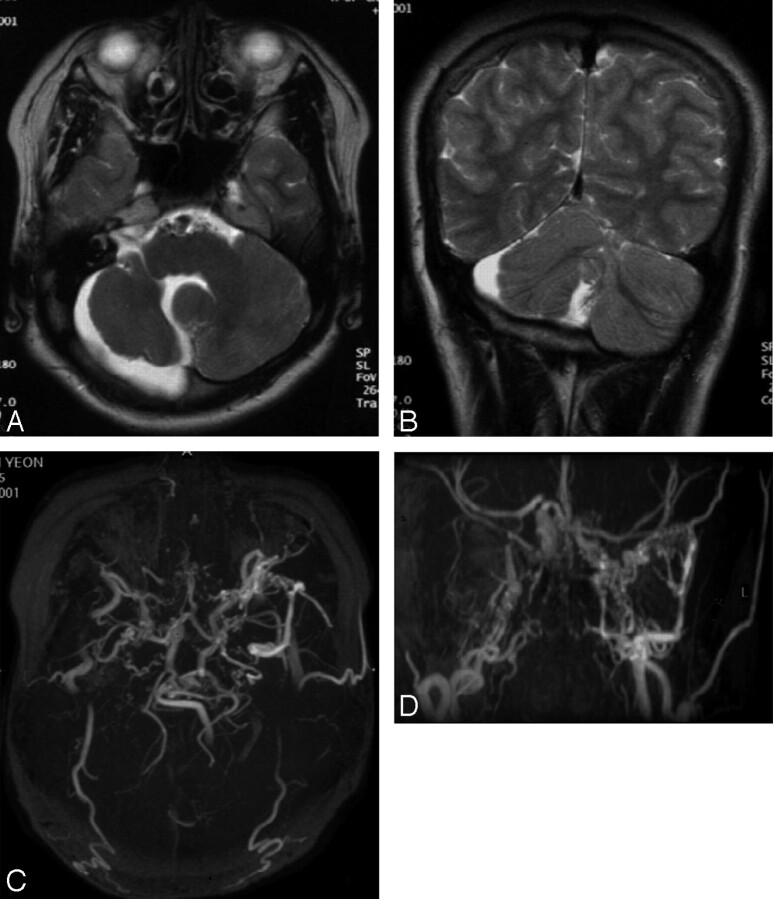
A 14-year-old girl was admitted with decreased visual acuity. She was born to nonconsanguineous healthy parents and presented with no family history of brain abnormalities or angiomas. Gestation and delivery were uneventful. She had a large facial hemangioma on the right eyelid and cheek that appeared soon after birth and regressed as she grew. She could open her right eye fully only after 7 years of age. Only a small scar was present on the right upper eyelid at admission. She had no abnormal findings on physical examination. Blood pressure was in the normal range. On neurologic examination, she had intermittent nonspecific headache and low visual acuity in her right eye and could only count fingers at a 3- to 4-m distance. On MR imaging and MR angiography, abnormality of the posterior fossa, no flow of the internal carotid arteries on both sides, and abnormality of the vertebrobasilar artery were noted (Fig 1). The right cerebellar hemisphere was hypoplastic, and the inferior cerebellar vermis was absent, with a normally preserved shape and size of the fourth ventricle. These features could be explained as Dandy-Walker variants.
Fig 1.
On axial (A) and coronal (B) T2-weighted images, hypoplasia of the right cerebellar hemisphere and absence of the inferior cerebellar vermis are present. Note that the abnormally dilated basilar artery with numerous small signal intensity void structures corresponds with the collateral networks at the prepontine cistern. On MR angiography (C, -D), both internal carotid arteries are not seen, and the distal portion of internal carotid arteries is reconstituted from the external carotid arteries.
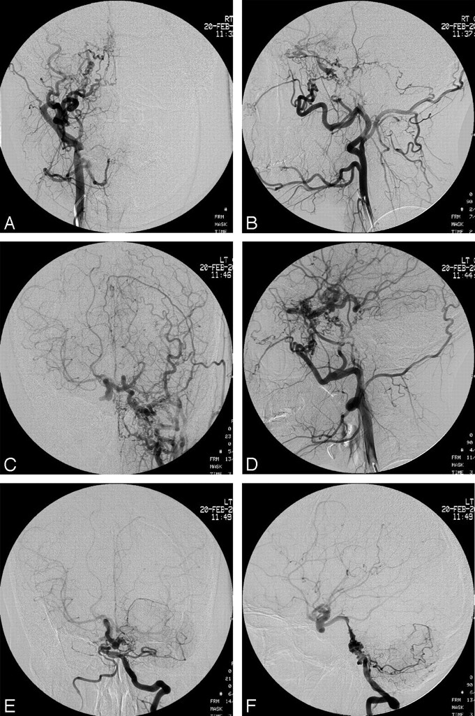
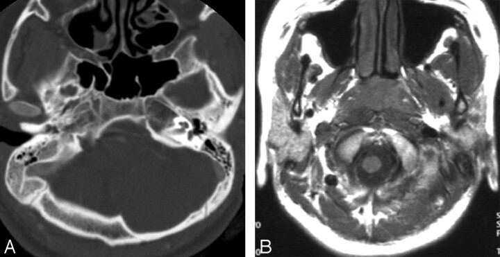
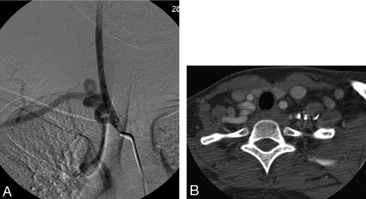
Digital angiography showed complete occlusion of the internal carotid artery from the cervical portion on right side and at the origin of left internal carotid artery, with transdural collaterals from the external carotid arteries forming multiple channels to reconstitute the distal internal carotid arteries (Fig 2). Injection of the right external carotid artery and left external carotid artery demonstrated the reconstitution of the distal internal carotid artery by branches of the internal maxillary arteries, middle meningeal arteries, deep temporal arteries, the artery of the foramen rotundum, ascending pharyngeal arteries, and collaterals of the occipital artery. There was also the discontinuation between the vertebral arteries and the abnormally dilated proximal basilar artery with collateral branches mainly from the left vertebral artery. Normal internal carotid arteries were not seen in the carotid spaces, and no carotid canals were noted on the brain CT and MRI (Fig 3). The right subclavian artery twisted like a screw from just distal to the origin of right common carotid artery (Fig 4). There were no demonstrable anomalies of the aorta and the heart.
Fig 2.

Injection of right external carotid artery (A and B) and left external carotid artery (C and D) demonstrates the reconstitution of the distal internal carotid artery by branches of the internal maxillary arteries, middle meningeal arteries, deep temporal arteries, artery of the foramen rotundum, ascending pharyngeal arteries, and collaterals of the occipital artery. Injection of the left vertebral artery (E and F) shows the discontinuation between the vertebral arteries and the abnormally dilated proximal basilar artery with collateral branches mainly from the left vertebral artery.
Fig 3.
Normal internal carotid arteries are not seen in the carotid spaces, and no carotid canals are noted on the bone window setting of the brain CT (A) and T1-weighted brain MRI (B).
Fig 4.
Injection of the innominate artery (A) and CT scan obtained at the level of thoracic inlet (B) demonstrate the unusual subclavian artery turning like a screw.
Discussion
This patient presented a spectrum of abnormalities, but not the full spectrum of the PHACE syndrome: a large facial hemangioma at birth, posterior fossa anomaly of Dandy-Walker variants, agenesis of the internal carotid artery, agenesis of vertebrobasilar artery, and deformation of right innominate artery. However, all previous studies have stressed the heterogeneity of the syndrome and the absence of one or more components (4). Hemangiomas are the most common benign tumors of infancy, occurring in up to 10% of children younger than 1 year of age and seem to be the unifying feature of the PHACE syndrome. Most patients are initially referred because of the complex nature and/or large size of their facial hemangiomas (4). As shown in this patient, the large hemangioma at the area of right forehead and cheek eventually regressed and all that remained was a small scar on the upper eyelid. The trigeminal division V1 is the most commonly affected region, and the extent and/or distribution of cutaneous involvement correlates with the type and severity of complications in some instances (4). Hemangiomas are typically bulky plaquelike lesions, not confined by their boundaries. Hemangiomas in PHACE syndrome are no different from sporadic lesions, but they show a female dominance of 9:1 (female–male ratio), compared with the 3:1 ratio for the sporadic lesions (5). Many reports recognize the association of cervicofacial hemangiomas with vascular anomalies and congenital heart disease (6, 7), and hemangiomas have also been associated with the Dandy-Walker syndrome (8). Thus the PHACE syndrome might be considered in infants presenting with a large plaquelike facial hemangioma, especially girls.
Classic Dandy-Walker malformation and the variants and cerebellar hypoplasia and arachnoid cysts or cortical dysgenesis can be associated with cutaneous hemangiomas and PHACE syndrome. Associated posterior cranial fossa malformations are present in 32%–74% of patients with PHACE syndrome (1, 6, 9). The causes of the Dandy-Walker malformation, however, are poorly understood, and a variety of theories and timings have been suggested for the provoking insult (10). These theories imply that certain insults of embryonic development at a specific critical time may give rise to very similar developmental outcomes, such as the PHACE syndrome and other neurocutaneous syndromes. In our patient, the right cerebellar hemisphere was hypoplastic and the inferior cerebellar vermis was absent, with normally preserved shape and size of the fourth ventricle. These features could be explained by Dandy-Walker variants.
Ophthalmologic findings are not as common as posterior fossa and arterial anomalies; however, quite a large number of abnormalities had been described, including colobomas, arcus corneae, optic nerve hypoplasia, increased retinal vascularity, and glaucoma (11). We could detect the decreased visual acuity in our patient’s right eye without any other structural abnormalities. No evidence of increased retinal vascularity was seen on angiography, and the normal choroidal blush was noted on both sides from collaterals of the external carotid arteries. The large facial hemangioma on the right eyelid and cheek and the lack of light stimuli might explain our patient’s low visual acuity; thus, she could open her right eye fully only after the age of 7 years. The eye needs to be stimulated by light to avoid this problem; if the eye can open slightly, a patch on the good eye might encourage the child to use the muscles of the bad eye optimally, helping to open the eye. Thus, early intervention would be warranted in the case of a hemangioma on the eyelid that interferes with opening the eye.
There are a wide variety and extent of vascular anomalies associated with this syndrome. Two types of arterial anomalies can occur most commonly: persistent embryonic arteries, such as the trigeminal artery, always ipsilateral to the cutaneous hemangioma, and agenesis of major arteries, such as the internal carotid and vertebral arteries, which are usually ipsilateral to the cutaneous lesion, as in our patient (3, 9). In our patient, the cervical segments of both internal carotid arteries were absent, with transdural collaterals from external carotid arteries forming multiple channels to reconstitute distal internal carotid arteries.
The internal carotid arteries originate from the dorsal aorta and the third aortic arch at approximately the 4- to 5-mm embryonic stage, with full development of the internal carotid artery by 6 weeks (12). Some investigators argue that both the proximal internal carotid artery and the external carotid artery arise jointly from the third aortic arch; thus, agenesis of the internal carotid artery should be accompanied by absence of the ipsilateral external carotid artery, or the external carotid artery and the common carotid artery can develop normally in the setting of internal carotid artery agenesis, because the former arises independently from the aortic sac (13, 14).
Others describe the internal carotid artery as related to the third aortic arch, where the external carotid artery corresponds to the ventral aorta. The existence of both systems is unrelated, as is the existence of each internal carotid artery segment (15). The latter seems more plausible because numerous cases of internal carotid artery agenesis with normally developed external carotid artery systems are reported in the literature. As Le Douarin and Kalcheim (16) have shown in avian embryos, neural crest cells forming the walls of the internal and external carotid arteries and their branches arise from the same metameric levels as the developing cerebellum; certain insults before migration of the neural crest and mesodermal cells may give rise to very similar developmental outcomes and the widespread lesions, as seen in the PHACE syndrome. However, a lesion of the neural crest (or even the crest and adjacent neural plate for cerebellar involvement) alone may not explain the spectrum of abnormalities in the PHACE syndrome. The putative insult must occur before migration of the neural crest and mesodermal cells to account for the widespread lesions.
All blood vessels are composed of an inner layer of endothelial cells and an immediately adjacent layer of pericytes. The outer wall layers, though originating from the mesoderm in the body, can be made by neural crest cells in the head (16). Cephalic neural crest cells contribute to the muscular and connective walls of large arteries derived from the branchial arches (17), including the cardiac septum that separates the aorta from the pulmonary artery trunk (18). The cephalic neural crest cells from the posterior diencephalons contribute to part of the wall of the internal carotid artery and neural crest cells from the upper part of the rhombencephalon to the proximal maxillary artery, the stapedial artery, and the common carotid artery. Neural crest cells from the lower part of the rhombencephalon are distributed within the musculoconnective wall of the large arteries of great vessels (19). Because of damaged neural crest cells in each site or both, abnormalities of the internal carotid arteries, common carotid arteries, and great vessels, including the aorta and the heart, could be present. Thus, damaged neural crest cells in the diencephalons and the rhombencephalon could lead to anomalies of the posterior fossa and the arteries, as a complete phenotypic expression of PHACE syndrome (5). Posterior diencephalons and more rostral neural crest cell involvement may not have the features of anomalies in the aorta, heart, and sternum as a spectrum of the PHACE syndrome as in our patient.
Extradural arteries are segmental, and the segments of the fully developed internal carotid artery correspond to embryonic structures. These are autonomous from an embryologic viewpoint, and each may be present or absent independently or in association with the adjacent ones (20). As shown in our patient, each of the embryonic arteries can be a potential source of reconstitution of the distal flow in internal carotid artery segmental agenesis. These vascular networks, the rete mirabile, are rare in humans but can be observed in some vertebrates such as cobaye, cat, cow, and sheep. The first few cases of rete mirabile reported by Minagi and Newton (21) and Danziger et al (22) were associated with a hypoplastic cervical and petrous internal carotid artery; a rich anastomotic intradural network at the base of the skull and a cavernous internal carotid artery bypassed the absent segments. In our patient, the reconstitution of the distal internal carotid artery probably occurred by forming rete mirabile from branches of the internal maxillary arteries, middle meningeal arteries, deep temporal arteries, artery of the foramen rotundum, ascending pharyngeal arteries, and collaterals of the occipital artery. The discontinuation between the vertebral arteries after joining of the large left and small right vertebral arteries and the abnormally dilated proximal basilar artery with collateral branches mainly from the left vertebral artery suggests the agenesis at the level of the proximal basilar artery.
In conclusion, we report a case of bilateral internal carotid artery agenesis with transcranial collaterals from the external carotid arteries and vertebrobasilar artery agenesis in a PHACE syndrome. The extent of the cephalic neural crest cells insulted genetically or by other causes at a certain time during the development of the embryo might explain the variable phenotypic expression of PHACE syndrome.
Acknowledgments
We thank Professor Pierre Lasjaunias (L’hôpital Kremlin-Bicêtre, Paris) for his important suggestions and valuable comments.
References
- 1.Afifi AK, Godersky JC, Menezes A, Smoker WR, Bell WE, Jacoby CG. Cerebral hemiatrophy, hypoplasia of internal carotid artery, and intracranial aneurysm: a rare association occurring in an infant. Arch Neurol 1987;44:232–235 [DOI] [PubMed] [Google Scholar]
- 2.Chen CJ, Chen ST, Hsieh FY, Wang LJ, Wong YC. Hypoplasia of the internal carotid artery with intercavernous anastomosis. Neuroradiology 1998;40:252–254 [DOI] [PubMed] [Google Scholar]
- 3.Frieden IJ, Reese V, Cohen D. PHACE syndrome: the association of posterior fossa brain malformations, hemangiomas, arterial anomalies, coarctation of the aorta and cardiac defects, and eye abnormalities. Arch Dermatol 1996;132:307–311 [DOI] [PubMed] [Google Scholar]
- 4.Metry DW, Dowd CF, Barkovich AJ, Frieden IJ. The many faces of PHACE syndrome. J Pediatr 2001;139:117–123 [DOI] [PubMed] [Google Scholar]
- 5.Bhattacharya JJ, Luo CB, Alvarez H, Rodesch G, Pongpech S, Lasjaunias PL. PHACES syndrome: a review of eight previously unreported cases with late arterial occlusions. Neuroradiology 2004;46:227–233 [DOI] [PubMed] [Google Scholar]
- 6.Pascual-Castroviejo I. Vascular and nonvascular intracranial malformation associated with external capillary hemangiomas. Neuroradiology 1978;16:82–84 [DOI] [PubMed] [Google Scholar]
- 7.Mizuno Y, Kurokawa T, Numaguchi Y, Goya N. Facial hemangioma with cerebrovascular abnormalities and cerebellar hypoplasia. Brain Dev 1982;4:375–378 [DOI] [PubMed] [Google Scholar]
- 8.Hirsch JF, Pierre-Kahn A, Renier D, Sainte-Rose C, Hoppe-Hirsch E. The Dandy-Walker malformation: a review of 40 cases. J Neurosurg 1984;61:515–522 [DOI] [PubMed] [Google Scholar]
- 9.Pascual-Castroviejo I, Viano J, Moreno F, et al. Hemangiomas of the head, neck and chest with associated vascular and brain anomalies: a complex neurocutaneous syndrome. AJNR Am J Neuroradiol 1996;17:461–471 [PMC free article] [PubMed] [Google Scholar]
- 10.Golden JA, Rorke LB, Bruce DA. Dandy-Walker syndrome and associated anomalies. Pediatr Neurosci 1987;13:38–44 [DOI] [PubMed] [Google Scholar]
- 11.Coats DK, Paysse EA, Levy ML. PHACE: a neurocutaneous syndrome with important ophthalmologic implications: case report and literature review. Ophthalmology 1999;106:1739–1741 [DOI] [PubMed] [Google Scholar]
- 12.Padget DH. The development of the cranial arteries in the human embryo. Carnegie Institution of Washington Publication 575; Contributions to Embryology 1948;32:205–261 [Google Scholar]
- 13.Lie TA. Congenital anomalies of the carotid arteries. Excerpta Medica 1968;35–51
- 14.Quint DJ, Boulos RS, Spera TD. Congenital absence of the cervical and petrous internal carotid artery with intercavernous anastomosis. AJNR Am J Neuroradiol 1989;10:435–439 [PMC free article] [PubMed] [Google Scholar]
- 15.Lasjaunias P, Berenstein A, TerBrugge KG. Surgical neuroangiography. Vol1 , 2nd ed. Berlin: Springer-Verlag;2001. :393–425 [Google Scholar]
- 16.Le Douarin NM, Kalcheim C. Developmental and cell biology series: the neural crest. 2nd ed. Cambridge, UK: University Press;1999
- 17.Le Lievre CS, Le Douarin NM. Mesenchymal derivatives of the neural crest: analysis of chimaeric quail and chick embryos. J Embryol Exp Morphol 1975;34:125–154 [PubMed] [Google Scholar]
- 18.Waldo K, Miyagawa-Tomita S, Kumiski D, Kirby ML. Cardiac neural crest cells provide new insight into septation of the cardiac outflow tract: aortic sac to ventricular septal closure. Dev Biol 1998;196:129–144 [DOI] [PubMed] [Google Scholar]
- 19.Etchevers HC, Vincent C, Le Douarin NM, Couly GF. The cephalic neural crest provides pericytes and smooth muscle cells to all blood vessels of the face and forebrain. Development 2001;128:1059–1068 [DOI] [PubMed] [Google Scholar]
- 20.Mahadevan J, Batista L, Alvarez H, Bravo-Castro E, Lasjaunias P. Bilateral segmental regression of the carotid and vertebral arteries with rete compensation in a Western patient. Neuroradiology 2004;46:444–449 [DOI] [PubMed] [Google Scholar]
- 21.Minagi H, Newton TH. Carotid rete mirabile in man: a case report. Radiology 1966;86:100–102 [DOI] [PubMed] [Google Scholar]
- 22.Danziger J, Bloch S, Hefer AG. Bilateral rete carotids in man. S Afr Med J 1972;46:1487–1488 [PubMed] [Google Scholar]


