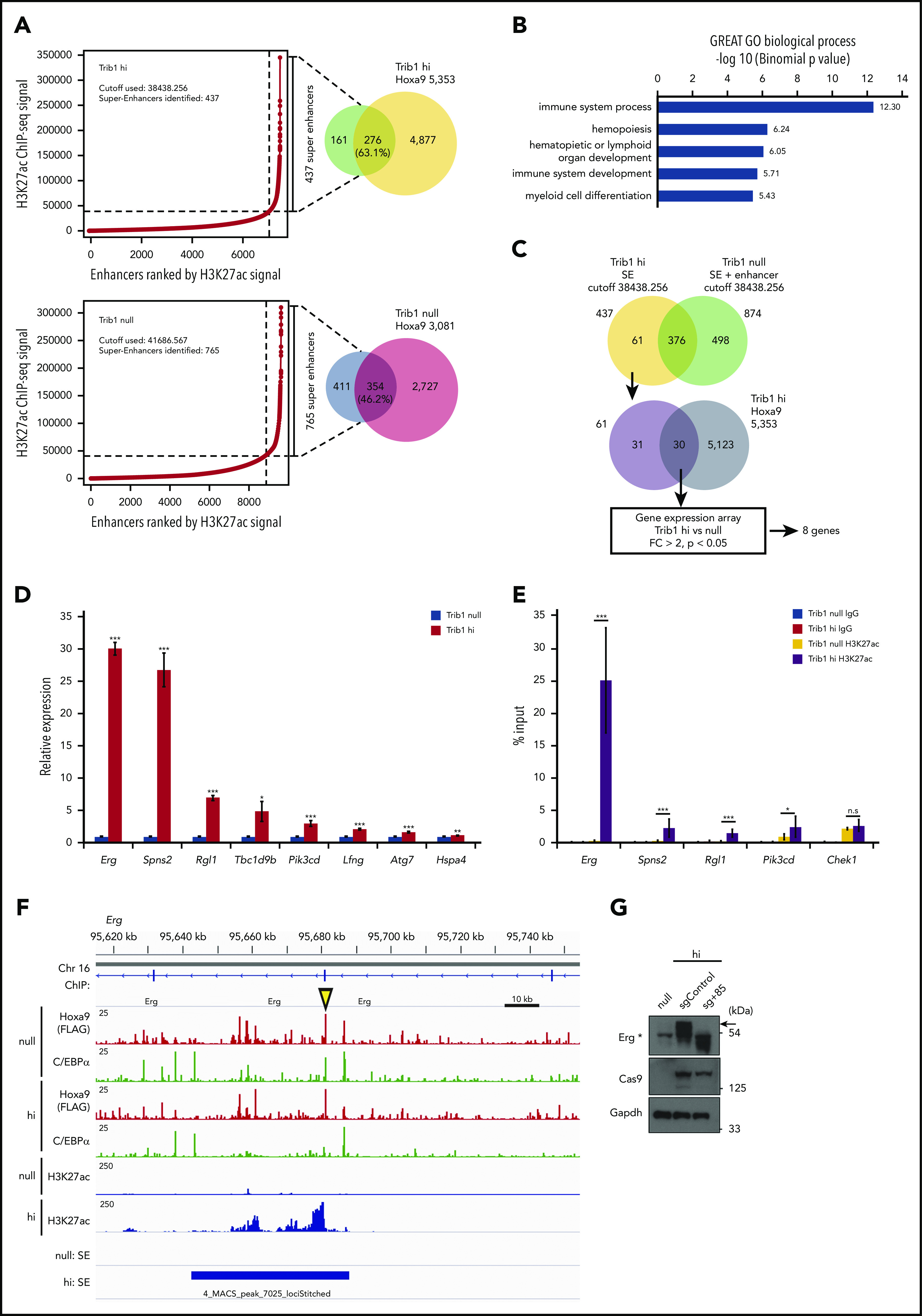
Figure 3.

Different super-enhancer distribution between Trib1 hi and null cells identifies Hoxa9/Trib1 target genes. (A) Enhancers were ranked by increasing H3K27ac ChIP-seq signals in Trib1 hi (left, top) and null (left, bottom) cells. Using the ROSE algorithm, 437 and 765 enhancers were defined as super-enhancers in Trib1 hi and null cells, respectively. Overlap between super-enhancers and Hoxa9 DNA-binding peaks are shown in Venn diagrams (right). (B) Enrichment of Gene Ontology biological process for Trib1 hi super-enhancer loci. (C) Schematic diagram for target gene identification. SE, super-enhancer. (D) Quantitative RT-PCR shows increased expression of Hoxa9/Trib1 target genes in Trib1 hi cells. (E) Quantitative ChIP-PCR of H3K27ac signals for super-enhancers of Erg, Spns2, Rgl1, and Pik3cd. The Chek1 locus is shown as a negative control. Three distinct loci for each super-enhancer were examined, and the average accumulation in these 3 loci is indicated. (F) Density plots for ChIP-seq reads of C/EBPα, Hoxa9, and H3K27ac in Trib1 hi and null cells at the +85 enhancer of Erg. The yellow arrowhead indicates the position of the sgRNA for the +85 enhancer. (G) Immunoblotting shows significant decrease of Erg protein expression (arrow) by knockout of the Erg enhancer using a sgRNA for the +85 enhancer. The asterisk in immunoblotting indicates nonspecific bands. *P < .05, **P < .01, ***P < .001; n.s, not significant.
