Abstract
BACKGROUND AND PURPOSE: Dyslexia is a language disorder in which reading ability is compromised because of poor phonologic skills. The purpose of this study was to measure the effect of a phonologically driven treatment for dyslexia on brain lactate response to language stimulation as measured by proton MR spectroscopic imaging.
METHODS: Brain lactate metabolism was measured at two different time points (1 year apart) during four different cognitive tasks (three language tasks and one nonlanguage task) in dyslexic participants (n = 8) and in control participants (n = 7) by using a fast MR spectroscopic imaging technique called proton echo-planar spectroscopic imaging (1 cm3 voxel resolution). The age range for both dyslexic and control participants was 10 to 13 years. Between the first and second imaging sessions, the dyslexic boys participated in an instructional intervention, which was a reading/science workshop.
RESULTS: Before treatment, the dyslexic boys showed significantly greater lactate elevation compared with a control group in the left anterior quadrant (analysis of variance, P = .05) of the brain during a phonologic task. After treatment, however, brain lactate elevation was not significantly different from that of the control group in the left anterior quadrant during the same phonologic task. Behaviorally, the dyslexic participants improved in the phonologic aspects of reading.
CONCLUSION: Instructional intervention that improved phonologic performance in dyslexic boys was associated with changes in brain lactate levels as measured by proton echo-planar spectroscopic imaging.
We have previously reported that dyslexic boys show a greater area of brain lactate elevation compared with a control group during a phonologic task in the left anterior quadrant of the brain (1). Dyslexia is a language disorder characterized by poor reading due to a phonologic deficit (1, 2). We used a fast spectroscopic imaging technique called proton echo-planar spectroscopic imaging to measure brain lactate changes associated with language activation noninvasively. Functional MR spectroscopy using the proton echo-planar spectroscopic imaging technique is an alternative approach for detecting regional brain activation. It measures tissue-based lactate changes (a direct measure of metabolism) produced by a temporary mismatch of oxygen delivery and consumption in response to neuronal activation (3). After completion of the previous functional MR spectroscopy study, the dyslexic boys entered into a treatment program designed to improve their phonologic abilities. Both the dyslexic and the control participants underwent subsequent imaging a year after the initial imaging session. We hypothesized that a positive response to therapy would be reflected by changes in the distribution of brain lactate activation as measured by functional MR spectroscopy. The purpose of this study was to test the effect of this instructionally based treatment for dyslexia on the brain lactate response during reading-related language tasks.
Methods
Study Design
Eight dyslexic and seven nondyslexic (control) boys underwent proton echo-planar spectroscopic imaging (4) while they performed four different cognitive tasks. Only male subjects were chosen for this study to minimize the variance between participants, especially considering that Shaywitz et al (5) showed large functional MR imaging differences between the sexes during language processing. The dyslexic boys underwent imaging before and after a 3-week phonologically driven treatment (plus follow-up studies) for dyslexia, as described by Berninger (6). The control group also underwent imaging at the same two time points as the dyslexic group but did not receive treatment because the control participants were already reading well above grade level and age expectations. Subsequent imaging of the control participants performed at the same time that the dyslexic participants underwent subsequent imaging permitted evaluation of reasons for changes in the dyslexic participants; ie, maturation versus instruction. The imaging protocol and the language stimuli were identical across repeated images and were equally spaced for the two groups. The boys did not undergo subsequent imaging immediately after treatment because we wanted to evaluate whether the behavioral gain observed immediately after treatment would result in a long-lasting change in lactate brain activity. The dyslexic and control groups were well matched in age, intelligence quotient, and head size (number of total voxels) but not in reading skills, for which they showed marked differences at the first time point, as will be described. The experimental tasks were designed to activate phonologic and lexical access functions of the brain, whereas a tone task was used to activate auditory nonlanguage functions of the brain. Two control tasks (listening to imager noise and passive listening to word lists) were used to subtract out low-level acoustic-stimulus effects. The phonologic and lexical access tasks engage additional linguistic processes beyond that required for passive listening of stimulus words. Task-specific activation was assessed by subtracting the passive listening condition from the phonologic and lexical access tasks. This method provides a means to identify activation related to processing phonologic information or accessing word meanings, independent of brain activation due to the characteristics of word stimuli. The component of brain activation related to auditory processing not specific to language was assessed by subtracting out the imager noise from the tone judgments.
MR Imaging and Spectroscopy
Conventional MR imaging and proton echo-planar spectroscopic imaging were performed on a clinical 1.5-T Signa MR imaging system (General Electric, Waukesha, WI) equipped with version 5.7 software and a custom-built RF head coil developed by Hayes and Mathis (7). MR images were acquired in the sagittal plane (600/20 [TR/TE]) and also in the axial plane (2000/35/80 [TR/TEfirst/TEsecond]). The custom-designed head coil was necessary to acquire MR spectroscopic data with high enough signal-to-noise ratios to detect the small lactate peak and also to maintain a reasonably short acquisition time to avoid motion and habituation artifacts from these young participants. This custom-built RF head coil has been measured to produce an MR signal with 35% better signal-to-noise ratios than the standard General Electric RF head coil (product hardware delivered with the imager) (7). The coordinates of the sylvian fissure and surrounding language-related structures were determined on the sagittal images, which were coregistered with the axial images for both MR and spectroscopic imaging. The areas sampled with proton echo-planar spectroscopic imaging were based on the work presented by Ojemann (8) that invasively showed language activation in the anatomic region encompassing the sylvian fissure and adjacent opercula. The single proton echo-planar spectroscopic imaging section was oriented to encompass the frontal operculum and the posterior portion of the superior temporal gyrus. Deeper subcortical structures were also included that are associated (through neuronal connectivity) with the cortical areas. Proton spectra were acquired using proton echo-planar spectroscopic imaging, a spin-echo pulse sequence developed by Posse et al (4) that allows fast spectroscopic imaging that is 32 times faster than conventional hydrogen spectroscopic imaging for the same spatial resolution. Proton echo-planar spectroscopic imaging is a method that is somewhat demanding on gradient amplifiers and may not function properly on most conventional imagers without echo-planar capability. Proton echo-planar spectroscopic imaging was chosen in our experiment over a single-voxel technique such as point-resolved spectroscopy because we needed to map the lactate distribution over the whole brain section to define the regional metabolic activation. The parameters for data acquisition included 4000/272, two averages, 32 × 16 spatial matrix (zero-filled to 32 × 32 for reconstruction), 512 echoes in the echo-planar acquisition, 32 complex points per echo, full echo acquisition, 24-cm field of view, and 20-mm section thickness. The voxel size was approximately 1 cm3, and the acquisition time for each proton echo-planar spectroscopic image was approximately 4.5 minutes. Five proton echo-planar spectroscopic images were acquired at each session during the following instances: 1) baseline (no extra sounds); 2) phonologic task; 3) lexical access task; 4) tone discrimination task; and 5) passive listening. The data were processed as described previously (9). The metabolites were integrated using the following procedures: 1) magnetic field inhomogeneity shifts (B0 shifts) were corrected by finding the maximum point of the N-acetylaspartate (NAA) peak and resetting the ppm scale to 2.0 ppm for each spectrum; 2) the average baseline was determined from 32 points to the right of 0.0 ppm; 3) the maximum intensity point of the peak was determined within a set spectral range (NAA = 2.0 ± 0.07, lactate = 1.3 ± 0.1 ppm); and 4) integration was performed by summing the spectral intensities for the NAA and lactate for the ppm ranges specified in step 3.
Subject Characterization
The University of Washington Human Subjects Institutional Review Board approval was obtained for this study, and each participant (as well as parent/guardian) provided written, informed consent. All participants were right-handed (90−100% on the Edinburgh Handedness Scale [10]). The control participants had a history of learning to read easily and were reading at a level above normal for their age (average was 1 SD above mean for age using the Woodcock Reading Mastery Test-Revised [11]). The dyslexic participants had a developmental history of extreme difficulty in learning to read despite many forms of extra assistance at school and also had a family history of multi-generational dyslexia, which was confirmed in a concurrent family genetics study (W. Raskind, personal communication) conducted at our center. At the time of the initial imaging, the dyslexic boys were reading an average of 1.66 SD below the mean for their age on the Woodcock test (11). In addition, all the dyslexic boys had deficits in three skills that are predictive of ease of learning to read and response to intervention: phonologic (phoneme segmentation, memory for spoken nonwords), rapid automated naming, and orthographic facility (coding written words in short-term and long-term memory) (12). Based on independent t tests, the control participants (M = 127.3, SD = 10.8) and dyslexic participants (M = 124.3, SD = 11.1) did not differ in age in months at the time of the initial imaging (t(11) = 0.49, P = .637). Likewise, at the time of the initial imaging, the control participants (M = 15.6, SD = 3.2) and dyslexic participants (M = 13.2, SD = 1.6) did not differ in age-corrected Weschsler Intelligence Scale for Children-III vocabulary scores (t(11) = 1.68, P = .12), which provide the best estimate of full-scale intelligence quotient. At the time of the initial imaging, however, the control and dyslexic participants did differ significantly in age-corrected standard scores for reading real words on the word identification subtest of the Woodcock Reading Mastery Test-Revised and for reading pseudowords on the word attack subtest of the Woodcock Reading Mastery Test-Revised: t(11) = 6.81, P < .001 on the word identification subtest and t(10) = 6.02, P < .001 on the word attack subtest. The differences for both real-word reading (word identification, control group: M = 115.1, SD = 9.2; word identification, dyslexic group: M = 75.5, SD = 11.8) and pseudoword reading (word attack, control group: M = 110.2, SD = 6.8; word attack, dyslexic group: M = 79.0, SD = 10.7) were large as well as statistically significant.
Phonologically Driven Instructional Treatment
Treatment consisted of 15 2-hour group sessions in a 3-week reading/science workshop. To motivate the boys who had a long struggle learning to read, they were told that they were Einstein's Ninja turtles. The instructor told them the story of how Albert Einstein had had learning disabilities in reading that he overcame (13). She also explained that gene mutations can confer special advantages, such as the Ninja turtles have, as well as disadvantages, such as trouble learning to read. She used the fable of the tortoise and the hare to convince them that despite a slow start in learning to read, they could finish the race as skilled readers. A systems approach to intervention was used in the treatment in which instruction was aimed at all levels of language (subword, word, and text [6]) during the first hour of each instructional session. Each session began with sound games to remediate their deficits in phonologic processing. High-interest, polysyllabic words taken from science texts were presented orally. The boys counted the number of syllables in the spoken word and used colored counters to represent each phoneme in the syllables. Only after they analyzed the phonologic structure of each word did they see the same words in written form. Next, they were taught to decode the words by using syllabic patterns of written English derived from Anglo-Saxon, Latin/Romance, and Greek origin (14) and correspondences between one- and two-letter spelling units and phonemes (12). They then competed in a “reading bee” to decode transfer words that varied in predictability and complexity but contained the most common syllabic patterns and spelling-phoneme correspondences of English. Finally, they took turns orally reading science texts that contained words practiced at the beginning of the session and that covered subjects about the human brain (week 1), astronomy (week 2), and endangered species (week 3). After this phonologically driven instruction, the last hour of each session was devoted to hands-on science activities. Guest speakers who were scientists provided lectures and information regarding famous scientists as part of this training. Follow-up sessions over several months were held to facilitate and maintain the improvements. Additional procedural details are reported by Berninger (6).
Brain Stimulation Tasks during Proton Echo-Planar Spectroscopic Imaging
During repeated MR imaging, the same brain stimulation tasks were used. The children were asked to listen to aurally presented words, nonwords, and tone pairs at a rate of one stimulus pair every 4 seconds. Language stimuli were composed of four groupings of word pairs, crossed for lexical status (word versus nonword) and sound similarity (rhyming versus nonrhyming), resulting in four sets of stimuli: word/word:nonrhyming (eg, FLY-CHURCH); word/word:rhyming (eg, FLY-EYE); word/nonword:nonrhyming (eg, CROW-TREEL); word/nonword:rhyming (eg, MEAL-TREEL). Nonwords such as TREEL allow assessment of sound processing without any meaning cues. The presenting order of word pair types were counterbalanced, and the ordering effects were thus controlled. During rhyming (phonologic task), participants listened to the word pairs and judged whether they rhymed or did not rhyme; whether words were real was irrelevant. During the lexical access condition, participants listened to the same word pairs and judged whether the word pairs contained two real words or contained a nonword; whether the words rhymed was irrelevant. Thus, the same stimulus lists were used for lexical access and rhyming; only the task instructions were changed. Participants indicated their rhyme and lexical decisions by raising cards held in the right and left hands (the hand used to signal a “yes” response was counterbalanced across participants). During passive listening, participants listened to the same stimuli but were instructed to raise the left and right hand alternately without making any judgments on the stimuli. For the tone judgment task, five pure tones (329.6, 350, 415.3, 440, and 523 Hz) were grouped into pairs of identical tones or different tones. Participants were asked to raise one hand if the tones were identical and to raise the opposite hand if the tones were different. The participants were tested for accuracy of their responses for all tasks during a preimaging training session and during the actual MR imaging. For the tone subtraction, the baseline imager noise was used instead of passive listening. The proton echo-planar spectroscopic imaging acquisition time for each task for approximately 4.5 minutes, and a recovery period of 5 minutes was allowed between tasks based in part on the time for lactate recovery measured by Frahm et al (3).
Data Analysis
To evaluate focal brain activation from proton echo-planar spectroscopic images affirmatively, z-score maps were created from the lactate:NAA ratios based on the following equation: [lactate/NAA (task) − lactate/NAA (passive listening)]/[SD of lactate/NAA (passive listening)], where (task) in the equation refers to the task given during imaging which was either phonologic, lexical access, or tone differentiation. The SD of the lactate/NAA was calculated for each participant by using all valid spectra of the control task (either passive listening or imager noise). This z score was calculated for all voxels that contained valid spectra for each language condition (9). Definition of the threshold for lactate elevation indicating brain activation was based on z scores greater than 2.0 on a voxel-by-voxel basis. The cross-sectional proton echo-planar spectroscopic imaging section of the brain was divided into four quadrants (described below), and the number of voxels with elevated lactate within each quadrant was counted for each participant.
The proton echo-planar spectroscopic imaging data were analyzed to sum the number of activated voxels (with elevated lactate above the threshold) in each of the four regions of the brain. Because regional specificity of lactate response is not well established and also because of the large variability between participants in the spatial location of the lactate response, we divided the spectroscopic imaging section into four quadrants. The brain was divided into four quadrants based on the following: 1) left to right, brain midline defined on the axial MR image; and 2) anterior to posterior, using the midpoint of the thalamus as a landmark. Inferential statistics were used to compare relative activation for each group in each brain quadrant on each task. Analysis of variance was used to test for differences in the number of activated voxels between the control and dyslexic groups. The number of valid voxels for the dyslexic group was not significantly different from that for the control group (control group: M = 160.6, SD = 6.9; dyslexic group: M = 166, SD = 19, t(11) = 0.76, P < .48).
The lactate:NAA ratio was used to normalize for RF inhomogeneity and variable CSF contribution and also to standardize the lactate signal across participants. An automated computer software mask was applied to the spectra to ensure that the MR lactate signal was not contaminated with scalp lipid signal (1). In this software, a true lactate peak was differentiated from lipid contamination based on MR frequency, which was determined from an in vitro lactate measurement (the lipid peak MR frequency was not the same as lactate frequency). Additionally, in previous work, we varied the TE to assess J-coupling properties of the peak at 1.3 ppm, which identified this peak to be lactate (15).
Results
Behavioral Improvement
Immediately after the intervention, the boys improved an average of 8.9 points on age-corrected standard scores (0.6 SD unit) of a measure of phonologic decoding (word attack subtest of the Woodcock Reading Mastery Test-Revised [11]). On a criterion measure of oral reading of text, three were at the grade level just completed, three were above the grade level just completed, and one was still below the grade level for oral reading but was at the grade level just completed for silent reading. Eight months later (approximately 1 year after the initial MR and proton echo-planar spectroscopic images were obtained), all except two had age-appropriate phonologic awareness. The group showed a relative gain of 0.9 standard score units in phonologic memory since the initial assessment that was conducted before the first imaging session, and the group maintained a relative gain of 8.7 points on age-corrected standard scores (0.6 SD unit) on phonologic decoding since first assessed before undergoing neuroimaging (Table 1). Their phonologic decoding (word attack) was now at the border between low average and average (M = 89.7, SD = 8.5 [6]). Additional details are reported by Berninger (6).
TABLE 1:
Phonological skills of the dyslexic boys

MR Spectroscopic Imaging Changes
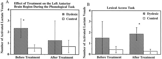
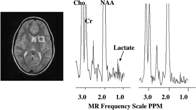
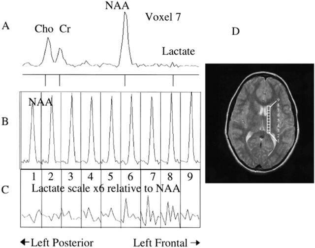
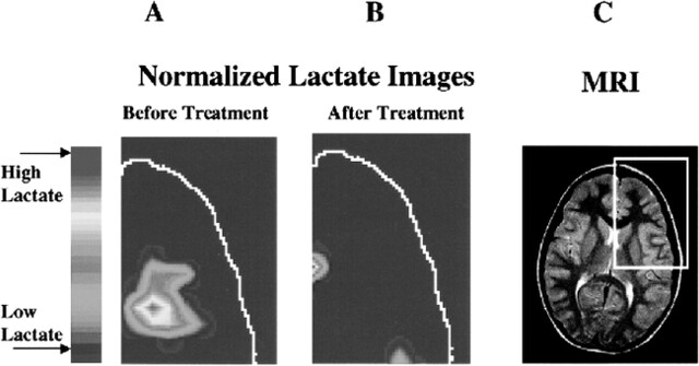
Before treatment, dyslexic boys had significantly more brain voxels with elevated MR lactate levels (2.33 ± SE 0.843) compared with the control group (0.57 ± SE 0.30) during a phonologic task in the left anterior quadrant (Table 2, analysis of variance, F(1,11) = 4.41, P = .05). After treatment for the same phonologic task and in the same region of the brain, however, the dyslexic participants were not significantly different from the control participants, who were stable across repeated imaging (Table 2, Fig 1A). Figure 2 shows an example of spectra and the reduction in lactate from a dyslexic participant before and after treatment in the left anterior brain region during the phonologic task. Figure 3 shows an example of spectra from a dyslexic boy during the phonologic task and increased lactate in the midfrontal region of the brain compared with the posterior portion. Figure 4 shows an example of normalized lactate metabolite images of the left frontal region of the brain from a dyslexic boy before and after treatment. For the lexical access task, although there was no significant difference between dyslexic and control participants before treatment, there was a significant difference after treatment in the left anterior brain region (Table 2, Fig 1B). Again, the control participants were stable across repeated imaging during the lexical access task (Table 2, Fig 1B). Examining the pre- and posttreatment data indicates that at each time point, dyslexic participants had overall elevated rates for the lexical access task. The variability observed for this task during the first imaging session, however, likely obscured the significance of this effect. For the lexical access task, the intervention had reduced the variance in this dyslexic group, as can be seen in Figure 1B.
TABLE 2:
Activated lactate voxel counts for the left anterior brain region
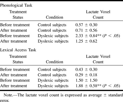
fig 1.
Bar graph of the number of average activated voxels (as defined by MR spectroscopy lactate increases) in the left anterior brain quadrant for both dyslexic and control participants. Within each graph, the data on the left were obtained before treatment, and the data on the right were obtained after treatment. Error bars indicate the standard error of the mean. The asterisk indicates dyslexic versus control comparisons that were significantly different. The data were collected using 4000/272 proton echo-planar spectroscopic imaging. A, Data obtained during the phonologic task from the left anterior brain. B, Data obtained during the lexical access task also from the left anterior brain region
fig 2.
MR image and proton spectra from an activated brain region of one dyslexic participant. The spectroscopic data were collected using 4000/272 proton echo-planar spectroscopic imaging. The MR image was collected using a 2000/80 fast spin-echo pulse sequence. The intensity axis of the spectra is scaled so that the lactate can be visualized more easily; however, choline and NAA are scaled off the figure. Note the decrease in the lactate peaks for the posttreatment spectrum. Cho, choline; CR, creatine. A, MR image with white box indicating the brain region measured with MR spectroscopy. B, Proton MR spectrum from the white box brain region before treatment (the lactate resonance has a signal-to-noise ratio of 2.2). C, Proton MR spectrum from the white box brain region after treatment (lactate has a signal-to-noise ratio of 1.6).
fig 3.
Proton MR spectra and image from a dyslexic boy before treatment during the phonologic task. A, Spectrum from voxel 7 shows choline (CHO), creatine (Cr), NAA, and lactate. B, Nine spectra from nine adjacent voxels (position is shown in D), zoomed into the NAA region (1.7−2.3 ppm). C, Nine spectra from nine adjacent voxels, zoomed into the lactate region (1.10−1.33 ppm). The vertical scale of this set was increased six times higher than the NAA set so that lactate could be easily visualized. An increase in lactate can be clearly seen in voxels 6 through 8 compared with voxels 1 through 4. D, MR image shows the location of the nine voxels shown in A through C.
fig 4.
Example of normalized lactate metabolite images of the left frontal region of the brain from a dyslexic boy before and after treatment. The data were collected using 4000/272 proton echo-planar spectroscopic imaging. A, Normalized lactate image from the left anterior quadrant of the section shown in the MR image of a dyslexic boy created from the proton echo-planar spectroscopic imaging spectra before the phonologically driven instructional treatment. B, Image obtained after treatment. C, MR image shows the middle slice of the proton echo-planar spectroscopic imaging anatomic section location. The white box on the MR image shows the area that is displayed in the lactate images. This is the brain region that had a significant difference between dyslexic and control groups. The lactate was normalized according to this equation: (lactate/NAA)phonologic − (lactate/NAA)passive
As can be seen in Figure 1 and Table 2, the control participants showed very consistent activated voxel counts at the two time points for both the phonologic and lexical access tasks. For the tone task, there was no significant difference between dyslexic and control participants at any time point (either before or after treatment).
Discussion
Our main finding was that, after treatment, the dyslexic children had a reduction in the regional distribution of metabolic activation, characterized by the number of voxels with elevated lactate in the left anterior quadrant during the phonologic task. This reduction may be an indication that the treatment was effective in reducing the amount of lactate produced during metabolic activation required to perform phonologic judgments. Concurrently, collected behavioral data verified that the participants were attentively listening to the auditory stimuli. The behavioral data also showed that the dyslexic children were less accurate than the control children for the phonologic task before treatment. The left anterior quadrant, where we observed this change after treatment, is an area that encompasses portions of the frontal operculum, inferior frontal gyrus, and anterior temporal lobe, which are areas associated with motor speech. It also encompasses portions of the frontal lobe that are known to be associated with executive functioning of the brain (16). Our findings suggest that these areas are involved with dyslexia and also that treatment is affecting lactate metabolism in these areas. That control averages for the number of activated voxels remained constant across the treatment period supports the validity of the observed change in the dyslexic children. At the same time, lactate metabolic activation increased during the lexical access task for the dyslexic participants compared with the control participants. Apparently, once these boys were phonologically aware, it was more difficult for them to attend to meaning but ignore phonology when they were asked to judge between words and nonwords. Despite the reduction in lactate activation during the phonologic task after phonologic training, a brain signature may remain in which some linguistic processes are still difficult for people with dyslexia.
Although the present measure is not well suited for highly resolved spatial localization, it does provide new evidence for lactate metabolic changes after behavioral intervention. Importantly, this technique permits assessment of neuronal activation changes that are physiologically different from what is currently obtained from imaging studies that are reliant on hemodynamic properties (as measured by functional MR imaging). Thus, the present study adds to a growing number of studies that have shown functional imaging changes associated with behavioral changes or reorganization of the brain after recovery from language deficits (17, 18).
Conclusion
Our findings are important because they show that treatment is accompanied by lactate changes in the brain as well as changes in performance on behavioral tasks in young children with a developmental rather than an acquired disorder. This change was in the direction of a decreased area of lactate activation. We, however, recognize that lactate changes can be interpreted by a number of different mechanisms, including alterations in blood flow or mitochondrial dysfunction. The change in lactate metabolism may be a brain substrate for functional verbal efficiency (19), which is decreased in poor readers to some degree but may increase after partial remediation. Our results are also important because they suggest that the brain is not only an independent variable that can cause a language disorder, such as dyslexia, but is also a dependent variable that can be modified by instructional intervention from the environment (20).
Acknowledgments
The authors thank Drs. Martin Kushmerick and James Nelson for encouragement and support of this study and Bryn Floyd and K. C. Stegbauer for help with data analysis.
Footnotes
This work was funded by a special multidisciplinary learning disabilities center grant from the National Institutes of Health (National Institute of Child Health and Human Development), P50 HD33812.
Address reprint requests to Todd L. Richards, Radiology Department, Box 357115, University of Washington, Seattle, WA 98195.
References
- 1.Richards TL, Dager SR, Corina D, et al. Dyslexic children have abnormal brain lactate response to reading-related language tasks. AJNR Am J Neuroradiol 1999;20:1393-1398 [PMC free article] [PubMed] [Google Scholar]
- 2.Wagner R, Torgesen J. The nature of phonological processing and its role in the acquisition of reading skills. Psychol Bull 1987;101:192-212 [Google Scholar]
- 3.Frahm J, Krueger G, Merboldt KD, Kleinschmidt A. Dynamic NMR studies of perfusion and oxidative metabolism during focal brain activation. Adv Exp Med Biol 1997;413:195-203 [DOI] [PubMed] [Google Scholar]
- 4.Posse S, Dager SR, Richards TL, et al. In vivo measurement of regional brain metabolic response to hyperventilation using magnetic resonance proton echo planar spectroscopic imaging (PEPSI). Magn Reson Med 1997;37:858-865 [DOI] [PubMed] [Google Scholar]
- 5.Shaywitz BA, Shaywitz SE, Pugh KR, et al. Sex differences in the functional organization of the brain for language [comments]. Nature 1995;373:607-609 [DOI] [PubMed] [Google Scholar]
- 6.Berninger VW. Dyslexia: the invisible, treatable disorder: the story of Einstein's ninja turtles. Learn Disabil Q (in press)
- 7.Hayes CE, Mathis CM. Improved brain coil for fMRI and high resolution imaging. Paper resented at: Fourth Annual Meeting of the International Society for Magnetic Resonance in Medicine, 1996; Berkeley, CA
- 8.Ojemann G. Localization of language in frontal cortex. Adv Neurol 1992;57:361-368 [PubMed] [Google Scholar]
- 9.Richards TL, Dager SR, Panagiotides HS, et al. Functional MR spectroscopy during language activation: a preliminary study using proton echo-planar spectroscopic imaging (PEPSI). Int J Neuroradiol 1997;3:490-495 [Google Scholar]
- 10.Oldfield RC. The assessment and analysis of handedness: The Edinburgh Inventory. Neuropsychologia 1971;9:97-113 [DOI] [PubMed] [Google Scholar]
- 11.Woodcock R. Woodcock Reading Mastery Test-Revised. Circle Pines: American Guidance Service; 1987
- 12.Berninger V. Process Assessment of the Learner: Guides for Intervention. San Antonio: The Psychological Corporation; 1998
- 13.Patten B. Visual mediated thinking: a report of the case of Albert Einstein. J Learn Disabil 1973;6:15-20 [Google Scholar]
- 14.Henry M. Words: Integrated Decoding and Spelling Instruction Based on Word Origin and Word Structure.. Austin: Pro-Ed; 1990 [DOI] [PubMed]
- 15.Dager SR, Marro KI, Peterson J, Richards TL. Applications of magnetic resonance spectroscopy to investigate panic disorder. In: Nasrallah H, Pettegrew J, eds. NMR Spectroscopy in Psychiatric Brain Disorders. Washington: American Psychiatric Press, Inc. 1995 147-177
- 16.Leskela M, Hietanen M, Kalska H, et al. Executive functions and speed of mental processing in elderly patients with frontal or nonfrontal ischemic stroke. Eur J Neurol 1999;6:653-661 [DOI] [PubMed] [Google Scholar]
- 17.Small SL, Flores DK, Noll DC. Different neural circuits subserve reading before and after therapy for acquired dyslexia. Brain Lang 1998;62:298-308 [DOI] [PubMed] [Google Scholar]
- 18.Thulborn KR, Carpenter PA, Just MA. Plasticity of language-related brain function during recovery from stroke. Stroke 1999;30:749-754 [DOI] [PubMed] [Google Scholar]
- 19.Perfetti C. Reading Ability. New York: Oxford University Press; 1985
- 20.Berninger VW, Corina D. Making cognitive neuroscience educationally relevant: creating bi-directional collaborations between educational psychology and cognitive neuroscience. Educ Psychol Rev 1998;10:343-354 [Google Scholar]


