Abstract
Summary: A patient with a rare variation of the fronto-orbital artery is presented. In this case, the fronto-orbital artery arises from the A1 segment of the contralateral anterior cerebral artery. The angiographic findings in this case are illustrated and discussed. The importance of recognizing this variant is related to the planning of surgery or endovascular therapy in the anterior cerebral artery region.
The anatomy of the anterior cerebral artery (ACA) is known to be highly variable. Variations in the origin of Heubner's artery and in the anterior communicating artery (ACoA) complex are quite common and are well described (1). Variations in the fronto-orbital artery (FOA), an artery arising in the same region, are not as well described in the literature.
Case Report
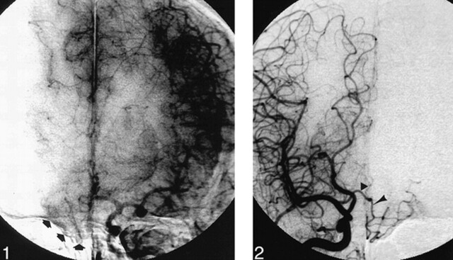
A 40-year-old woman presented with a long-standing history of intermittent headaches and recent onset of burning pain in her left occiput and left arm. CT, MR imaging, and MR angiography showed a left-sided sylvian fissure mass consistent with a middle cerebral artery bifurcation aneurysm. There was no evidence of subarachnoid hemorrhage. The physical examination, past medical history, and family history were unremarkable. The patient underwent preoperative cerebral angiography, which confirmed a 7-mm left middle cerebral artery bifurcation aneurysm as well as an unsuspected 2-mm right ophthalmic, internal carotid artery–junction aneurysm. On the left carotid injection, incidental note was made of the lack of filling in the left fronto-orbital artery territory (Fig 1). This occurred despite dense opacification of both A2 segments of the ACAs and the ACoA. Filling of the right FOA occurred normally from the right pericallosal artery. Injection of the right carotid artery showed right A1 segment hypoplasia, flash filling of the A2 segments, and dense opacification of the left FOA from a right A1 segment origin (Fig 2). Surgical clipping of the left middle cerebral artery aneurysm was performed without complication. There was no exploration of the region of the anomalous vessel.
fig 1.
Late arterial phase of left carotid arteriogram shows filling of both A2 territories, including right fronto-orbital artery (small arrows). The left fronto-orbital artery does not opacify with this injection.
fig 2. Frontal view of right common carotid injection shows hypoplastic right A1 segment (triangle) and opacification of the left fronto-orbital artery (arrowhead)
Discussion
The fronto-orbital artery is the first cortical branch of the ACA and normally arises from the ipsilateral pericallosal artery but can share a common trunk with the frontopolar artery or callosal marginal artery (2). The course of the fronto-orbital artery is typically along the inferior or medial surface of the frontal cerebral pole (3). The typical supply of the FOA is to the gyrus rectus, medial gyrus olfactorus, olfactory bulb and tract, and anterior part of the superior frontal lobe gyrus (2–3).
Frequent variation and abnormalities of the A1 segment of the ACA have been described on the basis of angiographic, operative, and autopsy findings (4). Variant anatomies of the ACA consist of hypoplasia, fenestration, infraoptic course of the A1, variant A1 perforators including Heubner's artery, arterial duplication and myriad variations of the anterior communicating artery. Nevertheless, there have been few reports of anomalies of the fronto-orbital artery. According to Markinkovic's results, the incidence of FOA origin from the ipsilateral A1 is 4% (5). Tulleken noted that Heubner's artery arises symmetrically from FOAs originating from the A1s when azygous pericallosal anatomy is present (6). A ruptured aneurysm arising from the junction of A1 and anomalous FOA was reported by Hong (7).
Conclusion
We have reported an unusual variant of the fronto-orbital artery arising from a hypoplastic contralateral ACA A1 segment. Recognizing and reporting this variant could be helpful in preventing complications of surgery or intervention in the ACA-ACoA region. Although injury to the fronto-orbital artery itself is unlikely to result in clinical deficit, precise knowledge of the vascular anatomy, including such variants, is essential to minimizing complications. This is especially true of the open operative procedure with limited field of view and landmarks.
Footnotes
Address reprint requests to James D. Eastwood, MD, Department of Radiology, Duke University Medical Center, Box 3808, Durham, 27710-3808.
References
- 1.Perlmutter D, Rhoton AL. Microsurgical anatomy of anterior cerebral-anterior communicating-recurrent artery complex. J Neurosurg 1976;45:259-272 [DOI] [PubMed] [Google Scholar]
- 2.Newton TH, Potts DG. Radiology of the Skull and Brain Angiography.. St. Louis, Mo: Mosby; 1974:1414
- 3.Huber P, Bosse G. Cerebral Angiography.. New York: Thieme-Stratton 1982:85-88 [Google Scholar]
- 4.Mäuer J, Mäuer E, Perneczky A. Surgically verified variations in the A1 segment of the anterior cerebral artery. J Neurosurg 1991;75:950-953 [DOI] [PubMed] [Google Scholar]
- 5.Marinkovic S, Milisavljevic M, Kovacevic M. Anatomical bases for surgical approach to the initial segment of the anterior cerebral artery. Microanatomy of the Heubner's artery and perforating branches of the anterior cerebral artery. Surg Radiol Anat 1986;8:7-18 [DOI] [PubMed] [Google Scholar]
- 6.Tulleken CA. A study of the anatomy of the anterior communicating artery with the aid of the operating microscope. Clin Neurol Neurosurg 1978;80:169-173 [DOI] [PubMed] [Google Scholar]
- 7.Hong SK. Ruptured proximal anterior cerebral artery (A1) aneurysm located at an anomalous branching of the fronto-orbital artery—a case report. J Korean Med Sci 1997;12:576-580 [DOI] [PMC free article] [PubMed] [Google Scholar]


