Abstract
BACKGROUND AND PURPOSE: Acute thromboembolic stroke complicated by ipsilateral carotid occlusion may present both mechanical and inflow-related barriers to effective intracranial thrombolysis. We sought to review our experience with a novel method of mechanical thrombectomy, in such cases, using the Possis AngioJet system, a rheolytic thrombectomy device.
METHODS: A review of our interventional neuroradiology database revealed three patients in whom an occluded cervical internal carotid artery was encountered during endovascular treatment for acute stroke and in whom thrombectomy was attempted, using the 5F Possis AngioJet thrombectomy catheter. The medical records and radiographic studies of these patients were reviewed.
RESULTS: Three patients were identified (ages, 52–84 years). Two patients had isolated occlusion of the internal carotid artery; in one patient, thrombus extended down into the common carotid artery. Treatment was initiated within 190 to 360 minutes of stroke onset. Thrombectomy of the carotid artery was deemed necessary because of poor collateral flow to the affected hemisphere (chronic contralateral internal carotid artery occlusion [one patient] and thrombus extending to the carotid “T” [one patient]) or inability to pass a microcatheter through the occluded vessel (one patient). Adjunctive therapy included pharmacologic thrombolysis with tissue plasminogen activator (all patients), carotid angioplasty and stenting (two patients), and middle cerebral artery angioplasty (one patient). Patency of the carotid artery was reestablished in two patients, with some residual thrombus burden. In the third patient, the device was able to create a channel through the column of thrombus, allowing intracranial access.
CONCLUSION: Rheolytic thrombectomy shows potential for rapid, large-burden thrombus removal in cases of internal carotid artery thrombosis, allowing expedient access to the intracranial circulation for additional thrombolytic therapy.
Reestablishing cerebral blood flow in a timely fashion during an acute stroke has shown efficacy in improving outcomes (1–3). In cases of stroke complicated by acute carotid occlusion, access to the intracranial circulation is more difficult and intracranial thrombolysis can be delayed or prevented. Among patients with poor collateral flow across the circle of Willis, inflow from the carotid artery can be critical to sustain the patency of a thrombolysed intracranial territory. Therefore, it is sometimes necessary to open an occluded carotid artery before an intracranial thrombus can be treated. Reestablishing patency of an occluded carotid artery can be a technical challenge because, in addition to dealing with any underlying stenosis, control of a large amount of clot is needed. Currently, there are no widely accepted techniques for large-burden clot removal in the internal carotid artery (ICA). Rheolytic thrombectomy with a commercially available device, the Possis AngioJet, has shown efficacy for thrombus removal in clotted dialysis fistula grafts (4), the peripheral circulation (arterial and venous) (5–7), the coronary system (8, 9), and cases of dural sinus thrombosis (10, 11). We report a small series of patients in whom this device was used in an attempt to reestablish patency of an occluded ICA in the setting of an acute stroke.
Methods
We retrospectively reviewed cases of intraarterial thrombolysis performed for acute stroke at two institutions between January 1997 and December 1999. A total of 78 patients were treated. We identified three patients in whom the Possis AngioJet catheter was used in an attempt to recanalize an ipsilaterally occluded ICA. The radiographic and medical records of these patients were analyzed.
Results
Patient 1
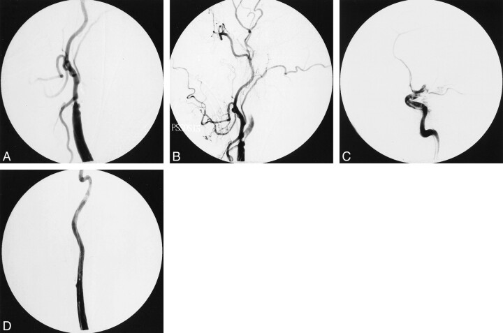
A 69-year-old right-handed woman presented to the emergency department 2 hours 30 minutes after the acute onset of aphasia and right hemiplegia. Her National Institutes of Health Stroke Scale score was 23. Emergent CT and CT angiography showed no evidence of hemorrhage or clearly defined infarct but did show bilateral carotid occlusion with thrombus extending into the left middle cerebral artery (MCA). Emergent angiography, performed with the patient under general anesthesia, verified bilateral ICA occlusion. The right ICA occlusion was presumed chronic because of well-developed external-to-internal collaterals and posterior cerebral leptomeningeal collaterals, which reconstituted the right MCA territory. However, there was little filling of the left anterior cerebral artery territory and essentially no filling of the left MCA territories. The neurologic deficit indicated a large left MCA territory at risk. Therefore, the left ICA occlusion needed to be opened to obtain access and inflow to the left MCA territory. At 3 hours 10 minutes after symptom onset, a 5F Possis AngioJet catheter was passed through a 6F Envoy MPC guide catheter (Cordis) into the ICA. The device was advanced over a Transcend EX .014-in microguidewire (Boston Scientific) up to the level of the skull base, while thrombectomy was performed during a period of a few minutes. Slow to-and-fro movements in the cervical carotid artery were performed with the device engaged to promote thrombolysis. These maneuvers removed most of the clot within the cervical left ICA and revealed an underlying high-grade stenosis of the proximal ICA. Because of the inherent stiffness of this 5F device, we were unable to pass it into the petrous or cavernous carotid artery, and chemical thrombolysis with tissue plasminogen activator was performed in the distal ICA as well as in the left MCA. A RapidTransit microcatheter (Cordis) was placed over a Transcend EX .014-in microwire through the carotid siphon and into the distal M1 segment of the MCA. During slow withdrawal of the microcatheter, 1 mg of tissue plasminogen activator was instilled into the clotted segment. This maneuver was repeated multiple times during a 20-minute period for total instillation of 12 mg of tissue plasminogen activator, resulting in some success (Fig 1). Because of residual M1 and supraclinoid carotid thrombus, however, we elected to perform balloon angioplasty of these vessels. An Endeavor 4 × 10-mm balloon (Boston Scientific) was navigated into the M1 segment of the MCA and the cavernous and supraclinoid ICA, and gentle balloon angioplasty was performed. After establishing some intracranial runoff, we addressed the high-grade (probable) culprit ICA stenosis with angioplasty and stenting. An 8F Shuttle sheath (Cook) was placed into the common carotid artery, and the stenosis was crossed with an exchange length .014-in microwire. Predilation was performed with a Symmetry 5-mm balloon (Boston Scientific), after which a 10 × 42-mm Wallstent (Boston Scientific) was deployed. Final control angiography revealed a widely patent left and anterograde filling of left anterior cerebral artery and M1 branches, with remaining occlusion of the superior M1 division and multiple distal anterior cerebral artery branch occlusions (Fig 1). At the time of discharge, the patient remained mute and hemiplegic but was able to follow simple commands.
fig 1.
Patient 1.
A, Lateral-view angiogram of the left common carotid artery shows occlusion of the ICA at its origin.
B, After one pass with the Possis AngioJet, there is partial reconstitution.
C, After repeated passes with the Possis AngioJet, the cervical ICA has reconstituted, but there is residual thrombus in the cavernous and supraclinoid ICA.
D, After angioplasty and stenting, wide patency of the ICA is reestablished.
Patient 2
An 84-year-old right-handed woman presented to the emergency department 50 minutes after the acute onset of aphasia and right hemiplegia. Her National Institutes of Health Stroke Scale score was 24. Emergent CT showed no evidence of hemorrhage or completed infarction. CT angiography revealed an occlusion of the left ICA, with thrombus extending into the left M1 segment. Emergent angiography confirmed the left ICA occlusion, extending from just above the level of the carotid bifurcation. Initial attempts to navigate the occlusion with a microcatheter failed; we were unable to maneuver the microcatheter/microwire combination (a RapidTransit microcatheter and a Boston Scientific Fast Dasher 014-in wire) into the intracranial circulation. As such, we decided to decrease clot burden in the ICA in an attempt to help achieve intracranial access. This was initiated 4 hours 30 minutes after symptom onset. The 5F Possis AngioJet was used in the cervical ICA in a coaxial fashion through a 6F guide catheter and over a Transcend .014-in wire and was advanced into the high cervical carotid. Slow to-and-fro movements in the artery were performed with the device engaged to promote thrombolysis. This achieved partial thrombectomy and allowed us to place the guide catheter (an Envoy 6F MPC guide) higher in the cervical ICA, allowing microcatheter access to the MCA. Intracranially, extensive parenchymal staining and luxury perfusion were observed at the time of microcatheter MCA injection, suggesting completion of infarction of a large portion of the MCA territory. Because of concerns regarding increased risk of intracranial hemorrhage, the procedure was aborted and no thrombolysis occurred. At the long-term follow-up examination, the patient was hemiplegic, mute, and did not follow commands.
Patient 3
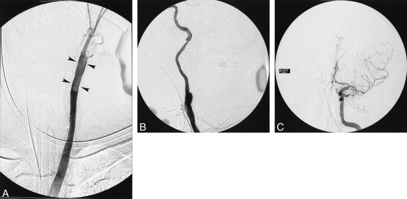
A 52-year-old man presented mute, confused, and with right hemiplegia 3 hours 20 minutes after ictus. His National Institutes of Health Stroke Scale score was 25. Emergent CT showed no evidence of hemorrhage. CT angiography revealed a left carotid T-type lesion extending from the cervical carotid bifurcation into both the left A1 and M1 arteries. At angiography, thrombus was seen extending from the common carotid artery, 4 to 5 cm below the carotid bifurcation, through the entirety of the ICA to the ICA bifurcation. Considering the magnitude of the clot, the Possis AngioJet was used to primarily debulk the clot in two passes. A 6F Envoy MPC guide catheter was placed into the mid-common carotid artery, through which the AngioJet was placed over a Transcend .014-in wire. Two slow passes through the region of thrombus were made. Next, the intracranial clot was lysed using a microcatheter infusion of 10 mg of tissue plasminogen activator for 20 minutes, with repeated passes made through the thrombus (in a fashion similar to that used for patient 1). At the time of completion of thrombolysis, 2 hours after initiation of the procedure, the ICA was patent, as was the bulk of the MCA territory (Fig 2). However, infarction of the majority of the left MCA territory occurred and the patient died as a result of cerebral edema and herniation 2 days later.
fig 2.
Patient 3.
A, Initial lateral-view angiogram of the left common carotid artery shows ICA occlusion and extensive thrombus (arrows) extending into the distal common carotid artery.
B, After using the Possis AngioJet in the distal common carotid artery and ICA, wide ICA patency is reestablished.
C, This allowed access to the intracranial circulation, which was then treated with intraarterial thrombolysis, leading to a good angiographic result.
Discussion
In many cases of acute stroke with concurrent carotid occlusion, conventional management has included deferred treatment of the carotid lesion to allow more timely thrombolysis of intracranial lesions (12). This is often the case when there is good collateralization through the circle of Willis, and inflow from the ipsilateral carotid artery is not necessary to maintain the patency of a reopened MCA (13). In this situation, the therapeutic options available for access to intracranial thrombus include navigation across the anterior communicating artery from the contralateral side and navigation through the occluded segment of the ipsilateral ICA (14). The first may be technically difficult because of anatomic considerations, and thus may be time consuming, and the microcatheter (in the anterior communicator) may be partially occlusive in an important collateral pathway. In addition, this route of catheterization places the normal hemisphere at risk for complications. The second option, although direct, carries the theoretical risk of artery-to-artery embolus because of the necessary navigation through a column of thrombus, which is not always possible, as was the case with patient 2. Rethrombosis of a lysed M1 segment can also occur if inadequate inflow via collaterals exists.
In certain instances, however, when the circle of Willis is inadequate, it is vital to reestablish ICA patency before intracranial thrombolysis can be attempted. In contrast to intracranial thrombolysis, this usually entails large clot volume removal, the bulk of which is usually fresh clot secondary to in situ thrombosis with a culprit high-grade carotid stenosis (15). Tissue plasminogen activator is less than ideally suited for this purpose because the prolonged infusion times necessary for lysis of a large clot burden do not leave time for treatment of the intracranial occlusion.
The Possis AngioJet is a mechanical thrombectomy device that uses high-pressure, pulsed saline to create a low-pressure Venturi effect that fragments and “sucks” thrombus into the catheter lumen. Its mechanism of action has been more fully described elsewhere (3–10). The use of this device has been well documented in the peripheral interventional radiology literature in association with peripheral arterioocclusive disease and dialysis fistula graft maintenance. There are also reports of the device being used to declot a coronary vein graft, in an occluded superior vena cava, and in the superior sagittal sinus in cases of dural sinus thrombosis.
Mechanical thrombectomy holds several theoretical advantages over chemical thrombolysis in the treatment of acute stroke. The ability to reduce the amount of lytic agent administered, thereby reducing the risk of hemorrhagic conversion, is one hypothetical consideration. In addition, the procedure can be performed in a relatively expeditious fashion, which is extremely important, considering the inherent time constraints when dealing with acute ischemic stroke. In our cohort of patients, when the preoperative CT angiographic studies suggested carotid occlusion, the Possis machine was made readily available while the patient was prepared for angiography. This minimized additional delay. The rheolytic thrombectomies were rapid (typically requiring only a few minutes) and effectively provided a pathway for intracranial access.
The natural history of MCA occlusion coupled with acute ICA occlusion is poor, with estimates of 16% to 55% mortality and 40% to 69% significant morbidity (16). This is reflected in our small series, with one death and two significantly morbid outcomes. Unfortunately, the patients who are dependent on reestablished inflow from an occluded carotid artery, who become AngioJet candidates, constitute a group with poor collateral flow who would be expected to have larger amounts of brain tissue at risk, with a shorter window of opportunity for rescue. Our results reflect this. Although AngioJet thrombectomy was technically feasible in our patient group, it may well be that the poor natural history of this cohort precludes effective intervention, at least considering the current status of neurologic protection in cases of acute stroke.
Conclusion
In summary, rheolytic thrombectomy of the occluded ICA is technically feasible and can be performed in selected patients who have suffered acute stroke, with only small additional time expenditures. Although this small cohort of patients is insufficient to determine whether this maneuver will improve patient outcomes, its use should be considered in patients in whom rapid reestablished patency of the ICA is deemed integral to success of intracranial thrombolysis.
Footnotes
Presented at the 38th Annual Meeting of the American Society of Neuroradiology, Atlanta, GA, April 2000.
Address reprint requests to Richard J. Bellon, MD, Department of Interventional Neuroradiology, GRB 289, Massachusetts General Hospital, 55 Fruit Street, Boston, MA 02114.
References
- 1. The National Institute of Neurological Disorders and Stroke rt-PA Stroke Study Group. Tissue plasminogen activator for acute ischemic stroke. N Engl J Med 1995;333:1581-1587 [DOI] [PubMed] [Google Scholar]
- 2.del Zoppo GJ, Higashida RT, Furlan AJ, Pessin MS, Rowley HA, Gent M. PROACT: a phase II randomized trial of recombinant pro-urokinase by direct arterial delivery in acute middle cerebral artery stroke: PROACT investigators: prolyse in acute cerebral thromboembolism. Stroke 1998;29:4-11 [DOI] [PubMed] [Google Scholar]
- 3.Furlan A, Higashida R, Wechsler L, et al. Intra-arterial prourokinase for acute ischemic stroke: The PROACT II Study: a randomized controlled trial: prolyse in acute cerebral thromboembolism. JAMA 1999;282:2003-2011 [DOI] [PubMed] [Google Scholar]
- 4.Vesely TM, William D, Weiss M, et al. Comparison of the angiojet rheolytic catheter to surgical thrombectomy for the treatment of thrombosed hemodialysis grafts: Peripheral AngioJet Clinical Trial. J Vasc Interv Radiol 1999;10:1195-1205 [DOI] [PubMed] [Google Scholar]
- 5.Silva JA, Ramee SR, Collins TJ, et al. Rheolytic thrombectomy in the treatment of acute limb-threatening ischemia: immediate results and six-month follow-up of the multicenter AngioJet registry: Possis Peripheral AngioJet Study AnjioJet Investigators. Cathet Cariovasc Diagn 1998;45:386-393 [DOI] [PubMed] [Google Scholar]
- 6.Mathie AG, Bell SD, Saibil EA. Mechanical thromboembolectomy in acute embolic peripheral arterial occlusions with use of AngioJet Rapid Thrombectomy System. J Vasc Interv Radiol 1999;10:583-590 [DOI] [PubMed] [Google Scholar]
- 7.Dick A, Neuerburg J, Schmidt-Rode T, et al. Stenting after thrombectomy with the AngioJet catheter for acute myocardial infarction. Cathet Cardiovasc Diagn 1998;33:91-97 [DOI] [PubMed] [Google Scholar]
- 8.Nakagawa Y, Matsuo S, Yokoi H, et al. Stenting after thrombectomy with AngioJet catheter for acute myocardial infarction. Cathet Cardiovasc Diagn 1998;43:327-330 [DOI] [PubMed] [Google Scholar]
- 9.Nakagawa Y, Matsuo S, Kimura T, et al. Thrombectomy with AngioJet catheter in native coronary arteries for patients with acute or recent myocardial infarction. Am J Cardiol 1999;83:994-999 [DOI] [PubMed] [Google Scholar]
- 10.Opatowsky MJ, Morris PP, Regan JD, Mewborne JD, Wilson JA. Rapid thrombectomy of superior sagittal sinus and transverse sinus thrombosis with a rheolytic catheter device. AJNR Am J Neuroradiol 1999;20:414-417 [PMC free article] [PubMed] [Google Scholar]
- 11.Scarrow AM, Williams RL, Jungreis CA, Yonas H, Scarrow MR. Removal of a thrombus from sigmoid and transverse sinuses with a rheolytic thrombectomy catheter. AJNR Am J Neuroradiol 1999;20:1467-1469 [PMC free article] [PubMed] [Google Scholar]
- 12.Spearman MP, Jungreis CA, Wechsler LR. Angioplasty of occluded internal carotid artery. AJNR Am J Neuroradiol 1995;16:1791-1796 [PMC free article] [PubMed] [Google Scholar]
- 13.Connors JJ, Wojak JC. Interventional Neuroradiology: Strategies and Practical Techniques.. Philadelphia: W.B. Saunders Co. 1999:725-728 [Google Scholar]
- 14.Nesbit GM, Clark WM, Oisin OR, Barnwell SL. Intracranial intraarterial thrombolysis facilitated by microcatheter navigation through an occluded cervical internal carotid artery. J Neurosurg 1996;84:387-392 [DOI] [PubMed] [Google Scholar]
- 15.Endo S, Kuwayama N, Akai T, Akai T, Nishijima M, Takaku A. Results of urgent thrombolysis in patients with major stroke and atherothrombotic occlusion of the cervical internal carotid artery. AJNR Am J Neuroradiol 1998;19:1169-1175 [PMC free article] [PubMed] [Google Scholar]
- 16.Meyer FB, Piepgras DG, Sandok BA, Sundt TM Jr, Forbes G. Emergency carotid endarterectomy for patients with acute carotid occlusion and profound neurological deficits. Ann Surg 1986;203:82-89 [DOI] [PMC free article] [PubMed] [Google Scholar]


