Abstract
Summary: The most frequent cause of an aggressive mass in the larynx is squamous cell carcinoma (SCC). Rheumatoid arthritis is known to affect the larynx but does not usually produce an aggressive mass. We present a case of rheumatoid arthritis in a 63-year-old woman who presented with acute upper airway obstruction. On CT scans, an erosive mass on the right cricoid cartilage with significant destruction of the surrounding structures was presumed to be an aggressive SCC. Surgical biopsies revealed rheumatoid arthritis of the cricoarytenoid joint. When a patient with rheumatoid arthritis presents with a mass in the larynx, cricoarytenoid rheumatoid arthritis should be ruled out even in the face of an aggressive lesion appearance at CT.
A laryngeal mass has many potential etiologies, including trauma (e.g., intubation and bronchoscopy), infection (e.g., streptococcus, gonococcus, mumps, diphtheria, syphilis, and tuberculosis), benign tumors (e.g., polyps, hemangiomas, and paragangliomas), and cancer. The most frequent (95%) cause of a mass in the larynx is squamous cell carcinoma (SCC; 1). Several risk factors are associated with SCC: age >55 years, male sex, history of smoking, and alcohol intake. Symptoms of laryngeal SCC include hoarseness or voice changes, difficulty breathing, cough, dysphagia, and otalgia. Pain and weight loss usually occur late in the disease process (2). Laryngoscopy with biopsy and CT are used to stage and plan the treatment of laryngeal SCC (2).
Cross-sectional imaging plays an integral role in the assessment of an abnormal mass of the larynx because laryngoscopy is limited to direct visualization of the mucosa without providing adequate information about deep extension within and around the larynx. Submucosal lesions and submucosal extent of mucosal lesions can be imaged with either CT or MR imaging (3). The differential diagnosis of a submucosal mass includes collagen vascular disease (e.g., relapsing polychondritis), infection (e.g., tuberculosis), cyst, laryngocele, benign tumor, and malignancy (e.g., chondrosarcoma). SCC, although most frequently a mucosal mass, still makes up 54% of all submucosal masses (4).
The cricoarytenoid joint is a true diarthrodial articulation formed by the cricoid and arytenoid cartilages on the upper lateral aspect of the cricoid cartilage. As in other diarthrodial joints, synovial membranes line the surfaces and synovial fluid fills the space enclosed by the fibrous joint capsule. Clinical manifestations of rheumatoid arthritis in the cricoarytenoid joint are rare. Although clinical cases of laryngeal involvement with rheumatoid arthritis are well documented (5–12), imaging studies (6–8) of this disease process are limited. This report describes a large, aggressive mass in the larynx that was interpreted as SCC on CT imaging but was found to be rheumatoid arthritis. To the best of our knowledge, a mass of this size and aggressiveness has not been previously described as a feature of rheumatoid arthritis of the larynx.
Case Report
A 63-year-old woman with known rheumatoid arthritis and hypertension presented with a 2-year history of progressive shortness of breath when lying supine. The patient denied any history of alcohol use but had a long history of smoking and remained an active smoker. During initial evaluation, the patient had a sudden dyspneic episode with severe upper airway symptoms, including stridor. The patient was admitted to the hospital and underwent an emergency tracheostomy. In the operating room, esophagoscopy and direct laryngoscopy revealed a postcricoid submucosal mass near the right arytenoid cartilage. The pathology report of the intraoperative biopsy was equivocal.
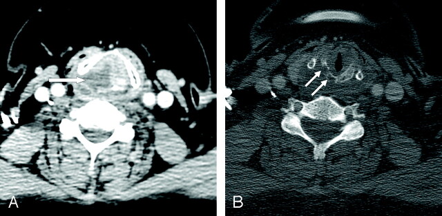
CT of the neck revealed a 3-cm mass in the posterior aspect of the larynx with central hypoattenuation and thick, irregular, enhancing walls (Fig). The tumor was centered on the right cricoid cartilage with erosion of the right cricoid cartilage and both arytenoid cartilages. The margins of the mass were ill defined. There was extensive extralaryngeal spread inferiorly along the medial aspect of the right lobe of the thyroid. No abnormal lymph nodes were observed. The CT scan was interpreted as SCC, predominantly on the basis of the destructive nature of the lesion and its location. The results of the laryngoscopy, including the submucosal appearance of this lesion, were not available at the time of initial CT interpretation.
Fig 1.
Cricoarytenoid rheumatoid arthritis mimicking SCC. A, Axial 3-mm-thick contrast-enhanced CT demonstrates an aggressive, erosive mass of the posterior larynx (arrow). The mass has lower attenuation centrally, and the borders are ill defined. The thyroarytenoid gap is widened, and the mass extends posteriorly toward the right pyriform sinus. B, CT with bone algorithm at a slightly lower level shows erosion of the cricoid cartilage (arrows). The scattered locules of air are from a recent tracheostomy.
A few days later, the patient once again underwent direct laryngoscopy with biopsies to confirm the presence of SCC in the larynx. Laryngectomy and bilateral neck dissections were planned, on the assumption of a confirmatory biopsy. The mass was found to be full of thick yellow fluid. Three biopsies were sent to the pathology department for intraoperative consultation, which indicated rheumatoid nodules and benign, inflamed squamous mucosa. All additional surgery was deferred. The patient was diagnosed with laryngeal rheumatoid arthritis and was discharged on increased doses of anti-inflammatory medications. On several follow-up visits to the otolaryngology service, the patient was doing well with no clinical evidence of laryngeal mass and no shortness of breath.
Discussion
Involvement of the larynx in rheumatoid arthritis was first described by Sir Morell MacKenzie in 1880 (5). The incidence of rheumatoid arthritis of the larynx is higher in female (65%) than in male (20%) patients with rheumatoid arthritis (6). On laryngoscopy, the prevalence of rheumatoid arthritis of the larynx has varied between 32–75% (7–8). The prevalence varies between 54% and 72% on CT scans (7, 8). Although involvement of the larynx is frequent, only 26% of patients with rheumatoid arthritis have laryngeal symptoms; hoarseness and foreign body sensation are the most common symptoms (9). Other symptoms include dyspnea, odynophagia, coughing, sore throat, stridor, and acute airway obstruction causing inspiratory difficulties (6, 7, 10–12). Most patients with rheumatoid arthritis of the larynx have minor symptoms or are asymptomatic.
Laryngoscopy and CT imaging have both been used to diagnose rheumatoid arthritis of the larynx. In the acute stage, laryngoscopy demonstrates edema or erythema of the vocal cords, bowing of vocal cords during inspiration, or tenderness on palpation of the larynx. In the chronic stage, the larynx initially appears to be normal. On careful observation, however, focal vocal cord lesions, changes in arytenoid symmetry, bowing of the true vocal cords, or decreased mobility of the true vocal cord can be seen (10). Abnormalities seen on CT imaging include the presence of cricoarytenoid erosion, cricoarytenoid luxation, cricoarytenoid prominence, and abnormal position of the true vocal cord (8). Laryngoscopy and CT are complementary studies in the diagnosis of cricoarytenoid rheumatoid arthritis.
The treatment of rheumatoid arthritis in the larynx depends on the patient’s presentation. Evaluation to determine the degree of airway obstruction is required. Patients with mild symptoms can be given high-dose systemic corticosteroids (6, 10). If systemic therapy fails, corticosteroid injection of the cricoarytenoid joint is advocated (11). Surgical intervention with tracheostomy, arytenoidectomy, or arytenoidopexy (13) is necessary only if the patient presents with acute airway obstruction or progression of airway obstruction despite medical treatment.
Laryngoscopy is used to differentiate SCC from rheumatoid arthritis. In addition to showing the distinctive features of rheumatoid arthritis at the cricoarytenoid joint, laryngoscopy is used to distinguish between mucosal and submucosal lesions. Submucosal lesions are much less likely to be SCC. When a submucosal lesion is observed, CT is employed to analyze the mass and define its extent. Malignant tumors of the larynx are invasive masses, often with central necrosis. Cartilage erosion evident on CT scans strongly suggests the presence of cancer (14). Unfortunately, these CT findings are not specific for SCC. As this case demonstrates, benign masses can provide a CT appearance identical to that of malignant SCC.
Conclusion
When a patient with a history of rheumatoid arthritis presents with a submucosal mass in the larynx, cricoarytenoid rheumatoid arthritis should be strongly considered. Communication with the referring physician is important to obtain a complete history and to make the radiologist aware of the mucosal or submucosal nature of the disease. Failure to diagnose rheumatoid arthritis of the larynx may subject the patient to unnecessary surgery and anxiety about a potentially lethal disease.
References
- 1.American Cancer Society. Laryngeal and hypopharyngeal cancer. [accessed November 2003. ]
- 2.National Cancer Institute. What you need to know about cancer of the larynx: information about detection, symptoms, diagnosis, and treatment of laryngeal cancer. NIH publication no. 02–1568; http://www.nci.nih.gov/cancerinfo/wyntk/larynx [accessed November 2003. ]
- 3.Yousem DM, Tufano RP. Laryngeal imaging. Magn Reson Imaging Clin North Am 2002;10:451–65 [DOI] [PubMed] [Google Scholar]
- 4.Saleh EM, Mancuso AA, Stringer SP. CT of submucosal and occult laryngeal masses. J Comput Assist Tomogr 1992;16:87–93 [DOI] [PubMed] [Google Scholar]
- 5.MacKenzie M. Disease of the pharynx, larynx, and trachea. New York: William Wood; 1880:347 [Google Scholar]
- 6.Jurik AG, Pedersen U. Rheumatoid arthritis of the crico-arytenoid and crico-thyroid joints: a radiological and clinical study. Clin Radiol 1984;35:233–236 [DOI] [PubMed] [Google Scholar]
- 7.Lawry GV, Finerman ML, Hanafee WN, et al. Laryngeal involvement in rheumatoid arthritis: a clinical, laryngoscopic, and computerized tomographic study. Arthritis Rheum 1984;27:873–882 [DOI] [PubMed] [Google Scholar]
- 8.Brazeau-Lamontagne L, Charlin B, Levesque RT, Lussier A. Cricoarytenoiditis: CT assessment in rheumatoid arhtirits. Radiology 1986;158:463–466 [DOI] [PubMed] [Google Scholar]
- 9.Lofgren RH, Montgomery WW. Incidence of laryngeal involvement in rheumatoid arthritis. N Engl J Med 1962;267:193–195 [DOI] [PubMed] [Google Scholar]
- 10.Leicht MJ, Harrington TM, Davis DE. Cricoarytenoid arthritis: a cause of laryngeal obstruction. Ann Emerg Med 1987;16:885–888 [DOI] [PubMed] [Google Scholar]
- 11.Dockery KM, Sismanis A, Abedi E. Rheumatoid arthritis of larynx: the importance of early diagnosis and corticosteroid therapy. South Med J 1991;84:95–96 [DOI] [PubMed] [Google Scholar]
- 12.Kolman J, Morris I. Cricoarytenoid arthritis: a cause of acute upper airway obstruction in rheumatoid arthritis. Can J Anesth 2002;49:729–732 [DOI] [PubMed] [Google Scholar]
- 13.Whicker JH, Devine KD. Long-term results of Thornell arytenoidectomy in the surgical treatment of bilateral vocal cord paralysis. Laryngoscope 1982;11:361–364 [DOI] [PubMed] [Google Scholar]
- 14.Curtin HD. The larynx. In: Som PM, Curtin HD, eds. Head and neck imaging 4th ed. St Louis: Mosby;2003:1595–1699


