Abstract
SUMMARY: Primary intraosseous lytic meningiomas are rare tumors, with only 16 cases described in the literature. We present a case in which CT and MR imaging with contrast agent helped diagnose preoperatively an enlarging skull mass as a primary intraosseous lytic meningioma in a 70-year-old woman. Radiographic findings revealed a lytic mass centered on the coronal suture line that separated and thinned both the outer and inner tables of the frontal bone.
Meningiomas arise from arachnoid cap cells of the arachnoid cap villi. Primary extradural meningiomas are rare lesions, accounting for less than 2% of all meningiomas.1 Intraosseous meningiomas are increasingly rare, occurring in 14% of cases of ectopic or primary extradural meningiomas.2 Osteoblastic or mixed osteoblastic-osteolytic lesions compose most intraosseous meningiomas, with purely lytic lesions the least common.3 In 2004, Rosahl et al4 reported 16 cases of primary intraosseous osteolytic meningiomas without evidence of soft-tissue invasion.
We report the case of a primary calvarial osteolytic intraosseous meningioma in a 70-year-old woman.
Case Presentation
A 70-year-old right-handed woman presented with a progressively enlarging painless mass over her left frontal area that had been present for 2 years. Physical examination revealed a left frontotemporal bony protuberance with no neurologic or other significant findings. Noncontrast CT of the head was performed (Fig 1A, -B). The lesion was further characterized by using pre- and postcontrast MR imaging (Fig 2A, -B). The radiologic preoperative diagnosis was likely an intraosseous lytic meningioma. Percutaneous biopsy results from an outside facility in Southeast Asia were not diagnostic.
Fig 1.
A, Noncontrast CT axial projection of a 41-mm minimally hyperattenuated homogeneous lytic mass centered at the coronal suture line with the inner and outer bony tables thinned and partially interrupted. B, Bone window exhibiting an osteolytic lesion with anterior hyperostosis. C, Axial T1-weighted precontrast MR image demonstrating an isointense biconvex mass. D, Axial T1-weighted postcontrast MR image demonstrating intense homogeneous enhancement with a mild dural reaction.
Fig 2.
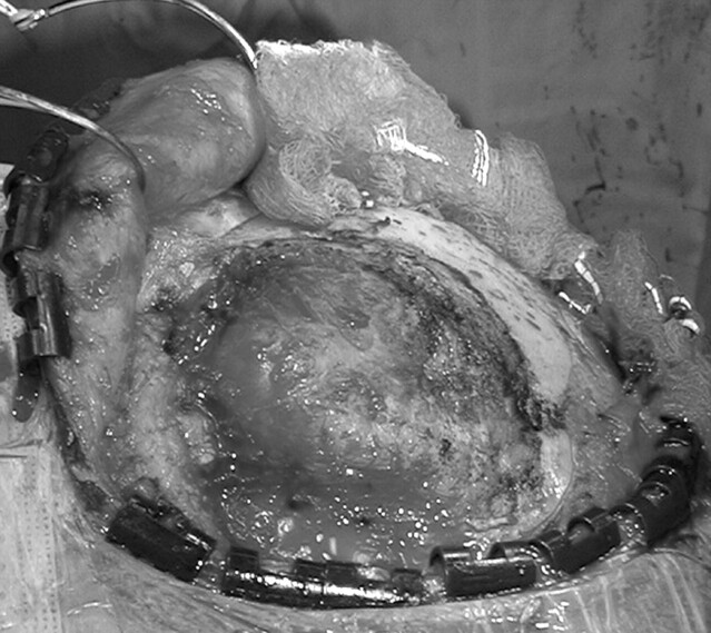
Intraoperative image demonstrating an exophytic relatively vascular skull mass.
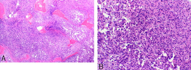
The patient underwent microsurgical resection of her lesion (Fig 2). Intraoperatively, most of the mass was resected. However, because of anesthesia complications, the dural component was not resected. Pathologic examination revealed a hypercellular meningothelial neoplasm composed of small cells with high nuclear-to-cytoplasmic ratio, with scattered atypical nuclei with prominent nucleoli (Fig 3A, -B). Findings of pathology were consistent with a WHO grade I meningioma.
Fig 3.
A, Light microscopy (H&E; original magnification × 2) of resected tumor involving bone. B, Light microscopy (H&E; original magnification × 40) reveals sheets of small cells with uniform nuclei forming small whorls.
Precontrast CT of the head performed 4 months following surgical resection showed no evidence of tumor recurrence. However, a postcontrast scan showed a 4.5-mm-thick area of dural enhancement adjacent to the craniotomy site, partially effacing the Sylvian fissure.
Discussion
As reported by Rosahl et al,4 16 cases of primary osteolytic intraosseous meningiomas have been published. The differential diagnosis for a primary lytic calvarial lesion includes lytic metastasis, epidermoid tumor, multiple myeloma, eosinophilic granuloma, fibrous dysplasia, brown tumor, skull dermoid, lytic intraosseous meningioma, giant cell tumor, hemangiopericytoma, and hemangioma.4 Although this lesion involved the inner and outer bony tables, consistent with lesions such as eosinophilic granulomas and hemangiomas, the lesion did not exhibit radiologic findings completely consistent with these or other intraosseous lesions. Additionally, primary intraosseous meningiomas tend to present with hyperostosis, which was seen in this lesion.5 Patients with primary calvarial lesions commonly present with a lump in the scalp of long evolution with a relationship to the cranial sutures.6
Some dural invasion noted in surgery raised the question as to whether the tumor could be classified as a primary extradural meningioma. The review of Lang et al1 of all primary extradural meningiomas concluded that extracalvarial meningiomas that erode the inner table of the skull with dural reaction and/or invasion cannot be excluded from being classified as primary extradural lesions. A lesion demonstrating tumor growth with the center outside the dura can be classified as a primary extradural meningioma.1 Radiologic and pathologic findings in our patient supported this designation.
The proposed origin in our patient may have been due to trapping of arachnoid cells in a prior healed skull fracture or to embryologic rests of arachnoid cap cells within the developing calvaria. Therefore, primary intraosseous meningioma should be considered in the differential diagnosis when a solitary lytic calvarial lesion is seen near the suture line or is associated with a prior skull injury.
References
- 1.Lang FF, Macdonald K, Fuller GN, et al. Primary extradural meningiomas: a report of nine cases and review of the literature from the era of computerized tomography scanning. J Neurosurg 2000;93:940–50 [DOI] [PubMed] [Google Scholar]
- 2.Daffner RH, Yakulis R, Maroon JC. Intraosseous meningioma. Skeletal Radiol 1998;27:108–11 [DOI] [PubMed] [Google Scholar]
- 3.Levin M, Wertheim SE, Klein E, et al. Unusual lytic intraosseous meningioma. J Neuroimaging 1995;5:247–48 [DOI] [PubMed] [Google Scholar]
- 4.Rosahl SK, Mirzayan M-J, Samii M. Osteolytic intra-osseous meningiomas: illustrated review. Acta Neurochir (Wien) 2004;146:1245–49. Epub 2004 Sep 9 [DOI] [PubMed] [Google Scholar]
- 5.Lee HY, Prager J, Hahn Y, et al. Intraosseous meningioma: CT and MR appearance. J Comput Assist Tomogr 1992;16:1000–01 [PubMed] [Google Scholar]
- 6.Arana E, Diaz C, Latorre FF, et al. Primary intraosseous meningiomas. Acta Radiol 1996;37:937–42 [DOI] [PubMed] [Google Scholar]


