Abstract
Purpose of Review
The olive tree (Olea europaea L.) has featured as a significant part of medicinal history, used to treat a variety of ailments within folk medicine. The Mediterranean diet, which is rich in olive products, is testament to Olea europaeas positive effects on health, associated with reduced incidences of cancer and cardiovascular disease. This review aims to summarise the current literature regarding the therapeutic potential of Olea europaea products in cancer, detailing the possible compounds responsible for its chemotherapeutic effects.
Recent Findings
Much of the existing research has focused on the use of cell culture models of disease, demonstrating Olea europaea extracts, and specific compounds within these extracts, have efficacy in a range of in vitro and in vivo cancer models. The source of Olea europaeas cytotoxicity is yet to be fully defined; however, compounds such as oleuropein and verbascoside have independent cytotoxic effects on animal models of cancer.
Summary
Initial results from animal models are promising but need to be translated to a clinical setting. Treatments utilising these compounds are likely to be well tolerated and represent a promising direction for future research.
Keywords: Olive, Olea europaea, Chemotherapeutic, Cancer
Introduction
The olive tree (Olea europaea L.) and its products have been an important commodity throughout human history. Today, 98% of olive products are cultivated in the Mediterranean basin and are an important part of the economy of the region [1]. The value of the olive tree, however, extends past economics to its nutritional and medicinal properties [2]. Olive tree products were used in traditional medicines to treat a variety of ailments such as fever [3, 4]. In more recent times however, it has demonstrated possibilities in cancer prevention. Cancer rates in the Mediterranean are notably lower than other western countries, with cancer rates in Greece of 279.8 per 100,000 compared to 352.2 and 319.2 in the USA and UK respectively. It is expected that this protection is a result of dietary differences between these populations, as the Mediterranean diet is consistently associated with reduced incidences of cancer and cardiovascular disease [5, 6].
Academic studies of the beneficial properties of olives initiated in 1854 with the work of Daniel Hanbury, who noted that a decoction of olive leaf was effective in reducing fevers associated with malaria [4, 7]. Many pharmacological reports have since exhibited the potential of olive-based extracts to relieve arrhythmia, increase blood flow, lower blood pressure, inhibit viral activity and prevent intestinal muscle spasms [8, 9]. Scientific studies looking at the anti-proliferative components of certain flora, have recently demonstrated this potential within Olea europaea products, largely from in vitro studies using extracts in addition to testing isolated compounds from Olea europaea products [7]. This review will summarise the key studies that relate to Olea europaea extract’s anti-cancer effects and discuss compounds chosen for their high abundance within Olea europaea, revealing their anti-proliferation properties.
Olea europaea Extracts
The preponderance of research in this field has focussed on the use of cell culture models of disease, with efficacy in a range of these cancer models. Fares et al. [10] studied the effect of olive leaf extract (OLE) on human lymphoblastic leukaemia cell line, Jurkat. The results reported a 78% inhibition of Jurkat cell proliferation at a concentration of 4 μg/mL at 48 hours. However, the study could not elucidate the mechanism of apoptosis. OLE has demonstrated efficacy against the myelogenous leukaemic cell line, K562. After a 72-h treatment with 150 μg/mL of OLE, the cell proliferation was inhibited to 17%, whilst also demonstrating a significant decrease in viability [11].
Examination of OLE on a pancreatic cancer cell line (MiaPACa-2) demonstrated efficacy in culture [12]. At concentrations of 200 μg/mL, the OLE was able to reduce cell viability to less than 1% compared with controls. Coccia et al. [13] observed the cytotoxic effect of extra virgin olive oil extract on two different bladder cancer cell lines. It was demonstrated that oil significantly decreased proliferation in T24 and 5637 bladder cell lines in a dose dependent manner. Cell viability of T24 cells was decreased by up to 90% with 100 μg/mL oil, with an IC50 of ~32 μg/mL, with similar treatment efficacy in 5637 cells [13]. Furthermore, the cell cycle progression was monitored by flow cytometry, that observed a growth arrest at the G2/M phase after treatment in both cell lines. In the T24 cell line, a decrease was observed in the G0/G1 phase and an increase in the sub-G1 fraction, indicative of induction of apoptosis. Western blot analysis of pro-caspase-3, -9 and PARP-1 further demonstrated an apoptotic effect of oil [13]. Olive oil production generates waste materials often referred to as olive mill wastewater. This wastewater still contains many of the same compounds present in olives. Recently, Baci et al. [14•] tested this wastewater on PC-3 prostate cancer cells in culture. The results demonstrated wastewater was able to inhibit cell proliferation, adhesion, migration and invasion. At a molecular level, the wastewater was observed to inhibit NF-κB signalling in addition to reducing pro-angiogenic growth factors, VEGF, CXCL-8 and CXCL-12 production. The results demonstrated that even from the lowest dilution tested (1:5000), wastewater was able to inhibit PC-3 cell proliferation [14•].
Isleem et al. [15] demonstrated the importance of extraction techniques on OLE efficacy. In this study, OLE was mixed with either deionised water or 80% methanol, and these extracts were subsequently tested individually in a dose- and time-dependent manner on the human breast cancer cell line MCF-7. After a 48-h treatment, the deionised water extract had an IC50 value of 182 μg/mL whilst the methanol extract was 135 μg/mL, versus relevant controls. Furthermore, the source of the products are often not comparable, something which is supported by in vivo studies. For example, a study by Escrich, Moral and Solana [16] investigated how diets rich in extra virgin olive oil reduce the risk of breast cancer compared with diets high in corn oil. Olive oil induced molecular changes in tumours that resulted in higher rates of apoptosis, lower proliferation and lower DNA damage. Furthermore, Milanizadeh and Reza Bigdeli [17] demonstrated a reduction in mammary cancer weight and volume after a treatment of 150 and 225 mg/kg/day of OLE in a breast cancer mouse model. This reduced growth was proposed to be related to the polyphenol content of OLE, resulting in an increase of antioxidant enzyme activity including superoxide dismutase and catalase. The mechanisms responsible for inhibited tumour growth are likely to stem from OLE polyphenols blocking the cell cycle in G1/S through reducing COX2 and cyclin D1 expression, which results in the reduction of cell growth and proliferation [17, 18].
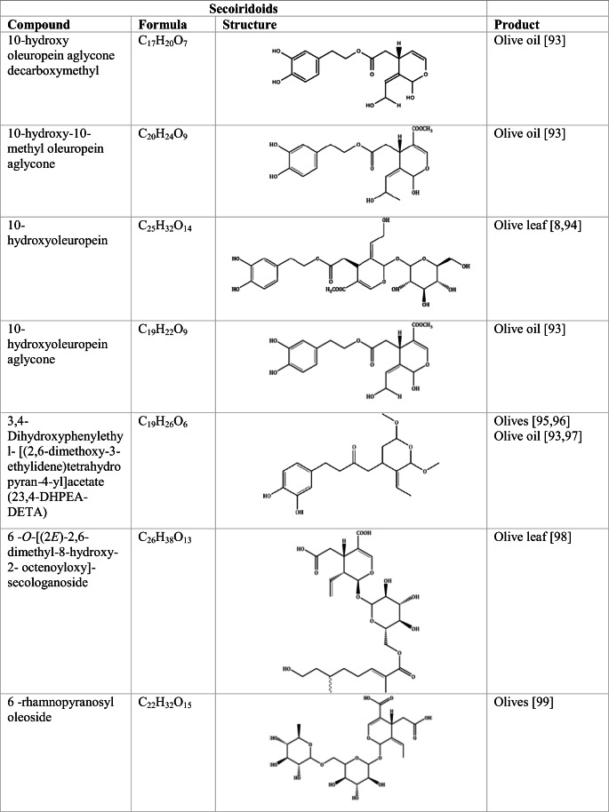
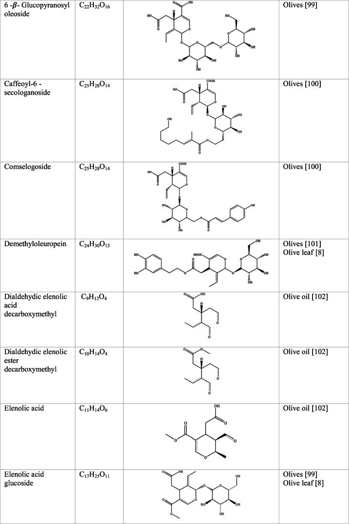
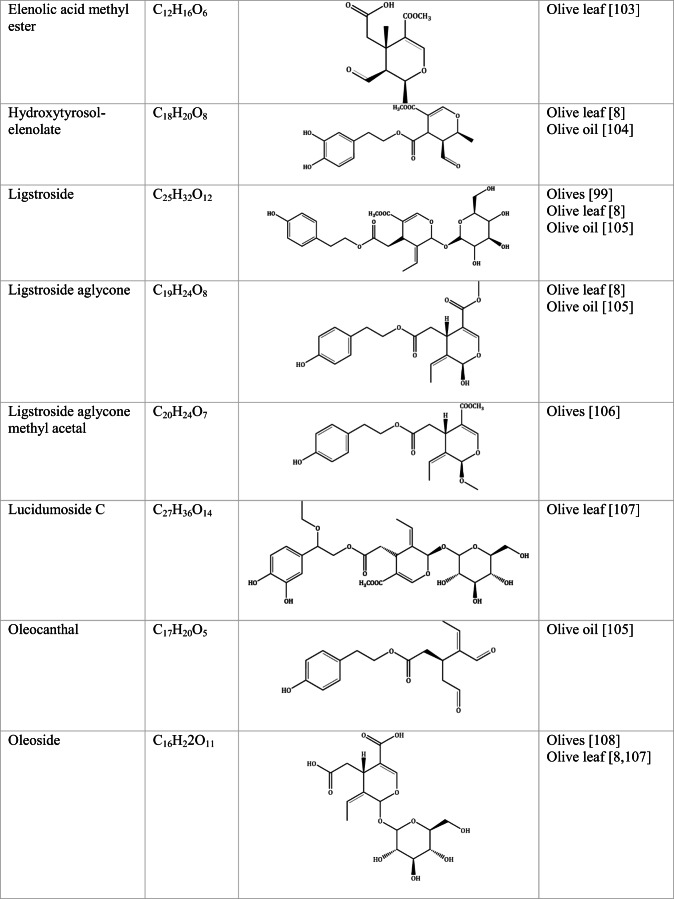
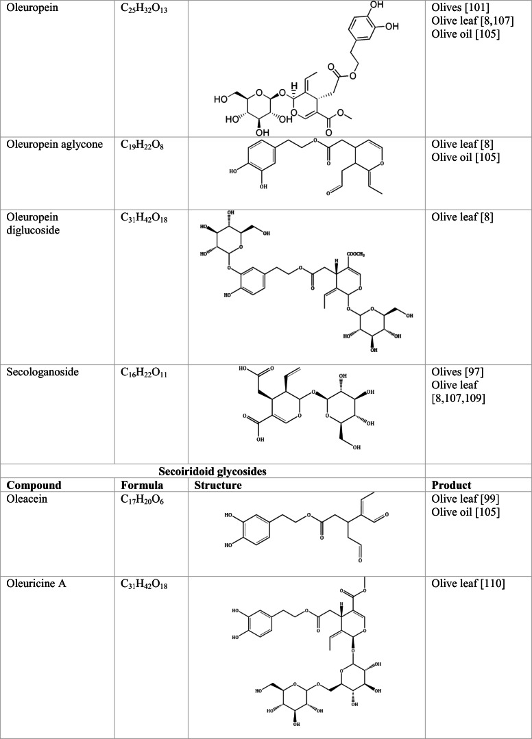
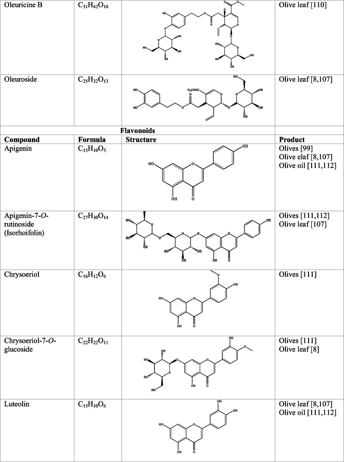
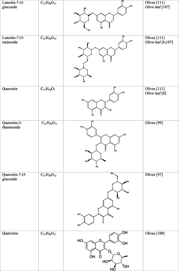
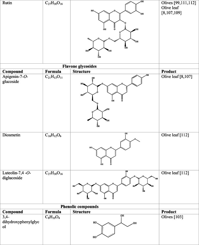
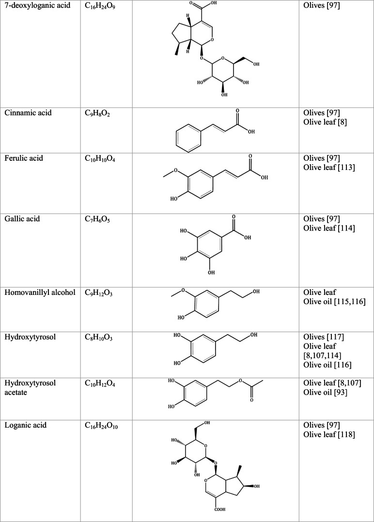
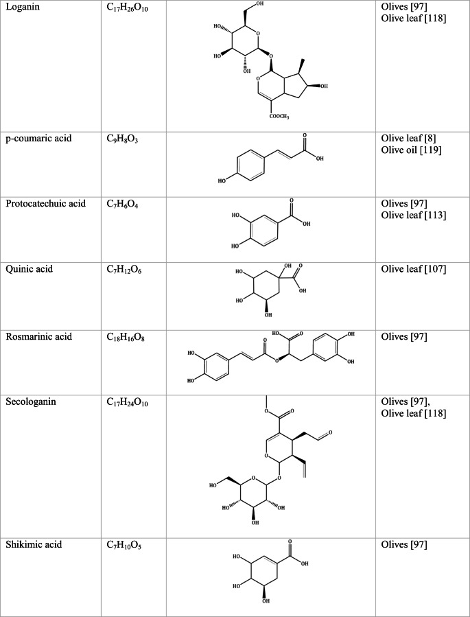
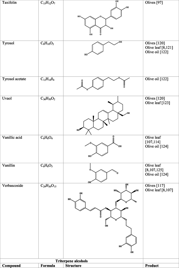
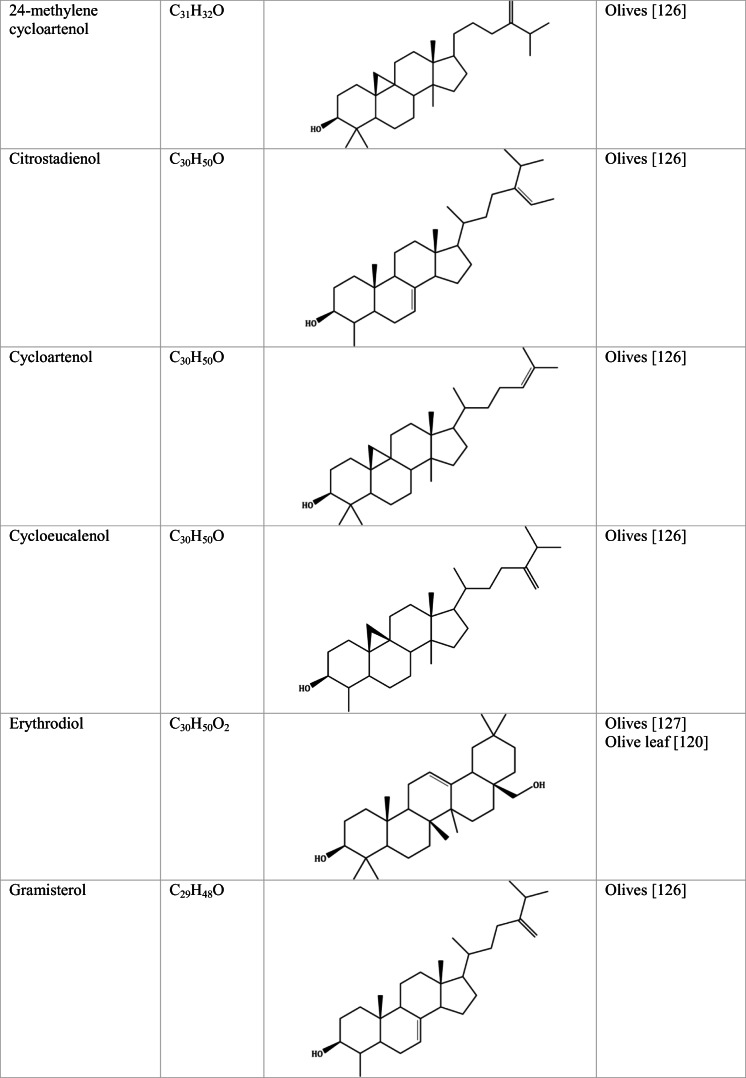
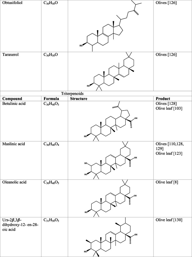
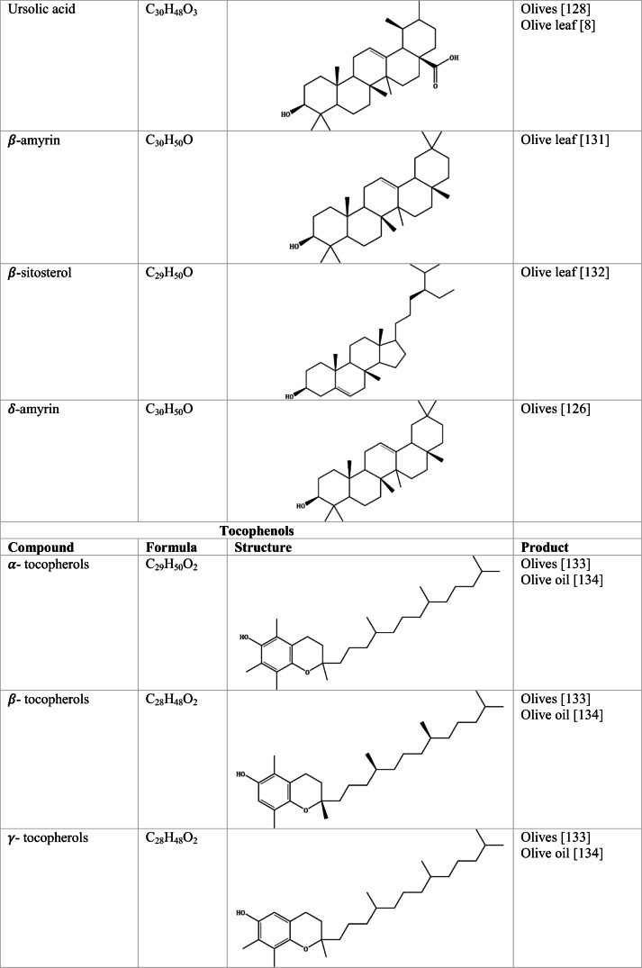
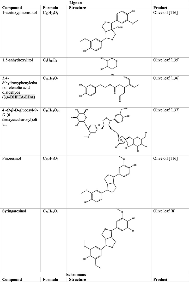
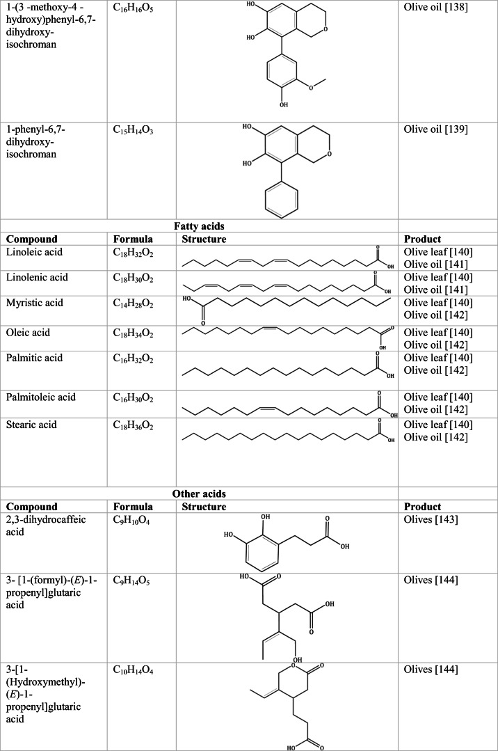
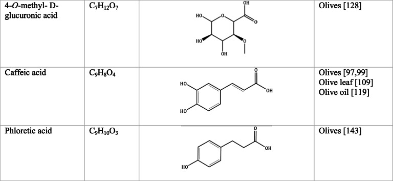
Whilst the source of cytotoxicity of OLE has yet to be fully defined, oleuropein is the most abundant compound in olive leaves, followed by hydroxytyrosol, luteolin, apigenin and verbascoside. Oleuropein is a heterosidic ester of elenolic acid and dihydroxyphenylethanol. Hydroxytyrosol (3,4-Dihydroxyphenyl ethanol), is the principal degradation product of oleuropein [19]. Within unprocessed olive fruit and leaves, oleuropein is more abundant, whereas hydroxytyrosol is present in higher amounts of processed olive fruit and leaves [20]. This change in concentration takes place due to chemical and enzymatic reaction that occurs during maturation of olive products or as a result of processing. One key component of olives and olive oil is their fatty acid composition. Olive oil largely consists of triacylglycerols (98-99%), a group of glycerol esters with varying fatty acids [21]. The main fatty acid in olive oil is oleic acid; however, it also contains linoleic acid, palmitic acid, palmitoleic acid, stearic acid and the triterpene maslinic acid. A variety of amphiphilic and lipophilic microconstituents exist in olive oil including, tocopherols, squalene, phytosterols, and phenolic compounds [22]. These compounds are summarised in Table 1.
Table 1.
Phytochemicals isolated from Olea europaea (Olive tree) products
Polyphenols
Polyphenols are a family of micronutrients, named due to the presence of multiple hydroxylated phenol rings in their structure. These compounds are broadly water soluble and include plant pigments and tannins [23]. The proposed mechanisms of action associated with polyphenols are largely related to their antioxidant activity, directly leading to a reduction in reactive oxygen species (ROS) [24, 25]. Phenols, oleuroepsides and flavonoids have proven to demonstrate significant antioxidant activity towards free radicals, because of the redox properties of their phenolic hydroxyl groups and the structural relationships within their chemical structure [9]. Other research has demonstrated the ability of polyphenols to modulate the human immune system, resulting in increased regulatory T cell and splenocyte production, in addition to reducing oxidative burst activity of neutrophils [26].
The concentration of polyphenols in olive products can depend on factors including climate, cultivation, maturity, rootstock, agricultural practices and the method of extraction, separation and quantification [27–29]. It was observed by Wang and Fordham [30] that the phenolic content of olives is season dependent, with olives harvested in the autumn having a higher polyphenol and carotenoid content, and therefore, a higher antioxidant capacity. Pereira [31] highlighted that black olives present a higher antioxidant capacity than their green counterpart, due to higher concentrations of phenolic compounds in black olives. Thus, the different variety of olives and processing result in polyphenol content varying significantly in different olives and consequently olive oil. Research studying the olive leaves has observed notable variety and quantity of polyphenols compared with olive oil, notably oleuropein and hydroxytyrosol [32]. Just as with olives, the chemical composition of olive leaves changes under conditions such as climate, country of origin, moisture, storage condition and soil content [33].
The reduced cancer rates associated with the Mediterranean diet and the history of Olea europaea products in traditional medicine have made olive tree products the focus of phytochemical research [34–36]. Studies have identified some compounds from olive tree products for a deeper investigation of their cytotoxic effect. The most ubiquitous compounds are the phenolic compounds, flavonoids, secoiridoids and secoiridoid glycosides. However, compounds such as flavanones, flavone glycosides, triterpenes, benzoic acid derivatives, biophenols, sterols, sugars, xylitol and isochromans have clear biological activities [28].
Secoiridoids
Secoiridoids are uniquely present in plants of Olearaceae family [37]. Secoiridoids are monoterpenoids based on the 7,8-secocyclopenta[c]-pyranoid skeleton. In plants, it is possible that they derive from iridoids that are cleaved with redox enzymes and subsequently undergo several secondary modifications (oxidation, epoxidation, esterification) of the generated hydroxyl groups within the main skeleton. This produces a group of compounds, which constitute the class [38].
Oleuropein
Oleuropein is recognised to have antiviral, antimicrobial and antifungal properties [39]. Moreover, oleuropein has noted hypoglycaemic and hypotensive properties, and is a powerful antioxidant [40, 41]. Oleuropein has proven to exert protective properties against cancer and heart disease, in addition to supporting immunoregulatory actions [42]. Oleuropein inhibits migration and proliferation of human tumour cells in culture. Hamadi and Castellon [43] demonstrated that oleuropein irreversibly rounds cancer cells, preventing motility, replication and invasiveness, whilst these effects were reversible upon treatment removal in normal cells. The study [43] demonstrated this reversibility through carrying out additional washing tests on Matrigel-free cultures of RPMI-7951 melanoma cells in addition to normal cells. After 48 h of extensive washing, normal cells flattened out and again became mobile, whilst cancer cells remained immobilized. This work was supported in vivo, when 1% oleuropein was administered to mice orally within drinking water leading to tumour regression in 9-12 days [43]. The cell rounding triggered by oleuropein is linked to the disruption of the actin cytoskeleton and actin filaments. In Hamadi and Castellon’s [43] study, it was established that this effect was somewhat offset by the addition of glucose in the culture media. Due to oleuropeins glucose moiety, it is likely that oleuropein enters the cell through glucose transporters (GLUTs). Removing oleuropeins glucose moiety with β-glucosidase decreases its anti-proliferative activity, demonstrating that entry of oleuropein into the cell is at least partially dependent on the glucose moiety [43]. Human malignancies are associated with elevated glucose uptake and enhanced expression of several GLUT isoforms [44]. This explains how normal cells can reverse the rounding effects of oleuropein treatment, as normal cells have low expression of GLUTs [45, 46].
Numerous studies have demonstrated oleuropeins efficacy against certain breast cancer cell lines. Sirianni et al. [47] demonstrated the inhibition of estradiol-dependent activation of extracellular regulated kinase 1/2 (ERK 1/2) by oleuropein in the MCF-7 cell line. This is significant as estradiol stimulates the growth of several breast tumours, through induced cellular proliferation. It is established that oleuropein can reduce cell proliferation of MCF-7 and T47D cells, this reduce proliferation is associated with the activation of autophagy and suppression of migration and invasion; this is achieved through p62 downregulation, in addition to Beclin-1 and LC3II/LC3I upregulation [48]. Oleuropein increases the level of ROS and induces apoptosis via modulating NF-κB activation cascade, within MDA-MB-231 and MCF-7 cell lines [49]. Elamin et al. [50], treated breast cancer xenografts (in nude mice) in vivo, with a combination of doxorubicin and oleuropein. This treatment combination resulted in a downregulation of NF-κB, Bcl-2 and survivin, triggering apoptosis. All treatments significantly reduced tumour volume compared to untreated controls (tumour volume, 173mm3). However, combination therapy of doxorubicin and oleuropein (48.7mm3) had greater efficacy than either doxorubicin (69mm3) or oleuropein (79mm3) alone. Oleuropein has further demonstrated efficacy in numerous cancer cell lines such as colorectal, thyroid and lung [51–54]. Future work will be needed to apply this research to the clinical environment.
Oleocanthal
Studies have outlined the potential of oleocanthal for cancer prevention in many cancer types. Akl et al. [55] demonstrated that oleocanthal can inhibit the growth of human breast cancer cell lines MCF-7, MDA-MB-231 and BT-474, whilst not affecting normal human cell growth of MCF10A. Possible mechanisms of action in these cell lines point to the blocking of cell migration, invasion and G1/S cell cycle progression. This mechanism occurs through the inhibition of Hepatocyte growth factor (HGF)-induced c-Met activation [55]. Oleocanthal was investigated for its antiproliferation activity in human melanoma cell lines, 501Mel and A375. It was determined that oleocanthal inhibits cyclooxygenase enzymes to exert important anti-inflammatory activities [56]. This study demonstrated that oleocanthal did not produce significant changes in human dermal fibroblast viability, signifying selective activity for cancer cells over normal cells. This selectivity has been associated with oleocanthals ability to induce lysosomal membrane permeabilization leading to apoptosis and/or necrosis. Cancer cells largely have weak lysosomal membranes compared to noncancerous cells, thus making them susceptible to cell death via lysosomotropic agents [57]. Fogli et al. [56] demonstrated oleocanthal induced cell growth inhibition in 501Mel and A375 cells in a concentration-dependent manner, with an IC50 of 13.6 and 20 μM respectively. It was demonstrated that oleocanthal downregulates Bcl-2, Erk1/2 and AKT signal transduction pathways [56, 58]. These pathways play a key role in oleocanthal-induced cytotoxicity in multiple myeloma cells. Moreover, the activation of the AKT pathway is closely associated with resistance to BRAF inhibitors in melanoma patients [59].
Flavonoids
Flavonoids are secondary metabolites corresponding to polyphenols that have a diverse structure, taking the form glycosides or aglycones in fruits and vegetables such as onions, berries, kale, and green tea [60]. Flavonoids have a chemical structure of 15 carbons, with a skeleton of phenyl-benzo-γ-pyran (C6–C3–C6), also known as nucleus flava, composed of two phenyl rings and a heterocyclic (pyran) ring. Flavonoids comprise of flavones, flavonols, flavonoids, flavanones, isoflavones and anthocyanidins [61].
Apigenin
A recent study investigated the anti-proliferative effects of apigenin, an abundant flavonoid, on the human cervical cancer cell line HeLa [62]. Initial research demonstrated an IC50 of 15 μM – similar to that observed for some common chemotherapeutics. Reduced viability was associated with an apoptotic profile as determined by annexin V and propidium iodide positivity. This was demonstrated by Western blot where apigenin increased the expression of Bax and decreased the expression of Bcl-2, commonly observed in apoptosis. The authors concluded that apigenin has apparent potential as a chemotherapeutic agent [62]. Erdogan et al. [63] observed apigenin reduce prostate cancer stem cell survival and migration, via treating PC3 and cancer stem cells with a series of μM concentrations of apigenin for 48 hours. In a dose-dependent manner, apigenin inhibited PC3 and cancer stem cell survival, that was associated with PI3K, AKT and NF-κB downregulation, and increases in p27 and p21. Apigenin significantly suppressed the migration rate of CD44+ cancer stem cells, as determined by a wound healing assay. This supressed migration was through downregulation of matrix metallopeptidases-2, -9, Snail and Slug. Thus, Erdogan et al. [63] highlighted apigenin to potentially prevent the proliferation and migration of cancer cells. Xu et al. [64] demonstrated apigenin to suppress colorectal cancer cell proliferation, migration and invasion through the inhibition of the Wnt/β-catenin signalling pathway. The occurrence of human tumorigenesis can be significantly affected by abnormal activation of the Wnt/β-catenin signalling pathway. Xu et al. [64] demonstrated antiproliferation effects of apigenin on HCT15 and SW480 colorectal cancer cell lines in vitro. It was determined that apigenin significantly reduced HCT15 and SW480 cell proliferation at an IC50 of 23.57 μM and 18.17 μM, respectively.
Luteolin-7-O-glucoside
Maatouk et al. [65••] investigated the protective role of luteolin-7-O-glucoside on oxidative stress in addition to DNA damage induced by cisplatin through comet assay. Balb/c mice were injected with 10 mg/kg of cisplatin following luteolin-7-O-glucoside treatment (40 mg/kg). The results demonstrated that luteolin-7-O-glucoside attenuates the genotoxicity associated with cisplatin [65••]. This included a reduction in markers of tissue damage (creatinine, interferon γ) and oxidative stress (malondialdehyde, catalase, glutathione peroxidase, superoxide dismutase, and glutathione). Work on the liver cancer cell line HepG2 demonstrated that exposure to luteolin 7-glucoside and apigenin 7-glucoside reduced cell viability, with an IC50 of 21 μg/mL and 17 μg/mL, respectively. These compounds reduced the expression of NF-κB, a key pathway in the chronic inflammation associated with hepatocellular carcinoma [66].
Hydroxytyrosol
Hydroxytyrosol was revealed to possess antibacterial, antioxidative, and anti-inflammatory properties [51]. Evidence further demonstrated effective chemotherapeutic properties through affecting several signalling pathways [67]; notably growth factor receptors [52, 68, 69], receptor support proteins [70, 71] and interleukin pathways [72]. Inhibition of cyclin D1 is core to hydroxytyrosol efficacy, resulting in cell cycle arrest at G1/S phase in the MCF-7 cell line [7]. Several studies have referenced cyclin D1 downregulation following hydroxytyrosol treatment in many cancer cell lines, including breast cancer (MCF-7, MB231) [7, 71], colon cancer (Caco-2) [73], and thyroid cancer (TPC-1, FB-2, WRO) [74].
Furthermore, hydroxytyrosol has exhibited protection to peripheral blood mononuclear cells from hydrogen peroxide-induced DNA damage and prevention of endoplasmic reticulum stress in hepatocellular carcinoma Hep G2 [75, 76]. Conversely, hydroxytyrosol has demonstrated ROS production, leading to apoptotic cell death and mitochondrial dysfunction in DLD1 colon cancer cells [77]. Moreover, evidence suggests that hydroxytyrosol can cause superoxide and hydrogen peroxide generation leading to induction of apoptosis in prostate cancer PC3 cells [78].
Zubair et al. [79] tested hydroxytyrosol on normal prostate epithelial cells (PWLE2 and RWPE1) alongside cancerous cells (LNCaP and C4-2), demonstrating inhibition of proliferation in a dose-dependent manner. The study revealed hydroxytyrosol inhibited cyclins D1/E and cyclin-dependent kinases cdk2/4 and induced the cell cycle inhibitors p21/p27, resulting in G1/S cell cycle arrest [79]. Hydroxytyrosol induced apoptosis, as demonstrated through caspase activation, PARP cleavage, and BAX/Bcl-2 ratio. Phosphorylation of Akt/STAT3 was inhibited and cytoplasmic retention of NF-kB was induced, which relates to the induction of apoptosis. Prostate cancers usually retain androgen receptor signalling and are normally dependent on activated Akt, NF-kB, and STAT3 signalling [80–82]. Hydroxytyrosol exhibits a pleiotropic activity against these signalling pathways leading to cell cycle arrest [79]. Terzuoli et al. [69] demonstrated that hydroxytyrosol significantly downregulates epidermal growth factor receptor expression in human colorectal adenocarcinoma cells (HT-29, WiDr and CaCo2). They concluded that hydroxytyrosol downregulated receptor expression through proteasomal and lysosomal degradation via receptor ubiquitination. This led Terzuoli et al. [69] to highlight the potential of hydroxytyrosol as a novel colon tumour treatment.
Verbascoside
Verbascoside has demonstrated anti-tumour effects in some human cancers. Apoptosis promotion by verbascoside is associated with HIPK2, p53, HIF-1α and Rac-1 in colorectal cancer cell lines [83, 84], in addition to downregulation of STAT3, epithelial-to-mesenchymal transition (EMT) markers (vimentin, snail and zeb1) and c-Met in glioblastomas [85, 86]. Hei et al. [85] further demonstrated the downregulation of the EMT markers and c-Met in an orthotopic glioblastoma xenograft mouse model. EMT is a fundamental hallmark of metastatic tumourigenesis and has an essential role in glioma aggressiveness [87]. Hei et al. [85] highlighted that verbascoside can bind directly to the c-Met protein, and that verbascoside causes c-Met protein degradation through the ubiquitination-proteasome pathway. Furthermore, verbascoside was able to suppress tumour growth and enhance survival in mice [85]. This model demonstrated that verbascoside exerted effects via the same mechanisms in vivo as it did in vitro, by suppressing c-Met-mediated EMT and inducing cancer death.
Triterpenoids
Triterpenoids are structurally varied organic compounds, made of a basic backbone modified in a multitude of ways, creating the formation of over 20,000 naturally occurring triterpenoids [88]. Triterpenoids are characterised by 30 carbon atoms, polymerised to form six isoprene units. Biosynthesis of triterpenoids occurs when its precursor squalene undergoes cyclization. Triterpenoids chemical structure is grouped from linear, through to pentacyclic [89]. Maslinic acid, oleanolic acid, erythrodiol and uvaol are the most abundant triterpenes in olive tree products [90].
Maslinic and Oleanolic Acid
Juan et al. [91] observed the effect of maslinic and oleanolic acid (two triterpenoids with similar structure) on HT-29 colon cancer cells. These compounds were examined for their effect on proliferation, necrosis and apoptosis. Maslinic acid inhibited cell growth with an IC50 of 101.2 μM, whilst oleanolic acid demonstrated lesser antiproliferation activity with an IC50 of 160.6 μM. Maslinic acid increased caspase-3-like activity that was associated with increased presence of mitochondrial ROS, whereas oleanolic acid cytotoxicity was not associated with either activated caspase-3 or ROS production [91]. Detection of increased DNA fragmentation and increase in plasma membrane permeability confirmed apoptosis by maslinic acid. Kim et al. [92•] investigated oleanolic acid-induced cancer cell death, apoptotic mechanisms, cell cycle status, and MAPK kinase signalling in MCF-7, DU145 (prostate cancer) and U87 (human glioblastoma) cell lines. The IC50 values for oleanolic acid-induced cytotoxicity were 132.29 in MCF-7, 112.57 in DU145 and 163.60 in U87 cells. At 100 μg/mL oleanolic acid, there was an increased number of apoptotic cells to 27.0% in MCF-7, 27.0% in DU145 and 15.7% in U87, when compared to control cells [92•]. This greater apoptosis was a result of increased p53, cytochrome c, Bax, PARP-1 and caspase-3 expression in the cell lines. Furthermore, the different cancer lines arrested at varying phases of the cell cycle, MCF-7 and U87 cells arrested in G1, whereas DU145 cells arrested in G2 [92•]. This suggests that oleanolic acid alters the expression of the cell cycle regulatory proteins inconsistently in different types of cancer cell lines.
Conclusion
Interest in Olea europaea products is increasingly researched for their beneficial effect on human health. The polyphenols detected in these products are of growing interest due to their effect on ROS production. However, this is not their only means of inhibiting cell proliferation in cancer. Natural plant polyphenols have demonstrated the ability to alter the level of ROS, either protecting biomolecules from oxidative damage (e.g., luteolin-7-O-glucoside) or inducing oxidative damage (e.g., maslinic acid). Oleuropein, hydroxytyrosol and triterpenoids are abundant in Olea europaea products. These polyphenolic compounds demonstrate powerful anti-oxidant, anti-angiogenic, chemotherapeutic and anti-inflammatory characteristics. Despite the continued positive results from in vitro studies on the beneficial properties of Olive europaea products, further in vivo investigation is needed. Initial results in animal models are promising but need to be translated to clinical setting. Treatments utilising these compounds are likely to be well tolerated, as initial animal experimentation has demonstrated [43, 50, 65, 85]. Nevertheless, initial investigation is encouraging in relation to the prevention and treatment of cancer.
Acknowledgements
The authors would like to thank Adam Thomas and Emily Watson for reviewing the manuscript prior to submission.
Abbreviations
- GLUT
glucose transporters
- IC50
half maximal inhibitory concentration
- OLE
olive leaf extract
- PARP
poly-ADP ribose polymerase
- ROS
reactive oxygen species
Declarations
Conflict of Interest
No conflicts present.
Footnotes
This article is part of the Topical Collection on Cancer
Publisher’s Note
Springer Nature remains neutral with regard to jurisdictional claims in published maps and institutional affiliations.
References
Papers of particular interest, published recently, have been highlighted as: • Of importance •• Of major importance
- 1.Ghanbari R, Anwar F, Alkharfy KM, Gilani AH, Saari N. Valuable nutrients and functional bioactives in different parts of olive (Olea europaea L.)—a review. Int J Mol Sci. 2012;13(3):3291–3340. doi: 10.3390/ijms13033291. [DOI] [PMC free article] [PubMed] [Google Scholar]
- 2.Muzzalupo I. Olive Germplasm: The Olive Cultivation, Table Olive and Olive Oil Industry in Italy: Books on Demand; 2012.
- 3.Altınyay Ç, Altun ML. HPLC analysis of oleuropein in Olea europaea L. Ankara universitesieczacılık fakültesi dergisi. 2006;35(1):1–11. [Google Scholar]
- 4.Hanbury D. On the febrifuge properties of the olive (Olea europea, L.) Pharm J Provincial Trans. 1854;353:354. [Google Scholar]
- 5.Estruch R, Ros E, Salas-Salvadó J, Covas MI, Corella D, Arós F, Gómez-Gracia E, Ruiz-Gutiérrez V, Fiol M, Lapetra J, Lamuela-Raventos RM. Primary prevention of cardiovascular disease with a Mediterranean diet. N Engl J Med. 2013;368(14):1279–1290. [Google Scholar]
- 6.Giacosa A, Barale R, Bavaresco L, Gatenby P, Gerbi V, Janssens J, Johnston B, Kas K, La Vecchia C, Mainguet P, Morazzoni P. Cancer prevention in Europe: the Mediterranean diet as a protective choice. Eur J Cancer Prev. 2013;22(1):90–95. doi: 10.1097/CEJ.0b013e328354d2d7. [DOI] [PubMed] [Google Scholar]
- 7.Bouallagui Z, Han J, Isoda H, Sayadi S. Hydroxytyrosol rich extract from olive leaves modulates cell cycle progression in MCF-7 human breast cancer cells. Food Chem Toxicol. 2011;49:179–184. doi: 10.1016/j.fct.2010.10.014. [DOI] [PubMed] [Google Scholar]
- 8.Fu S, Arráez-Roman D, Segura-Carretero A, Menéndez JA, Menéndez-Gutiérrez MP, Micol V, Fernández-Gutiérrez A. Qualitative screening of phenolic compounds in olive leaf extracts by hyphenated liquid chromatography and preliminary evaluation of cytotoxic activity against human breast cancer cells. Anal Bioanal Chem. 2010;397(2):643–654. doi: 10.1007/s00216-010-3604-0. [DOI] [PubMed] [Google Scholar]
- 9.Benavente-Garcıa O, Castillo J, Lorente J, Ortuño ADRJ, Del Rio JA. Antioxidant activity of phenolics extracted from Olea europaea L. leaves. Food Chem. 2000;68(4):457–462. [Google Scholar]
- 10.Fares R, Bazzi S, Baydoun SE, Abdel-Massih RM. The antioxidant and anti-proliferative activity of the Lebanese Olea europaea extract. Plant Foods Hum Nutr. 2011;66(1):58–63. doi: 10.1007/s11130-011-0213-9. [DOI] [PubMed] [Google Scholar]
- 11.Samet I, Han J, Jlaiel L, Sayadi S and Isoda H, 2014. Olive (Olea europaea) leaf extract induces apoptosis and monocyte/macrophage differentiation in human chronic myelogenous leukemia K562 cells: insight into the underlying mechanism. Oxidative Med Cell Longev. [DOI] [PMC free article] [PubMed]
- 12.Goldsmith CD, Vuong QV, Sadeqzadeh E, Stathopoulos CE, Roach PD, Scarlett CJ. Phytochemical properties and anti-proliferative activity of Olea europaea L. leaf extracts against pancreatic cancer cells. Molecules. 2015;20(7):12992–13004. doi: 10.3390/molecules200712992. [DOI] [PMC free article] [PubMed] [Google Scholar]
- 13.Coccia A, Mosca L, Puca R, Mangino G, Rossi A, Lendaro E. Extra-virgin olive oil phenols block cell cycle progression and modulate chemotherapeutic toxicity in bladder cancer cells. Oncol Rep. 2016;36(6):3095–3104. doi: 10.3892/or.2016.5150. [DOI] [PMC free article] [PubMed] [Google Scholar]
- 14.Baci D, Gallazzi M, Cascini C, Tramacere M, De Stefano D, Bruno A, Noonan DM, Albini A. Downregulation of pro-inflammatory and pro-angiogenic pathways in prostate cancer cells by a polyphenol-rich extract from olive mill wastewater. Int J Mol Sci. 2019;20(2):307. doi: 10.3390/ijms20020307. [DOI] [PMC free article] [PubMed] [Google Scholar]
- 15.Isleem RM, Alzaharna MM, Sharif FA. Synergistic anticancer effect of combining metformin with olive (Olea europaea L.) leaf crude extract on the human breast cancer cell line MCF-7. J Med Plants. 2020;8(2):30–37. [Google Scholar]
- 16.Escrich E, Moral R, Solanas M. Olive oil, an essential component of the Mediterranean diet, and breast cancer. Public Health Nutr. 2011;14(12A):2323–2332. doi: 10.1017/S1368980011002588. [DOI] [PubMed] [Google Scholar]
- 17.Milanizadeh S, Reza Bigdeli M. Pro-Apoptotic and Anti-Angiogenesis Effects of Olive Leaf Extract on Spontaneous Mouse Mammary Tumor Model by Balancing Vascular Endothelial Growth Factor and Endostatin Levels. Nutr Cancer. 2019;71(8):1374–1381. doi: 10.1080/01635581.2019.1609054. [DOI] [PubMed] [Google Scholar]
- 18.Milanizadeh S, Bigdeli MR, Rasoulian B, Amani D. The effects of olive leaf extract on antioxidant enzymes activity and tumor growth in breast cancer. Thrita. 2014;3(1):3–8. [Google Scholar]
- 19.Somova LI, Shode FO, Ramnanan P, Nadar A. Antihypertensive, antiatherosclerotic and antioxidant activity of triterpenoids isolated from Olea europaea, subspecies africana leaves. J Ethnopharmacol. 2003;84(2-3):299–305. doi: 10.1016/s0378-8741(02)00332-x. [DOI] [PubMed] [Google Scholar]
- 20.Tan HW, Tuck KL, Stupans I, Hayball PJ. Simultaneous determination of oleuropein and hydroxytyrosol in rat plasma using liquid chromatography with fluorescence detection. J Chromatogr B Anal Technol Biomed Life Sci. 2003;785(1):187–191. doi: 10.1016/s1570-0232(02)00855-3. [DOI] [PubMed] [Google Scholar]
- 21.Ramirez-Tortosa MC, Granados S, Quiles JL. Chemical composition, types and characteristics of olive oil. Oxford: Centre for agriculture and bioscience international publishing; 2006. pp. 45–62. [Google Scholar]
- 22.Boskou D, 2009. Other important minor constituents. Olive oil. Minorconstituents and health, pp.45-54.
- 23.Del Rio D, Rodriguez-Mateos A, Spencer JP, Tognolini M, Borges G, Crozier A. Dietary (poly) phenolics in human health: structures, bioavailability, and evidence of protective effects against chronic diseases. Antioxid Redox Signal. 2013;18(14):1818–1892. doi: 10.1089/ars.2012.4581. [DOI] [PMC free article] [PubMed] [Google Scholar]
- 24.Cicerale S, Lucas L, Keast R. Biological activities of phenolic compounds present in virgin olive oil. Int J Mol Sci. 2010;11(2):458–479. doi: 10.3390/ijms11020458. [DOI] [PMC free article] [PubMed] [Google Scholar]
- 25.Tripoli E, Giammanco M, Tabacchi G, Di Majo D, Giammanco S, La Guardia M. The phenolic compounds of olive oil: structure, biological activity and beneficial effects on human health. Nutr Res Rev. 2005;18(1):98–112. doi: 10.1079/NRR200495. [DOI] [PubMed] [Google Scholar]
- 26.John CM, Sandrasaigaran P, Tong CK, Adam A, Ramasamy R. Immunomodulatory activity of polyphenols derived from Cassia auriculata flowers in aged rats. Cell Immunol. 2011;271(2):474–479. doi: 10.1016/j.cellimm.2011.08.017. [DOI] [PubMed] [Google Scholar]
- 27.Carrasco-Pancorbo A, Cerretani L, Bendini A, Segura-Carretero A, Del Carlo M, Gallina-Toschi T, Lercker G, Compagnone D, Fernandez-Gutierrez A. Evaluation of the antioxidant capacity of individual phenolic compounds in virgin olive oil. J Agric Food Chem. 2005;53(23):8918–8925. doi: 10.1021/jf0515680. [DOI] [PubMed] [Google Scholar]
- 28.Visioli F, Poli A, Gall C. Antioxidant and other biological activities of phenols from olives and olive oil. Med Res Rev. 2002;22(1):65–75. doi: 10.1002/med.1028. [DOI] [PubMed] [Google Scholar]
- 29.Ryan D, Robards K. Critical review: phenolic compounds in olives. Analyst. 1998;123(5):31–44. [Google Scholar]
- 30.Wang S, Fordham I. Differences in chemical composition and antioxidant capacity among different genotypes of autumn olive (Elaeagnus umbellate Thunb.) Food Technol Biotechnol. 2007;45:402–409. [Google Scholar]
- 31.Pereira JA, Ferreira IC, Marcelino F, Valentao P, Andrade F, Seabra R, Estevinho L, Bento A, Pereira JA. Table olives from Portugal: phenolic compounds, antioxidant potential and antimicrobial activity. J Agric Food Chem. 2006;54:8425–8431. doi: 10.1021/jf061769j. [DOI] [PubMed] [Google Scholar]
- 32.Vogel P, Machado IK, Garavaglia J, Zani VT, de Souza D, Dal Bosco SM. Polyphenolsbenefits of olive leaf (Olea europaea L.) to human health. Nutr Hosp. 2015;31(3):1427–1433. doi: 10.3305/nh.2015.31.3.8400. [DOI] [PubMed] [Google Scholar]
- 33.Martin-Garcia A, Molina-Alcaide E. Effect of different drying procedures on the nutritive value of olive(Oleaeuropaeavar. europaea) leaves for ruminants. Anim Feed Sci Technol. 2008;142:317–329. [Google Scholar]
- 34.Kwan HY, Chao X, Su T, Fu X, Tse AKW, Fong WF, Yu ZL. The anticancer and anti-obesity effects of Mediterranean diet. Crit Rev Food Sci Nutr. 2017;57(1):82–94. doi: 10.1080/10408398.2013.852510. [DOI] [PubMed] [Google Scholar]
- 35.Dekanski D, Janićijević-Hudomal S, Tadić V, Marković G, Arsić I, Mitrović DM. Phytochemical analysis and gastroprotective activity of an olive leaf extract. J Serbian Chem Soc. 2009;74(4):367–377. [Google Scholar]
- 36.Saura-Calixto F, Goni I. Definition of the Mediterranean diet based on bioactive compounds. Crit Rev Food Sci Nutr. 2009;49(2):145–152. doi: 10.1080/10408390701764732. [DOI] [PubMed] [Google Scholar]
- 37.Servili M, Montedoro G. Contribution of phenolic compounds to virgin olive oil quality. Eur J Lipid Sci Technol. 2002;104(9-10):602–613. [Google Scholar]
- 38.Dinda B, Debnath S, Harigaya Y. Naturally occurring secoiridoids and bioactivity of naturally occurring iridoids and secoiridoids. A review, part 2. Chem Pharm Bull. 2007;55(5):689–728. doi: 10.1248/cpb.55.689. [DOI] [PubMed] [Google Scholar]
- 39.Park J, Min JS, Chae U, Lee JY, Song KS, Lee HS, Lee HJ, Lee SR, Lee DS. Anti-inflammatory effect of oleuropein on microglia through regulation of Drp1-dependent mitochondrial fission. J Neuroimmunol. 2017;306:46–52. doi: 10.1016/j.jneuroim.2017.02.019. [DOI] [PubMed] [Google Scholar]
- 40.Fabiani R, Rosignoli P, De Bartolomeo A, Fuccelli R, Morozzi G. Inhibition of cell cycle progression by hydroxytyrosol is associated with upregulation of cyclin-dependent protein kinase inhibitors p21(WAF1/Cip1) and p27(Kip1) and with induction of differentiation in HL60 cells. J Nutr. 2008;138(1):42–48. doi: 10.1093/jn/138.1.42. [DOI] [PubMed] [Google Scholar]
- 41.Rizzo M, Ventrice D, Giannetto F, Cirinnà S, Santagati NA, Procopio A, Mollace V, Muscoli C. Antioxidant activity of oleuropein and semisynthetic acetyl-derivatives determined by measuring malondialdehyde in rat brain. J Pharm Pharmacol. 2017;69(11):1502–1512. doi: 10.1111/jphp.12807. [DOI] [PubMed] [Google Scholar]
- 42.Nediani C, Ruzzolini J, Romani A, Calorini L. Oleuropein, a bioactive compound from Olea europaea L., as a potential preventive and therapeutic agent in non-communicable diseases. Antioxidants. 2019;8(12):578. doi: 10.3390/antiox8120578. [DOI] [PMC free article] [PubMed] [Google Scholar]
- 43.Hamadi HK, Castellon R. Oleuropein, a non-toxic olive iridoid, is an anti-tumor agent and cytoskeleton disrupter. Biochem Biophys Res Commun. 2005;334(3):769–778. doi: 10.1016/j.bbrc.2005.06.161. [DOI] [PubMed] [Google Scholar]
- 44.Barron CC, Bilan PJ, Tsakiridis T, Tsiani E. Facilitative glucose transporters: implications for cancer detection, prognosis and treatment. Metabolism. 2016;65(2):124–139. doi: 10.1016/j.metabol.2015.10.007. [DOI] [PubMed] [Google Scholar]
- 45.Canpolat T, Ersöz C, Uğuz A, Vardar MA, Altintaş A. GLUT-1 expression in proliferative endometrium, endometrial hyperplasia, endometrial adenocarcinoma and the relationship between GLUT-1 expression and prognostic parameters in endometrial adenocarcinoma. Turk J Pathol. 2016;32(3):141–147. doi: 10.5146/tjpath.2015.01352. [DOI] [PubMed] [Google Scholar]
- 46.Mendez LE, Manci N, Cantuaria G, Gomez-Marin O, Penalver M, Braunschweiger P, Nadji M. Expression of glucose transporter-1 in cervical cancer and its precursors. Gynecol Oncol. 2002;86(2):138–143. doi: 10.1006/gyno.2002.6745. [DOI] [PubMed] [Google Scholar]
- 47.Sirianni R, Chimento A, De Luca A, Casaburi I, Rizza P, Onofrio A, Pezzi V. Oleuropein and hydroxytyrosol inhibit MCF-7 breast cancer cell proliferation interfering with ERK1/2 activation. Mol Nutr Food Res. 2010;54:833–840. doi: 10.1002/mnfr.200900111. [DOI] [PubMed] [Google Scholar]
- 48.Lu HY, Zhu JS, Xie J, Zhang Z, Zhu J, Jiang S, Shen WJ, Wu B, Ding T and Wang SL. Hydroxytyrosol and Oleuropein Inhibit Migration and Invasion via Induction of Autophagy in ER-Positive Breast Cancer Cell Lines (MCF7 and T47D). Nutr Cancer, 2020 pp. 1-11. [DOI] [PubMed]
- 49.Liu L, Ahn KS, Shanmugam MK, Wang H, Shen H, Arfuso F, Chinnathambi A, Alharbi SA, Chang Y, Sethi G, Tang FR. Oleuropein induces apoptosis via abrogating NF-κB activation cascade in estrogen receptor–negative breast cancer cells. J Cell Biochem. 2019;120(3):4504–4513. doi: 10.1002/jcb.27738. [DOI] [PubMed] [Google Scholar]
- 50.Elamin MH, Elmahi AB, Daghestani MH, Al-Olayan EM, Al-Ajmi RA, Alkhuriji AF, Elkhadragy MF, 2017. Synergistic anti-breast-cancer effects of combined treatment with oleuropein and doxorubicin in vivo. Altern Ther Health Med, 23(7). [PubMed]
- 51.Bulotta S, Celano M, Lepore SM, Montalcini T, Pujia A. Beneficial effects of the olive oil phenolic components oleuropein and hydroxytyrosol: focus on protection against cardiovascular and metabolic diseases. J Transl Med. 2014;12:219. doi: 10.1186/s12967-014-0219-9. [DOI] [PMC free article] [PubMed] [Google Scholar]
- 52.Cardeno A, Sanchez-Hidalgo M, Rosillo MA, Alarcon de la Lastra C. Oleuropein, a secoiridoid derived from olive tree, inhibits the proliferation of human colorectal cancer cell through downregulation of HIF-1 alpha. Nutr Cancer. 2013;65:147–156. doi: 10.1080/01635581.2013.741758. [DOI] [PubMed] [Google Scholar]
- 53.Giner E, Recio MC, Ríos JL, Cerdá-Nicolás JM, Giner RM. Chemo-preventive effect of oleuropein in colitis-associated colorectal cancer in c57bl/6 mice. Mol Nutr Food Res. 2016;60(2):242–255. doi: 10.1002/mnfr.201500605. [DOI] [PubMed] [Google Scholar]
- 54.Mao W, Shi H, Chen X, Yin Y, Yang T, Ge M, Luo M, Chen D, Qian X. Anti-proliferation and migration effects of oleuropein on human A549 lung carcinoma cells. Lat Am J Pharm. 2012;31(8):1217–1221. [Google Scholar]
- 55.Akl MR, Ayoub NM, Mohyeldin MM, Busnena BA, Foudah AI, Liu YY, Sayed KAE. Olive phenolics as c-Met inhibitors:(-)-Oleocanthal attenuates cell proliferation, invasiveness, and tumor growth in breast cancer models. PLoS One. 2014;9(5):e97622. doi: 10.1371/journal.pone.0097622. [DOI] [PMC free article] [PubMed] [Google Scholar]
- 56.Fogli S, Arena C, Carpi S, Polini B, Bertini S, Digiacomo M, Gado F, Saba A, Saccomanni G, Breschi MC, Nieri P. Cytotoxic activity of oleocanthal isolated from virgin olive oil on human melanoma cells. Nutr Cancer. 2016;68(5):873–877. doi: 10.1080/01635581.2016.1180407. [DOI] [PubMed] [Google Scholar]
- 57.LeGendre O, Breslin PA, Foster DA. (-)-Oleocanthal rapidly and selectively induces cancer cell death via lysosomal membrane permeabilization. Molr Cell Oncol. 2015;2(4):e1006077. doi: 10.1080/23723556.2015.1006077. [DOI] [PMC free article] [PubMed] [Google Scholar]
- 58.Scotece M, Gómez R, Conde J, Lopez V, Gomez-Reino JJ, Lago F, Smith III, Gualillo O. Oleocanthal inhibits proliferation and MIP-1α expression in human multiple myeloma cells. Curr Med Chem. 2013;20(19):2467–2475. doi: 10.2174/0929867311320190006. [DOI] [PubMed] [Google Scholar]
- 59.Haarberg HE, Smalley KSM. Resistance to Raf inhibition in cancer. Drug Discov Today Technol. 2014;11:27–32. doi: 10.1016/j.ddtec.2013.12.004. [DOI] [PMC free article] [PubMed] [Google Scholar]
- 60.Rodríguez-García C, Sánchez-Quesada C, Gaforio JJ. Dietary flavonoids as cancer chemopreventive agents: An updated review of human studies. Antioxidants. 2019;8(5):137. doi: 10.3390/antiox8050137. [DOI] [PMC free article] [PubMed] [Google Scholar]
- 61.Hernández-Rodríguez P, Baquero LP and Larrota HR, 2019. Flavonoids: Potential Therapeutic Agents by Their Antioxidant Capacity. In Bioactive Compounds (pp. 265-288). Woodhead Publishing.
- 62.Yang J, Fa J, Li B. Apigenin exerts anticancer effects on human cervical cancer cells via induction of apoptosis and regulation of Raf/MEK/ERK signalling pathway. Trop J Pharm Res. 2018;17(8):1615–1619. [Google Scholar]
- 63.Erdogan S, Doganlar O, Doganlar ZB, Serttas R, Turkekul K, Dibirdik I, Bilir A. The flavonoid apigenin reduces prostate cancer CD44+ stem cell survival and migration through PI3K/Akt/NF-κB signalling. Life Sci. 2016;162:77–86. doi: 10.1016/j.lfs.2016.08.019. [DOI] [PubMed] [Google Scholar]
- 64.Xu M, Wang S, Song YU, Yao J, Huang K, Zhu X. Apigenin suppresses colorectal cancer cell proliferation, migration and invasion via inhibition of the Wnt/β-catenin signalling pathway. Oncol Lett. 2016;11(5):3075–3080. doi: 10.3892/ol.2016.4331. [DOI] [PMC free article] [PubMed] [Google Scholar]
- 65.Maatouk M, Abed B, Bouhlel I, Krifa M, Khlifi R, Ioannou I, Ghedira K, Ghedira LC. Heat treatment and protective potentials of luteolin-7-O-glucoside against cisplatin genotoxic and cytotoxic effects. Environ Sci Pollut Res. 2020;27:13417–11342. doi: 10.1007/s11356-020-07900-7. [DOI] [PubMed] [Google Scholar]
- 66.Witkowska-Banaszczak E, Krajka-Kuźniak V, Papierska K. The effect of luteolin 7-glucoside, apigenin 7-glucoside and Succisa pratensis extracts on NF-κB activation and α-amylase activity in HepG2 cells. Acta biochimica polonica. 2020;67(1):41–47. doi: 10.18388/abp.2020_2894. [DOI] [PubMed] [Google Scholar]
- 67.Bernini R, Merendino N, Romani A, Velotti F. Naturally occurring hydroxytyrosol: synthesis and anticancer potential. Curr Med Chem. 2013;20:655–670. doi: 10.2174/092986713804999367. [DOI] [PubMed] [Google Scholar]
- 68.Lamy S, Ouanouki A, Béliveau R, Desrosiers RR. Olive oil compounds inhibit vascular endothelial growth factor receptor-2 phosphorylation. Exp Cell Res. 2014;322(1):89–98. doi: 10.1016/j.yexcr.2013.11.022. [DOI] [PubMed] [Google Scholar]
- 69.Terzuoli E, Giachetti A, Ziche M, Donnini S. Hydroxytyrosol, a product from olive oil, reduces colon cancer growth by enhancing epidermal growth factor receptor degradation. Mol Nutr Food Res. 2016;60(3):519–529. doi: 10.1002/mnfr.201500498. [DOI] [PubMed] [Google Scholar]
- 70.Chimento A, Casaburi I, Rosano C, Avena P, De Luca A. Oleuropein and hydroxytyrosol activate GPER/ GPR30-dependent pathways leading to apoptosis of ERnegative SKBR3 breast cancer cells. Mol Nutr Food Res. 2014;58:478–489. doi: 10.1002/mnfr.201300323. [DOI] [PubMed] [Google Scholar]
- 71.Sarsour EH, Goswami M, Kalen AL, Lafin JT, Goswami PC. Hydroxytyrosol inhibits chemokine C-C motif ligand 5 mediated aged quiescent fibroblast-induced stimulation of breast cancer cell proliferation. Age (Dordr) 2014;36:9645. doi: 10.1007/s11357-014-9645-0. [DOI] [PMC free article] [PubMed] [Google Scholar]
- 72.Tutino V, Caruso MG, Messa C, Perri E, Notarnicola M. Antiproliferative, antioxidant and anti-inflammatory effects of hydroxytyrosol on human hepatoma HepG2 and Hep3B cell lines. Anticancer Res. 2012;32(12):5371–5377. [PubMed] [Google Scholar]
- 73.Corona G, Deiana M, Incani A, Vauzour D, Dessi MA, Spencer JP. Hydroxytyrosol inhibits the proliferation of human colon adenocarcinoma cells through inhibition of ERK1/2 and cyclin D1. Mol Nutr Food Res. 2009;53:897–903. doi: 10.1002/mnfr.200800269. [DOI] [PubMed] [Google Scholar]
- 74.Toteda G, Lupinacci S, Vizza D, Bonofiglio R, Perri E. High doses of hydroxytyrosol induce apoptosis in papillary and follicular thyroid cancer cells. J Endocrinol Investig. 2017;40:153–162. doi: 10.1007/s40618-016-0537-2. [DOI] [PubMed] [Google Scholar]
- 75.Rosignoli P, Fuccelli R, Sepporta MV, Fabiani R. In vitro chemo-preventive activities of hydroxytyrosol: the main phenolic compound present in extra-virgin olive oil. Food Funct. 2016;7:301–307. doi: 10.1039/c5fo00932d. [DOI] [PubMed] [Google Scholar]
- 76.Giordano E, Davalos A, Nicod N, Visioli F. Hydroxytyrosol attenuates tunicamycin-induced endoplasmic reticulum stress in human hepatocarcinoma cells. Mol Nutr Food Res. 2014;58:954–962. doi: 10.1002/mnfr.201300465. [DOI] [PubMed] [Google Scholar]
- 77.Sun L, Luo C, Liu J. Hydroxytyrosol induces apoptosis in human colon cancer cells through ROS generation. Food Funct. 2014;5(8):1909–1914. doi: 10.1039/c4fo00187g. [DOI] [PubMed] [Google Scholar]
- 78.Luo C, Li Y, Wang H, Cui Y, Feng Z. Hydroxytyrosol promotes superoxide production and defects in autophagy leading to anti-proliferation and apoptosis on human prostate cancer cells. Curr Cancer Drug Targets. 2013;13:625–639. doi: 10.2174/15680096113139990035. [DOI] [PubMed] [Google Scholar]
- 79.Zubair H, Bhardwaj A, Ahmad A, Srivastava SK, Khan MA, Patel GK, Singh S, Singh AP. Hydroxytyrosol induces apoptosis and cell cycle arrest and suppresses multiple oncogenic signalling pathways in prostate cancer cells. Nutr Cancer. 2017;69(6):932–942. doi: 10.1080/01635581.2017.1339818. [DOI] [PMC free article] [PubMed] [Google Scholar]
- 80.Das TP, Suman S, Alatassi H, Ankem MK, Damodaran C. Inhibition of AKT promotes FOXO3a-dependent apoptosis in prostate cancer. Cell Death Dis. 2016;7(2):e2111. doi: 10.1038/cddis.2015.403. [DOI] [PMC free article] [PubMed] [Google Scholar]
- 81.Jin R, Yamashita H, Yu X, Wang J, Franco OE, Wang Y, Hayward SW, Matusik RJ. Inhibition of NF-kappa B signalling restores responsiveness of castrate-resistant prostate cancer cells to anti-androgen treatment by decreasing androgen receptor-variant expression. Oncogene. 2015;34(28):3700–3710. doi: 10.1038/onc.2014.302. [DOI] [PMC free article] [PubMed] [Google Scholar]
- 82.Bishop JL, Thaper D, Zoubeidi A. The multifaceted roles of STAT3 signalling in the progression of prostate cancer. Cancers. 2014;6(2):829–859. doi: 10.3390/cancers6020829. [DOI] [PMC free article] [PubMed] [Google Scholar]
- 83.Zhou L, Feng Y, Jin Y, Liu X, Sui H, Chai N, Chen X, Liu N, Ji Q, Wang Y, Li Q. Verbascoside promotes apoptosis by regulating HIPK2–p53 signalling in human colorectal cancer. BioMed Central Cancer. 2014;14(1):1–11. doi: 10.1186/1471-2407-14-747. [DOI] [PMC free article] [PubMed] [Google Scholar]
- 84.Seyfi D, Behzad SB, Nabiuni M, Parivar K, Tahmaseb M, Amini E. Verbascoside attenuates Rac-1 and HIF-1α signalling cascade in colorectal cancer cells. Anti Cancer Agents Med Chem. 2018;18(15):2149–2155. doi: 10.2174/1871520618666180611112125. [DOI] [PubMed] [Google Scholar]
- 85.Hei B, Wang J, Wu G, Ouyang J, Liu RE. Verbascoside suppresses the migration and invasion of human glioblastoma cells via targeting c-Met-mediated epithelial-mesenchymal transition. Biochem Biophys Res Commun. 2019;514(4):1270–1277. doi: 10.1016/j.bbrc.2019.05.096. [DOI] [PubMed] [Google Scholar]
- 86.Jia WQ, Wang ZT, Zou MM, Lin JH, Li YH, Zhang L, Xu RX. Verbascoside inhibits glioblastoma cell proliferation, migration and invasion while promoting apoptosis through upregulation of protein tyrosine phosphatase SHP-1 and inhibition of STAT3 phosphorylation. Cell Physiol Biochem. 2018;47(5):1871–1882. doi: 10.1159/000491067. [DOI] [PubMed] [Google Scholar]
- 87.Boccaccio C, Comoglio PM. The MET oncogene in glioblastoma stem cells: implications as a diagnostic marker and a therapeutic target. Cancer Res. 2013;73(11):3193–3199. doi: 10.1158/0008-5472.CAN-12-4039. [DOI] [PubMed] [Google Scholar]
- 88.Petronelli A, Pannitteri G, Testa U. Triterpenoids as new promising anticancer drugs. Anti-Cancer Drugs. 2009;20(10):880–892. doi: 10.1097/CAD.0b013e328330fd90. [DOI] [PubMed] [Google Scholar]
- 89.Du JR, Long FY, Chen C. Research progress on natural triterpenoid saponins in the chemoprevention and chemotherapy of cancer. Enzymes. 2014;36:95–130. doi: 10.1016/B978-0-12-802215-3.00006-9. [DOI] [PubMed] [Google Scholar]
- 90.Sánchez-Quesada C, López-Biedma A, Warleta F, Campos M, Beltrán G, Gaforio JJ. Bioactive properties of the main triterpenes found in olives, virgin olive oil, and leaves of Olea europaea. J Agric Food Chem. 2013;61(50):12173–12182. doi: 10.1021/jf403154e. [DOI] [PubMed] [Google Scholar]
- 91.Juan ME, Planas JM, Ruiz-Gutierrez V, Daniel H, Wenzel U. Antiproliferative and apoptosis-inducing effects of maslinic and oleanolic acids, two pentacyclic triterpenes from olives, on HT-29 colon cancer cells. Br J Nutr. 2008;100(1):36–43. doi: 10.1017/S0007114508882979. [DOI] [PubMed] [Google Scholar]
- 92.Kim GJ, Jo HJ, Lee KJ, Choi JW, An JH. Oleanolic acid induces p53-dependent apoptosis via the ERK/JNK/AKT pathway in cancer cell lines in prostatic cancer xenografts in mice. Oncotarget. 2018;9(41):26370. doi: 10.18632/oncotarget.25316. [DOI] [PMC free article] [PubMed] [Google Scholar]
- 93.Pérez-Trujillo M, Gómez-Caravaca AM, Segura-Carretero A, Fernández-Gutiérrez A, Parella T. Separation and identification of phenolic compounds of extra virgin olive oil from Olea Europaea L. by HPLC-DAD-SPE-NMR/MS. Identification of a new diastereoisomer of the aldehydic form of oleuropein aglycone. J Agric Food Chem. 2010;58(16):9129–9136. doi: 10.1021/jf101847e. [DOI] [PubMed] [Google Scholar]
- 94.Kontogianni VG, Charisiadis P, Margianni E, Lamari FN, Gerothanassis IP, Tzakos AG. Olive leaf extracts are a natural source of advanced glycation end product inhibitors. J Med Food. 2013;16(9):817–822. doi: 10.1089/jmf.2013.0016. [DOI] [PMC free article] [PubMed] [Google Scholar]
- 95.Paiva-Martins F, Pinto M. Isolation and characterization of a new hydroxytyrosol derivative from olive (Olea europaea) leaves. J Agric Food Chem. 2008;56(14):5582–5588. doi: 10.1021/jf800698y. [DOI] [PubMed] [Google Scholar]
- 96.Paiva-Martins F, Rodrigues V, Calheiros R, Marques MP. Characterization of antioxidant olive oil biophenols by spectroscopic methods. J Sci Food Agric. 2011;91(2):309–314. doi: 10.1002/jsfa.4186. [DOI] [PubMed] [Google Scholar]
- 97.Peralbo-Molina A, Priego-Capote F, Luque de Castro MD. Tentative identification of phenolic compounds in olive pomace extracts using liquid chromatography–tandem mass spectrometry with a quadrupole–quadrupole-time-of-flight mass detector. J Agric Food Chem. 2012;60(46):11542–11550. doi: 10.1021/jf302896m. [DOI] [PubMed] [Google Scholar]
- 98.Karioti A, Chatzopoulou A, Bilia AR, Liakopoulos G, Stavrianakou S, Skaltsa H. Novel secoiridoid glucosides in Olea europaea leaves suffering from boron deficiency. Biosci Biotechnol Biochem. 2006;70(8):1898–1903. doi: 10.1271/bbb.60059. [DOI] [PubMed] [Google Scholar]
- 99.Savarese M, De Marco E, Sacchi R. Characterization of phenolic extracts from olives (Olea europaea cv. Pisciottana) by electrospray ionization mass spectrometry. Food Chem. 2007;105(2):761–770. [Google Scholar]
- 100.Jerman T, Trebše P, Vodopivec BM. Ultrasound-assisted solid liquid extraction (USLE) of olive fruit (Olea europaea) phenolic compounds. Food Chem. 2010;123(1):175–182. [Google Scholar]
- 101.Lo Scalzo R, Scarpati ML. A new secoiridoid from olive wastewaters. J Nat Prod. 1993;56(4):621–623. [Google Scholar]
- 102.Selvaggini R, Servili M, Urbani S, Esposto S, Taticchi A, Montedoro G. Evaluation of phenolic compounds in virgin olive oil by direct injection in high-performance liquid chromatography with fluorometric detection. J Agric Food Chem. 2006;54(8):2832–2838. doi: 10.1021/jf0527596. [DOI] [PubMed] [Google Scholar]
- 103.Olmo-García L, Kessler N, Neuweger H, Wendt K, Olmo-Peinado JM, Fernández-Gutiérrez A, Baessmann C, Carrasco-Pancorbo A. Unravelling the distribution of secondary metabolites in Olea europaea L.: exhaustive characterization of eight olive-tree derived matrices by complementary platforms (LC-ESI/APCI-MS and GC-APCI-MS) Molecules. 2018;23(10):2419. doi: 10.3390/molecules23102419. [DOI] [PMC free article] [PubMed] [Google Scholar]
- 104.Tsimidou MZ. In Handbook of Olive oil. Boston: Springer; 2013. Analytical methodologies: Phenolic compounds related to olive oil taste issues; pp. 311–333. [Google Scholar]
- 105.Karkoula E, Skantzari A, Melliou E, Magiatis P. Quantitative measurement of major secoiridoid derivatives in olive oil using qNMR. Proof of the artificial formation of aldehydic oleuropein and ligstroside aglycon isomers. J Agric Food Chem. 2014;62(3):600–607. doi: 10.1021/jf404421p. [DOI] [PubMed] [Google Scholar]
- 106.Bianco A, Chiacchio MA, Grassi G, Iannazzo D, Piperno A, Romeo R. Phenolic components of Olea europea: Isolation of new tyrosol and hydroxytyrosol derivatives. Food Chem. 2006;95(4):562–565. [Google Scholar]
- 107.Quirantes-Piné R, Lozano-Sánchez J, Herrero M, Ibáñez E, Segura-Carretero A, Fernández-Gutiérrez A. HPLC–ESI–QTOF–MS as a powerful analytical tool for characterising phenolic compounds in olive-leaf extracts. Phytochem Anal. 2013;24(3):213–223. doi: 10.1002/pca.2401. [DOI] [PubMed] [Google Scholar]
- 108.Ambra R, Natella F, Bello C, Lucchetti S, Forte V, Pastore G. Phenolics fate in table olives (Olea europaea L. cv. Nocellara del Belice) debittered using the Spanish and Castelvetrano methods. Food Res Int. 2017;100:369–376. doi: 10.1016/j.foodres.2017.07.027. [DOI] [PubMed] [Google Scholar]
- 109.Talhaoui N, Gómez-Caravaca AM, Leon L, De la Rosa R, Segura-Carretero A, Fernandez-Gutierrez A. Determination of phenolic compounds of Sikitita olive leaves by HPLC-DAD-TOF-MS. Comparison with its parents Arbequina and Picual olive leaves. Lebensmittel-wissenschaft Technol. 2014;58(1):28–34. [Google Scholar]
- 110.Wang XF, Li C, Shi YP, Di DL. Two new secoiridoid glycosides from the leaves of Olea europaea L. J Asian Nat Prod Res. 2009;11(11):940–944. doi: 10.1080/10286020903310979. [DOI] [PubMed] [Google Scholar]
- 111.Bouaziz M, Grayer RJ, Simmonds MS, Damak M, Sayadi S. Identification and antioxidant potential of flavonoids and low molecular weight phenols in olive cultivar Chemlali growing in Tunisia. J Agric Food Chem. 2005;53(2):236–241. doi: 10.1021/jf048859d. [DOI] [PubMed] [Google Scholar]
- 112.Meirinhos J, Silva BM, ValentÃo P, Seabra RM, Pereira JA, Dias A, Andrade PB, Ferreres F. Analysis and quantification of flavonoidic compounds from Portuguese olive (Olea europaea L.) leaf cultivars. Nat Prod Res. 2005;19(2):189–195. doi: 10.1080/14786410410001704886. [DOI] [PubMed] [Google Scholar]
- 113.Ghomari O, Sounni F, Massaoudi Y, Ghanam J, Kaitouni LBD, Merzouki M, Benlemlih M. Phenolic profile (HPLC-UV) of olive leaves according to extraction procedure and assessment of antibacterial activity. Biotechnol Rep. 2019;23:e00347. doi: 10.1016/j.btre.2019.e00347. [DOI] [PMC free article] [PubMed] [Google Scholar]
- 114.Brahmi F, Mechri B, Dhibi M, Hammami M. Variations in phenolic compounds and antiradical scavenging activity of Olea europaea leaves and fruits extracts collected in two different seasons. Ind Crop Prod. 2013;49:256–264. [Google Scholar]
- 115.Romani A, Ieri F, Urciuoli S, Noce A, Marrone G, Nediani C, Bernini R. Health effects of phenolic compounds found in extra-virgin olive oil, by-products, and leaf of Olea europaea L. Nutrients. 2019;11(8):1776. doi: 10.3390/nu11081776. [DOI] [PMC free article] [PubMed] [Google Scholar]
- 116.Christophoridou S, Dais P, Tseng LH, Spraul M. Separation and identification of phenolic compounds in olive oil by coupling high-performance liquid chromatography with post-column solid-phase extraction to nuclear magnetic resonance spectroscopy (LC-SPE-NMR) J Agric Food Chem. 2005;53(12):4667–4679. doi: 10.1021/jf040466r. [DOI] [PubMed] [Google Scholar]
- 117.Benincasa C, Muccilli S, Amenta M, Perri E, Romeo FV. Phenolic trend and hygienic quality of green table olives fermented with Lactobacillus plantarum starter culture. Food Chem. 2015;186:271–276. doi: 10.1016/j.foodchem.2015.02.010. [DOI] [PubMed] [Google Scholar]
- 118.Alañón ME, Ivanović M, Gómez-Caravaca AM, Arráez-Román D, Segura-Carretero A. Choline chloride derivative-based deep eutectic liquids as novel green alternative solvents for extraction of phenolic compounds from olive leaf. Arab J Chem. 2020;13(1):1685–1701. [Google Scholar]
- 119.Servili M, Sordini B, Esposto S, Urbani S, Veneziani G, Di Maio I, Selvaggini R, Taticchi A. Biological activities of phenolic compounds of extra virgin olive oil. Antioxidants. 2014;3(1):1–23. doi: 10.3390/antiox3010001. [DOI] [PMC free article] [PubMed] [Google Scholar]
- 120.Moreno-González R, Juan ME, Planas JM. Table olive polyphenols: A simultaneous determination by liquid chromatography–mass spectrometry. J Chromatogr A. 2020, 1609:460434. [DOI] [PubMed]
- 121.Ortega-García F, Peragón J. Phenol metabolism in the leaves of the olive tree (Olea europaea L.) cv. Picual, Verdial, Arbequina, and Frantoio during ripening. J Agric Food Chem. 2010;58(23):12440–12448. doi: 10.1021/jf102827m. [DOI] [PubMed] [Google Scholar]
- 122.Tsimidou MZ, Nenadis N, Mastralexi A, Servili M, Butinar B, Vichi S, Winkelmann O, García-González DL, Toschi TG. Toward a harmonized and standardized protocol for the determination of total hydroxytyrosol and tyrosol content in virgin olive oil (VOO). The pros of a fit for the purpose ultra-high performance liquid chromatography (UHPLC) procedure. Molecules. 2019;24(13):2429. doi: 10.3390/molecules24132429. [DOI] [PMC free article] [PubMed] [Google Scholar]
- 123.Martín-García B, Verardo V, León L, De la Rosa R, Arráez-Román D, Segura-Carretero A, Gómez-Caravaca AM. GC-QTOF-MS as valuable tool to evaluate the influence of cultivar and sample time on olive leaves triterpenic components. Food Res Int. 2019;115:219–226. doi: 10.1016/j.foodres.2018.08.085. [DOI] [PubMed] [Google Scholar]
- 124.Ricciutelli M, Marconi S, Boarelli MC, Caprioli G, Sagratini G, Ballini R, Fiorini D. Olive oil polyphenols: A quantitative method by high-performance liquid-chromatography-diode-array detection for their determination and the assessment of the related health claim. J Chromatogr A. 2017;1481:53–63. doi: 10.1016/j.chroma.2016.12.020. [DOI] [PubMed] [Google Scholar]
- 125.Lee OH, Lee BY, Lee J, Lee HB, Son JY, Park CS, Shetty K, Kim YC. Assessment of phenolics-enriched extract and fractions of olive leaves and their antioxidant activities. Bioresour Technol. 2009;100(23):6107–6113. doi: 10.1016/j.biortech.2009.06.059. [DOI] [PubMed] [Google Scholar]
- 126.Sakouhi F, Absalon C, Sebei K, Fouquet E, Boukhchina S, Kallel H. Gas chromatographic–mass spectrometric characterisation of triterpene alcohols and monomethylsterols in developing Olea europaea L. fruits. Food Chem. 2009;116(1):345–350. [Google Scholar]
- 127.López-López A, Rodríguez-Gómez F, Ruíz-Méndez MV, Cortés-Delgado A, Garrido-Fernández A. Sterols, fatty alcohol and triterpenic alcohol changes during ripe table olive processing. Food Chem. 2009;117(1):127–134. [Google Scholar]
- 128.Guinda A, Rada M, Delgado T, Gutierrez-Adanez P, Castellano JM. Pentacyclic triterpenoids from olive fruit and leaf. J Agric Food Chem. 2010;58(17):9685–9691. doi: 10.1021/jf102039t. [DOI] [PubMed] [Google Scholar]
- 129.Vlahov G, Rinaldi G, Del Re P, Giuliani AA. 13C nuclear magnetic resonance spectroscopy for determining the different components of epicuticular waxes of olive fruit (Olea europaea) Dritta cultivar. Anal Chim Acta. 2008;624(2):184–194. doi: 10.1016/j.aca.2008.06.049. [DOI] [PubMed] [Google Scholar]
- 130.Lim TK. In Edible Medicinal And Non-Medicinal Plants. Dordrecht: Springer; 2012. Olea europaea; pp. 82–105. [Google Scholar]
- 131.Stiti N and Hartmann MA, 2012. Nonsterol triterpenoids as major constituents of Olea europaea. J Lipids. [DOI] [PMC free article] [PubMed]
- 132.Ammar S, Kelebek H, Zribi A, Abichou M, Selli S, Bouaziz M. LC-DAD/ESI-MS/MS characterization of phenolic constituents in Tunisian extra-virgin olive oils: Effect of olive leaves addition on chemical composition. Food Res Int. 2017;100:477–485. doi: 10.1016/j.foodres.2016.11.001. [DOI] [PubMed] [Google Scholar]
- 133.Boskou, D., 2015. Olive fruit, table olives, and olive oil bioactive constituents. In Olive and olive oil bioactive constituents (pp. 1-30). American oil chemists’ society.
- 134.Bakre SM, Gadmale DK, Toche RB, Gaikwad VB. Rapid determination of alpha tocopherol in olive oil adulterated with sunflower oil by reversed phase high-performance liquid chromatography. J Food Sci Technol. 2015;52(5):3093–3098. doi: 10.1007/s13197-014-1309-7. [DOI] [PMC free article] [PubMed] [Google Scholar]
- 135.Campeol E, Flamini G, Cioni PL, Morelli I, D’Andrea F, Cremonini R. 1, 5-Anhydroxylitol from leaves of Olea europaea. Carbohydr Res. 2004;339(16):2731. doi: 10.1016/j.carres.2004.09.001. [DOI] [PubMed] [Google Scholar]
- 136.Paiva-Martins F, Gordon MH. Isolation and characterization of the antioxidant component 3, 4-dihydroxyphenylethyl 4-formyl-3-formylmethyl-4-hexenoate from olive (Olea europaea) leaves. J Agric Food Chem. 2001;49(9):4214–4219. doi: 10.1021/jf010373z. [DOI] [PubMed] [Google Scholar]
- 137.Schumacher B, Scholle S, Hölzl J, Khudeir N, Hess S, Müller CE. Lignans isolated from valerian: identification and characterization of a new olivil derivative with partial agonistic activity at A1 adenosine receptors. J Nat Prod. 2002;65(10):1479–1485. doi: 10.1021/np010464q. [DOI] [PubMed] [Google Scholar]
- 138.Bianco A, Coccioli F, Guiso M, Marra C. The occurrence in olive oil of a new class of phenolic compounds: hydroxy-isochromans. Food Chem. 2002;77(4):405–411. [Google Scholar]
- 139.Trefiletti G, Togna AR, Latina V, Marra C, Guiso M, Togna GI. 1-Phenyl-6, 7-dihydroxy-isochroman suppresses lipopolysaccharide-induced pro-inflammatory mediator production in human monocytes. Br J Nutr. 2011;106(1):33–36. doi: 10.1017/S0007114510005763. [DOI] [PubMed] [Google Scholar]
- 140.Cavalheiro CV, Picoloto RS, Cichoski AJ, Wagner R, de Menezes CR, Zepka LQ, Da Croce DM, Barin JS. Olive leaves offer more than phenolic compounds–Fatty acids and mineral composition of varieties from Southern Brazil. Ind Crop Prod. 2015;71:122–127. [Google Scholar]
- 141.Achat S, Rakotomanomana N, Madani K, Dangles O. Antioxidant activity of olive phenols and other dietary phenols in model gastric conditions: Scavenging of the free radical DPPH and inhibition of the haem-induced peroxidation of linoleic acid. Food Chem. 2016;213:135–142. doi: 10.1016/j.foodchem.2016.06.076. [DOI] [PubMed] [Google Scholar]
- 142.Kostik V, Memeti S, Bauer B. Fatty acid composition of edible oils and fats. J Hyg Eng Design. 2013;4:112–116. [Google Scholar]
- 143.Owen RW, Haubner R, Mier W, Giacosa A, Hull WE, Spiegelhalder B, Bartsch H. Isolation, structure elucidation and antioxidant potential of the major phenolic and flavonoid compounds in brined olive drupes. Food Chem Toxicol. 2003;41(5):703–717. doi: 10.1016/s0278-6915(03)00011-5. [DOI] [PubMed] [Google Scholar]
- 144.Gil M, Haïdour A, Ramos JL. Two glutaric acid derivatives from olives. Phytochemistry. 1998;49(5):1311–1315. [Google Scholar]


