Abstract
Tracheocutaneous fistula presents a challenge to the surgeon as different factors affect its formation and healing. A multidisciplinary approach and proper patient counseling, duration of cannulation, and comorbidities help in prognosis and outcome.
Keywords: fistula, pectoralis major musculocutaneous flap, thoracoacromial vessels, tracheostomy
Tracheocutaneous fistula presents a challenge to the surgeon as different factors affect its formation and healing. A multidisciplinary approach and proper patient counseling, duration of cannulation, and comorbidities help in prognosis and outcome.

1. CASE REPORT
Tracheostomy is a general surgical procedure performed by many surgeons on a routine basis. A tracheostomy orifice closes by secondary intention in many routine cases. The following case report is about a 54‐year‐old patient diagnosed with squamous cell carcinoma of the right vocal cord who underwent microlaryngeal excision with tracheostomy followed by radiation therapy. After 2 years, on decannulation, he had a persistent tracheocutaneous fistula. A tracheocutaneous fistula is commonly regarded as a pathological complication of temporary tracheostomy that results due to failure of spontaneous tracheostome closure post decannulation. Various factors present a challenge for the surgeon in managing such a complication such as chronic cough, infection, and other comorbidities. The need for a secondary closure is also warranted when the stoma does not close on itself within a specified time.
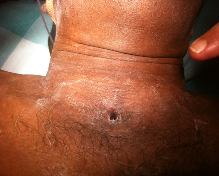
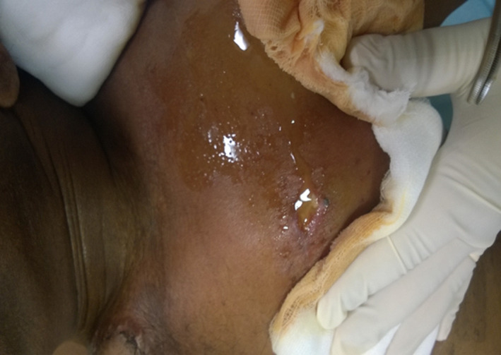
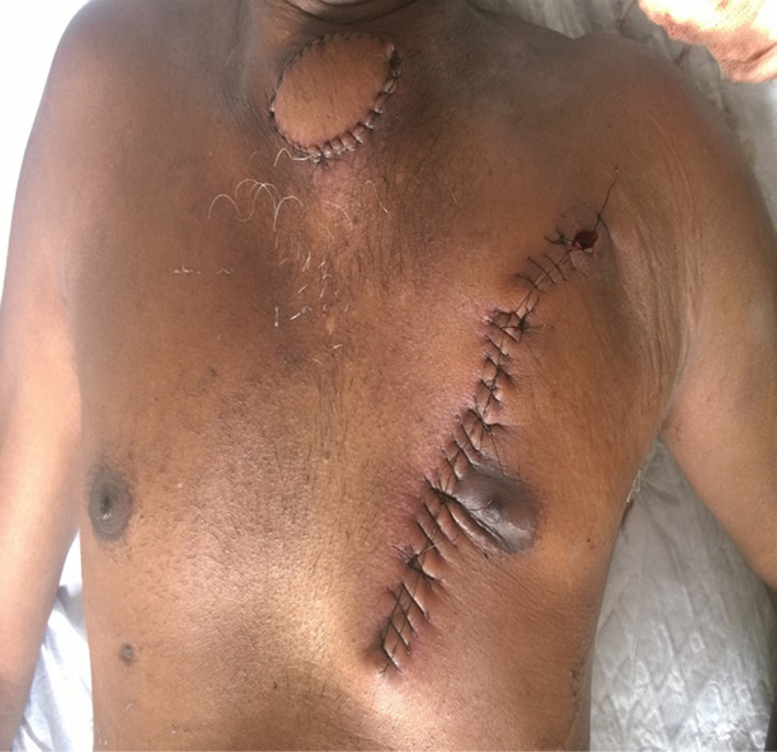
The patient was a 54‐year‐old man who was diagnosed with squamous cell carcinoma of the right vocal cord for which he had undergone microlaryngeal surgery along with tracheostomy for the same in July 2012. He then underwent intensity‐modulated radiotherapy. Since then, the patient has been on tracheostomy tube. Patient was decannulated following laryngoscopic assessment on April 2015. A strapping was done to seal the tracheostome. A month later, on follow‐up, the tracheostome was found to be still patent with an air leak (Figure 1). A secondary suturing was attempted to close the fistula. Despite this attempt, the fistula persisted due to his chronic cough. The patient was then posted for a flap cover to close the fistula. After admission for the same, he developed severe bronchitis for which he was treated medically. A bronchoscopy showed inflamed bronchus, and a bronchoalveolar lavage showed pseudomonas aeruginosa growth for which he was treated with culture sensitive antibiotics and steroids. During this course, he developed steroid‐induced hyperglycemia with a derangement of his renal parameters and was under the diabetologist and nephrologist care. Post recovery and after getting the medical clearance, he underwent a pectoralis major musculocutaneous flap cover in June 2015 (Figure 2). On the first post op day, he had an episode of transient unresponsiveness with stable vitals. He had no chest pain or hypoglycemic episodes. Examination revealed bilateral subcutaneous emphysema over the chest and face. Chest X‐ray showed no signs of pneumothorax. Screening ECHO showed a good LV systolic function with no pericardial effusion or evidence of acute coronary syndrome. Blood test for troponin T was negative. He was closely monitored in the Intensive Care Unit. He also received a unit of packed cells for his drop in hemoglobin post op. He was discharged from the hospital and was advised to follow‐up in the OPD for dressing. After a week from discharge, he came to the OPD with complaints of pain in the surgical donor site of the flap. Examination revealed an abscess at the surgical site which was drained, and the pus was sent for culture which later reported as carbapenem‐resistant enterobacteria (Figure 3). He was treated with IV antibiotics, and daily dressing of the surgical site wound was done. After 10 days, the wound healed well and was later discharged from the hospital.
FIGURE 1.

Tracheostoma
FIGURE 2.

Pus discharge from donor site
FIGURE 3.

Pectoralis major musculocutaneous flap and stoma closure
2. DISCUSSION
A tracheocutaneous fistula is commonly regarded as a pathological complication of temporary tracheostomy that results due to failure of spontaneous tracheostome closure post decannulation. Chronic TCF can impair the quality of life, vocalization, and the need for local hygiene with frequent hospital visits. 1 Although most of these fistulae close spontaneously after decannulation or after local debridement, a significant percentage do not and require some form of surgical closure. 2 Despite its clinical impact, there are few studies in the literature on its risk factors and pathogenesis. The incidence of fistula formation is known to be related to the cannulation time. Kulber et al reported that fistulas do not develop if the cannulation period is less than 16 weeks. 3 Incidences of TCF up to 70% have been reported when tracheostomies were maintained for more than 16 weeks. 3 In this case, the cannulation time exceeded 2 years. Furthermore, other factors such as previous irradiation of neck, previous tracheostomy, and obesity have been suggested to be risk factors for TCF. 4 Chronic cough and aspiration could play an important role favoring the onset of TCF, independent of decannulation timing, and may also influence the surgical failure and relapse rate. In this case, the patient being a known COPD case, the status of the lung functions was already compromised prior to tracheostomy and the same worsened due to the prolonged cannulation time. A multidisciplinary approach is hence needed for cases like this. TCF closure can be challenging since the laryngeal airway can be suboptimal and abnormal increases in subglottic pressure during the expiration phase can be present. 5 , 6 , 7
In conclusion, TCF presents a challenge to the surgeon as different pathogenic factors affect its formation and healing. A multidisciplinary approach is very much in need for a patient with different medical ailments. Proper patient counseling, duration of cannulation, assessment of risk factors, and management of the comorbidities all help in determining the incidence and prognosis of tracheocutaneous fistula.
3. SUMMARY
Tracheocutaneous fistula (TCF) is a serious complication associated with impaired quality of life. However, a successful TCF closure is difficult owing to the high incidence of recurrence. We utilized a pectoralis major musculocutaneous flap for closure of a TCF that occurred after failure of post tracheostomy decannulation. The pectoralis major is the workhorse flap for such reconstructions. It has reliable blood supply from the thoracoacromial vessels. It can provide large amount of tissue, which can fulfill the requirement of mucosal and skin defect. An advantage of musculocutaneous flaps is that the muscle lies in between the two skin paddles, which provides a waterproofing layer that reduces the chances of recurrent fistula. A successful closure of the fistula was achieved with this procedure.
CONFLICT OF INTEREST
The authors whose names are listed above certify that they have NO affiliations with or involvement in any organization or entity with any financial interest and no conflict of interest in the subject matter or materials discussed in this manuscript.
AUTHOR CONTRIBUTIONS
Author 1: Performed study conception, designed the study, collected the data, analyzed and interpreted the results, and prepared manuscripts. Author 2: Prepared the manuscript.
ACKNOWLEDGMENTS
I would like to express my special thanks of gratitude to all my colleagues and for their able guidance and support in completing my case report. Published with written consent of the patient.
Open access funding provided by Qatar National Library.
Chirakkal P, Al Hail ANIH. Tracheocutaneous fistula ‐ A surgical challenge. Clin Case Rep. 2021;9:1771–1773. 10.1002/ccr3.3901
Funding information
Open access funding provided by the Qatar National Library.
DATA AVAILABILITY STATEMENT
The Data that support the findings of this study are available from the corresponding author, upon reasonable request.
REFERENCES
- 1. Berbholz L, Vail S, Berlet A. Management of trachea cutaneous fistula. Arch Otolaryngol Head Neck Surg. 1992;118:869‐871. [DOI] [PubMed] [Google Scholar]
- 2. Eliashar R, Sichel JY, Eliachar I. A new surgical technique for primary closure of long‐term tracheostomy. Otolaryngol Head Neck Surg. 2005;132:15. [DOI] [PubMed] [Google Scholar]
- 3. Kulber H, Passy V. Trachea cutaneous closure and scar revision. Arch Otolaryngol. 1972;96:22‐26. [DOI] [PubMed] [Google Scholar]
- 4. Khaja SF, Fletcher AM, Hoffam HT. Local repair of persistent trachea cutaneous fistula. Ann Otol Rhinol Larnygol. 2001;120:622‐626. [DOI] [PubMed] [Google Scholar]
- 5. De Virgilio A, Fusconi M, Gallo A, et al. The oncological radicality of supracricoid partial laryngectomy with cricohyoidopexy in the treatment of advanced N0–N1 laryngeal squamous cell carcinoma. Laryngoscope. 2012;122:826‐833. [DOI] [PubMed] [Google Scholar]
- 6. Bussu F, Paludetti G, Almadori G, et al. Comparison of total laryngectomy with surgical and non‐surgical organ preservation modalities in advanced Laryngeal squamous cell carcinoma: a multi‐center retrospective analysis. Head Neck. 2013;35:554‐561. [DOI] [PubMed] [Google Scholar]
- 7. De Vincentiis M, De Virgilio A, Bussu F, et al. Oncological results of the surgical salvage of recurrent Laryngeal SCC in a multicentric retrospective series. The emerging role of supracricod partial laryngectomy. Head Neck Surg. 2010;142:873‐878. [DOI] [PubMed] [Google Scholar]
Associated Data
This section collects any data citations, data availability statements, or supplementary materials included in this article.
Data Availability Statement
The Data that support the findings of this study are available from the corresponding author, upon reasonable request.


