



Abstract
Background
Pleural effusion from patients with advanced non‐small cell lung cancer (NSCLC) has been proved valuable for molecular analysis, especially when the tissue sample not available. However, simultaneous detection of multiple driver gene alterations especially the fusions is still challenging.
Methods
In this study, 77 patients with advanced NSCLC and pleural effusion were enrolled, 49 of whom had matched tumor tissues. Supernatants, cell sediments, and cell blocks were prepared from pleural effusion samples for detection of driver alterations by a PCR‐based 9‐gene mutation detection kit.
Results
Mutations in EGFR, KRAS, and HER2 were detected in DNA and cfDNA, fusions in ALK was detected in RNA and cfRNA. Compared with matched tumor tissue, the supernatant showed the highest overall sensitivity (81.3%), with 81.5% for SNV/Indels by cfDNA and 80% for fusions by cfRNA, followed by cell blocks (71.0%) and the cell sediments (66.7%). Within the group of treatment‐naïve patients or malignant cells observed in the cell sediments, supernatant showed higher overall sensitivity (89.5% and 92.3%) with both 100% for fusions.
Conclusions
CfDNA and cfRNA derived from pleural effusion supernatant have been successfully tested with a PCR‐based multigene detection kit. Pleural effusion supernatant seems a preferred material for detection of multigene alterations to guide treatment decision of advanced NSCLC.
Keywords: ALK fusion, cfRNA, NSCLC, PCR, pleural effusion, supernatant
CfDNA and cfRNA derived from pleural effusion supernatant have been successfully tested with a PCR‐based multi‐gene detection kit. Pleural effusion supernatant seems a preferred material for detection of multi‐gene alterations to guide treatment decision of advanced NSCLC.

1. INTRODUCTION
Targeted therapies against driver gene alterations in patients with non‐small cell lung cancer (NSCLC) can significantly improve patient survival over conventional chemotherapy, 1 , 2 , 3 , 4 , 5 , 6 making molecular testing more important in standard clinical care. Today, therapies have been available for multiple actionable targets in NSCLC so that simultaneous detection of multigene alterations including both mutations and fusions has been routine clinical need. With the advancement of detection technology, detection of mutations based on tumor genomic DNA and detection of fusions based on tumor DNA or RNA can be achieved in a one‐shot test. 7 , 8 While tumor tissue sample for molecular profiling is the “gold standard” in clinical practice, it is not always available due to various reasons especially for patients with advanced clinical stage of the disease. 9 Liquid biopsy samples including plasma, pleural effusions, cerebrospinal fluids, urine, and sputum have become promising alternative sample types for molecular test. 10 , 11 , 12 , 13 Peripheral blood cfDNA is regarded as the main type of liquid biopsy sample which has been used as complementary or surrogate sample for detecting driver gene alterations in NSCLC. Blood‐based liquid biopsy has the advantage in overcoming inter/intra tumor heterogeneity for advanced/metastatic NSCLC, while it also has a main shortcoming of low sensitivity in detecting driver gene mutations, especially for detecting fusions due to limited amount of qualified cfRNA. 14 , 15 , 16
In clinic, a significant portion of patients with advanced NSCLC developed pleural effusions, especially when disease relapsed from prior treatments. 17 Pleural effusions contain both floating malignant cells and cell free nucleic acid, which can be collected for diagnosis purposes. 18 Multiple studies have reported that cell blocks derived from pleural effusions can be used to detect driver gene alterations as tumor tissue equivalent samples. 12 , 19 In addition, studies reported that the sensitivity of detecting EGFR mutations in the supernatant of pleural effusion were relatively independent from detectable malignant cells and blood cell contamination in pleural effusions. 20 , 21 , 22 , 23 However, supernatant of pleural effusions for multigene alterations testing with comparison to cell sediments, cell blocks, especially involving fusion detection of using cfRNA has not been fully investigated.
2. METHODS
2.1. Patients and samples
The pleural effusions from 77 patients with advanced NSCLC were collected between September 2019 and May 2020 at the Beijing Chest Hospital, Beijing, China, among which matched tumor tissue samples were available from 49 patients. The information of clinicopathological characteristics of these patients was collected. Each pleural effusion specimen was centrifuged at 3000 g for 10 min and further processed into three types of components: supernatant, cell sediment, and cell block (formalin fixed paraffin embedded sample derived from half of the cell sediment). The supernatants and cell sediments were stored at −80°C and cell blocks were stored at room temperature until use. All the cell sediment specimens underwent cytology evaluation and classified as malignant cells observed or no malignant cell observed. The study was approved by the Ethical and Institutional Review Boards for Human Investigation of the Beijing Chest Hospital (Number: BJXK‐2020–08). The study design and procedure are presented in Figure 1.
FIGURE 1.

Flowchart of the analysis for driver gene alterations. cfDNA, cell free DNA; cfRNA, cell free RNA; NSCLC, non‐small cell lung cancer
2.2. DNA/RNA extraction
Genomic DNA and total RNA were extracted from cell sediment and cell block samples using FFPE DNA/RNA Extraction Kit (Amoy Diagnostics) according to the manufacturer's instructions. The cfDNA and cfRNA were extracted from supernatant samples simultaneously using QIAamp Circulating Nucleic Acid Kit (Qiagen) following manufacturer's instructions (Figure S1).
2.3. Molecular analyses
DNA and RNA were required for next molecular analyses for every sample. The concentration of DNA should be greater than 2 ng/μl, the concentration of RNA should be between 10 and 500 ng/μl. And the OD260/OD280 of DNA and RNA should be between 1.8 and 2.1. The molecular analysis of all samples was carried out by using AmoyDx® Multi‐Gene Mutations Detection Kit (Amoy Diagnostics) according to the manufacturer's instructions. This kit contains DNA‐based mutation detection real‐time PCR assays and RNA‐based fusion detection real‐time PCR assays, which can detect 118 hotspot mutations/fusions in genes of EGFR, KRAS, BRAF, NRAS, HER2, PIK3CA, ALK, ROS1, and RET in a single test 8 (Supplementary Method and Table S1).
2.4. Statistics analysis
Analyses were performed using SPSS (version 19.0). McNemar's test was used to assess the difference of detection rate between pleural effusion samples and matched tumor samples, χ 2 or Fisher's exact tests was used to assess the detection rate between different sample types. p < 0.05 was considered statistically significant (two‐sided). Graphic analysis was performed using GraphPad Prism 8.0 software (GraphPad Software, Inc.).
3. RESULTS
3.1. Patient characteristics
A total of 77 patients with advanced NSCLC who provided pleural effusion specimens were enrolled in this study (Table 1). The median age of these patients was 63.1 years old. Relatively, more patients were male (57.1%), with adenocarcinoma (88.3%), and treatment naive (66.2%), and with distant metastases (50.6%) (Table 1).
TABLE 1.
Clinicopathological characteristics of 77 enrolled patients with advanced NSCLC
| Characteristics (N = 77) | |
|---|---|
| Age | No. of patients (%) |
| Median (range) | 63.1 ± 12.1 |
| Sex | |
| Male | 44 (57.1%) |
| Female | 33 (42.9%) |
| Pathological evaluation of paired tumor samples | |
| Adenocarcinoma | 68 (88.3%) |
| Squamous cell carcinoma | 5 (6.5%) |
| Other non‐small cell lung cancer | 4 (5.2%) |
| Patients’ treatment status | |
| Treatment naive | 51 (66.2%) |
| Relapsed from prior treatment | 22 (28.6%) |
| Unknown | 4 (5.2%) |
| Metastases status | |
| Without metastases | 9 (11.7%) |
| Local | 24 (31.2%) |
| Distant | 39 (50.6%) |
| Unknown | 5 (6.5%) |
3.2. Detection of driver gene alterations in different components of pleural effusion specimens from both treatment‐naïve patients and treatment relapsed patients
Driver gene alterations in EGFR, KRAS, HER2, and ALK were detected from pleural effusion specimens (for the details of alterations detected, see Table S2). Using driver gene alteration status in tumor tissue as reference, the supernatant, cell sediment, and cell block all showed specificity of 100%. The supernatant showed the highest overall sensitivity (81.3%, 95% CI 63.0%–92.1%), followed by cell block (71.0%, 95% CI 51.8%–85.1%), and the cell sediment (66.7%, 95% CI 48.1%–81.4%), although there was no statistically significant difference among the three groups (p = 0.273). For SNV/Indel mutations, supernatant cfDNAs demonstrated the highest sensitivity of 81.5% without significant difference from the tumor tissue samples, but the cell sediment DNAs and cell block DNAs exhibited similar sensitivities (68% vs. 66.7%) with significant difference from the tumor samples. For the detection of ALK fusions, the supernatant showed a sensitivity of 80% (95% CI 29.9%–98.9%) which was similar to that of cell block (83.3%) but superior to cell sediment (66.7%) (Table 2). EGFR T790M mutation was detected in supernatants from four patients with acquired resistance to 1st line EGFR‐TKI, among which only 1 cell block and 1 cell sediment were detected to be T790M positive (Table S3).
TABLE 2.
Concordance of driver gene alteration status between tumor tissue and supernatants, cell sediments, or cell blocks
| Cell block (N = 45) | Cell sediment (N = 48) | Supernatant (N = 47) | ||||
|---|---|---|---|---|---|---|
| Sensitivity | Specificity | Sensitivity | Specificity | Sensitivity | Specificity | |
| SNV/InDels | 68% (46.4%, 84.3%) | 100% (80.0%, 100%) | 66.7% (46.0%, 82.8%) | 100% (80.8%, 100%) | 81.5% (61.3%, 93.0%) | 100% (80.0%, 100%) |
| Fusions | 83.3% (36.5%, 99.1%) | 100% (88.8%, 100%) | 66.7% (24.1%, 94.0%) | 100% (89.6%, 100%) | 80% (29.9%, 98.9%) | 100% (89.6%, 100%) |
| Overall | 71.0% (51.8%, 85.1%) | 100% (73.2%, 100%) | 66.7% (48.1%, 81.4%) | 100% (74.7%, 100%) | 81.3% (63.0%, 92.1%) | 100% (74.7%, 100%) |
The supernatant samples achieved higher overall sensitivity of 89.5% in treatment naïve patients for the detection of mutations and fusions, compared with an overall sensitivity of 69.2% in patients relapsed from prior treatment, although there was no statistically significant difference between the two groups (p = 0.194). Cell block and cell sediment all showed similar overall sensitivities in treatment naïve patients and patients relapsed from prior treatment (70.6% vs. 71.4%, 64.3% vs. 69.2%). In treatment naïve patients, the sensitivities of SNV/Indels detection in cell block, sediment, and supernatant were 64.3%, 68.4%, and 88.2%, respectively, and the sensitivities of fusion detection in the three groups were all 100%. In patients relapsed from prior treatment, the sensitivities of SNV/Indels detection in cell block, sediment, and supernatant were 72.7%, 72.7%, and 70%, respectively, and the sensitivities of fusion detection in the three groups were all 66.7%, 33.3%, and 66.7%, respectively (Table 3).
TABLE 3.
Effects of treatment status on the detection sensitivities of gene alterations in different types of pleural effusion samples
| Cell block (N = 45) | Cell sediment (N = 48) | Supernatant (N = 47) | ||
|---|---|---|---|---|
| SNV/InDels | Treatment naive | 64.3% (35.6%, 86.0%) | 68.4% (43.5%, 86.4%) | 88.2% (62.3%, 97.9%) |
| Relapsed from prior treatment | 72.7% (39.3%, 92.7%) | 72.7% (39.3%, 92.7%) | 70% (35.4%, 91.9%) | |
| Fusions | Treatment naive | 100% (31.0%, 100%) | 100% (31.0%, 100%) | 100% (19.8%, 100%) |
| Relapsed from prior treatment | 66.7% (12.5%, 98.2%) | 33.3% (1.8%, 87.5%) | 66.7% (12.5%, 98.2%) | |
| Overall | Treatment naive | 70.6% (44.0%, 88.6%) | 64.3% (35.6%, 86.0%) | 89.5% (65.5%, 98.2%) |
| Relapsed from prior treatment | 71.4% (42.0%, 90.4%) | 69.2% (38.9%, 89.6%) | 69.2% (38.9%, 89.6%) | |
The false‐negative results of fusion detection were all in supernatants, cell sediments, and cell blocks from patients relapsed from prior TKI‐treatment. The false‐negative results of SNV/Indels detection in supernatant cfDNA were concentrated within patients relapsed from prior TKI treatment (3/5), while the false‐negative result of SNV/Indels detection in cell blocks and cell sediments were concentrated within treatment naïve patients (5/8, 6/9) (Table 3).
3.3. Effects of cytological evaluation results on the sensitivities of testing different types of pleural effusion specimens
In the samples with observed malignant cells, the sensitivities of SNV/Indels detection in cell block genomic DNA, sediment genomic DNA, and supernatant cfDNA were 85.0%, 85.7%, and 91.0%, respectively, and the sensitivities of fusion detection by testing RNAs from cell block and sediment and cfRNA from supernatant were all 100%. The overall sensitivities of testing cell block, sediment and supernatant for all alterations were 88%, 88%, and 92.3% in samples with observed malignant cells. In the samples without malignant cell, 40% sensitivity was achieved from testing supernatant for SNV/Indels detection but no positive result was achieved for all others (Table 4).
TABLE 4.
Effects of cytological evaluation results on the detection sensitivities of gene alterations in supernatants, cell sediments, and cell blocks (tumor tissue as reference)
| Cell block (N = 45) | Cell sediment (N = 48) | Supernatant (N = 47) | ||
|---|---|---|---|---|
| SNV/InDels | Malignant cells observed | 85% (61.1%, 96.0%) | 85.7% (62.6%, 96.2%) | 91.0% (69.4%, 98.4%) |
| No malignant cell | 0% (0%, 53.7%) | 0% (0%, 48.3%) | 40.0% (7.3%, 83.0%) | |
| Fusions | Malignant cells observed | 100% (46.3%, 100%) | 100% (39.6%, 100%) | 100% (39.6%, 100%) |
| No malignant cell | 0% (0%, 94.5%) | 0% (0%, 80.2%) | 0% (0%, 94.5%) | |
| Overall | Malignant cells observed | 88% (67.7%, 96.8%) | 88% (67.7%, 96.8%) | 92.3% (73.4%, 98.7%) |
| No malignant cell | 0% (0%, 48.3%) | 0% (0%, 40.2%) | 33.3% (6.0%, 75.9%) | |
3.4. The capability of molecular analysis for pleural effusion supernatant in patients with tumor tissue unavailable
Among 28 patients with no tissue samples available, 18 mutations (17 SNV/Indels, 1 fusion) could be detected using pleural effusion supernatant. And the detection rate of driver gene mutations in these patients was not significantly different from that of patients who provided the tumor tissue samples (Table 5).
TABLE 5.
Comparison of alteration detection rate between using tumor tissue and using pleural effusion supernatant
| Driver gene alterations detection rate | ||||
|---|---|---|---|---|
| Tumor tissue samples (N = 49) | Pleural effusion supernatants without paired tissue samples (N = 28) | All pleural effusion supernatants (N = 75) | p | |
| SNV/InDels | 28/49 (57.1%) | 17/28 (60.7%) | 39/75 (52%) | 0.802 |
| Fusions | 6/49 (12.2%) | 1/28 (3.6%) | 5/75 (6.7%) | 0.407 |
4. DISCUSSION
Tumor cells in patients with advanced lung cancer infiltrate pleural cavity via the hematogenous, direct, or lymphatic spread, resulting impaired lymphatic drainage and pleural effusion accumulation. 24 Cytological examination of pleural effusion showed that 95% of malignant serous effusions represented metastatic disease, mainly composed of adenocarcinomas (70%–77%). 25 In this study, 88.3% of the enrolled patients with lung adenocarcinoma and 81.8% of the patients with local or distant metastases, two characteristics that were similar to those reported in the literature. In addition, 36.4% (28/77) of the enrolled patients were diagnosed by pleural effusion cytology because tissue samples for diagnosis were not available. Therefore, pleural effusion samples are a valuable supplement for tumor tissue in pathological diagnosis. 26
Molecular testing based on pleural effusion cytology specimens has been widely recommended in clinical guidelines of NSCLC and has become a routine method used in clinical practice. 24 , 27 , 28 The mutation detection rates based on this kind of specimens are affected by tumor content. 21 , 22 Previous studies showed that pleural effusion supernatants contain more abundant tumor derived DNA (ctDNA) than pleural effusion sediments and plasma samples, and had higher detection rate of EGFR driver mutations especially for detection of T790M in EGFR‐TKI relapsed patients. 20 , 22 And several studies also showed that the somatic mutation detection sensitivity in supernatant cfDNA was independent of both tumor cellularity and pleural effusion appearance. 20 , 22 In our study, the SNV/Indels detection rate of supernatant was higher than that of cytology specimens (81.5% vs. 68.0% or 66.7%). A certain percentage of mutations were also detected in the supernatant of pleural effusion samples without malignant cells. However, in the group of patients with malignant cells observed in the pleural effusion, the SNV/Indels detection rate of cell blocks was increased from sensitivity of 68.0% to 85.0%, and the fusion detection rate was increased from 83.3% to 100%. Also, the SNV/Indels detection rate of cell sediment was increased from 65.7% to 85.7%, and the fusion detection rate was increased from 66.7% to 100%. This indicates the necessity of pathological assessment for molecular detection of pleural effusion cytology samples.
For the pleural effusion cfRNA detection, previous study showed that the pleural effusion cfRNA can successfully be extracted and achieving a 100% concordance rate of ALK fusion detection compared with cell blocks by PCR method. 29 However, this study did not obtain matched tissue specimens as reference. Another study also showed the success rates of pleural effusion cell free total nucleic acid extraction and NGS sequencing have been verified to be higher than that of cell blocks by Xiang et al. 22 In this study, we confirmed that fusions could be successfully detected in the cfRNA derived from MPE supernatant by using a PCR‐based method, achieving a concordance rate of 97.9% (46/47) with paired tissue samples. The results further strengthen the statement that the potential of pleural effusion specimens is superior to plasma specimens in detecting fusion variations. 30 , 31 In our study, more false‐negative mutations were detected in the supernatant of pleural effusion in patients who had previously received targeted therapy and progressed, including one patient with false‐negative in detection of ALK fusion. Although more EGFR T790M resistance mutations could be detected in the supernatant of pleural effusion specimens with malignant cells observed, suggesting that analysis may be necessary in combination with cytological evaluation for driver gene testing in patients relapsed from prior TKI treatment.
Use of cytology centrifuged supernatants of endobronchial ultrasound‐guided fine‐needle aspiration (EBUS‐guided FNA) could.improve cost and turnaround time for targeted next generation sequencing reported by Gokozan et al. 32 In our study, the traditional workflow using cell blocks for molecular detection went through at least five steps, the TAT was at least 3 days. If the cell sediment was directly used for molecular detection, the TAT was at least one day. The workflow using supernatants for molecular detection was through three steps before got mutation results, the TAT was only 4 hours, which made obtain mutation results within one day possible in clinical practice. Compared with the direct molecular detection of cell sediments, the decreased TAT by the supernatant detection was mainly due to the omission of cytological evaluation. And compared with the cell block, the reduction in time of supernatant detection came from the omission of cytological evaluation and formalin fixing and paraffin embedding. So supernatant would be the preferred pleural effusion type for molecular detection, when consider the turnaround time and experiment steps (Figure 2).
FIGURE 2.
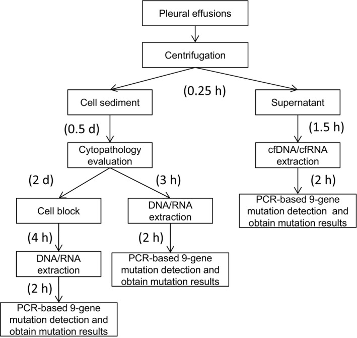
The workflow and TAT of different types of pleural effusion sample for driver gene alterations. cfDNA, cell free DNA; cfRNA, cell free RNA; d, days; h, hours; TAT, turnaround time
There are several limitations in this study. First, the sample size is from a single center and not large enough, especially for the fusion variation positive samples. Further investigation in multiple centers with larger sample size is under planning to validate the results of this work. Second, in this study, we did not compare with matched blood specimens, and follow‐up studies will be conducted to explore the differences in fusion variations between pleural effusion specimens and matched plasma specimens. Third, cfRNA profile in MPE supernatant needs to be further investigated.
In conclusion, our study indicated that supernatant of pleural effusion specimens can achieve cfDNA‐based SNV/Indels and cfRNA‐based fusion variations detection. Compared with cytology specimens, the supernatant has the highest overall detection sensitivity, and the TAT of molecular testing is the shortest, the mutation results can be obtained on the day the pleural effusion is obtained. The entire detection process of pleural effusion supernatant was easy to operate and with short TAT, which was more suitable for clinical application.
5. ETHICS STATEMENT
The study was approved by the Ethical and Institutional Review Boards for Human Investigation of the Beijing Chest Hospital (Number: BJXK‐2020–08).
CONFLICT OF INTEREST
Guanshan Zhu is the stock holder of Amoy Diagnostics.
Supporting information
Supplementary Material
Funding information
This study was funded by National Natural Science Foundation of China (Grant Number: 82072381) Beijing Municipal Science and Technology Project (Grant Numbers: Z191100006619079, Z181100001918027, D181100000418003, Z191100006619078) Beijing Tongzhou High Level Technique Talents Program (Grant Number: YHLD2018006).
DATA AVAILABILITY STATEMENT
Some or all data generated or used during the study are available from the corresponding author by request.
REFERENCES
- 1. Zhou C, Wu Y‐L, Chen G, et al. Erlotinib versus chemotherapy as first‐line treatment for patients with advanced EGFR mutation‐positive non‐small‐cell lung cancer (OPTIMAL, CTONG‐0802): a multicentre, open‐label, randomised, phase 3 study. Lancet Oncol. 2011;12:735‐742. 10.1016/s1470-2045(11)70184-x [DOI] [PubMed] [Google Scholar]
- 2. Solomon BJ, Mok T, Kim DW, et al. First‐line crizotinib versus chemotherapy in ALK‐positive lung cancer. N Engl J Med. 2014;371:2167‐2177. 10.1056/NEJMoa1408440 [DOI] [PubMed] [Google Scholar]
- 3. Janne PA, Yang JC, Kim DW, et al. AZD9291 in EGFR inhibitor‐resistant non‐small‐cell lung cancer. N Engl J Med. 2015;372:1689‐1699. 10.1056/NEJMoa1411817 [DOI] [PubMed] [Google Scholar]
- 4. Shaw AT, Gandhi L, Gadgeel S, et al. Alectinib in ALK‐positive, crizotinib‐resistant, non‐small‐cell lung cancer: a single‐group, multicentre, phase 2 trial. Lancet Oncol. 2016;17:234‐242. 10.1016/s1470-2045(15)00488-x [DOI] [PMC free article] [PubMed] [Google Scholar]
- 5. Yoh K, Seto T, Satouchi M, et al. Vandetanib in patients with previously treated RET‐rearranged advanced non‐small‐cell lung cancer (LURET): an open‐label, multicentre phase 2 trial. Lancet Respir Med. 2017;5:42‐50. 10.1016/s2213-2600(16)30322-8 [DOI] [PubMed] [Google Scholar]
- 6. Li J, Sasane M, Zhang J, et al. Is time to progression associated with post‐progression survival in previously treated metastatic non‐small cell lung cancer with BRAF V600E mutation? A secondary analysis of phase II clinical trial data. BMJ Open. 2018;8:e021642. 10.1136/bmjopen-2018-021642 [DOI] [PMC free article] [PubMed] [Google Scholar]
- 7. Tsoulos N, Papadopoulou E, Metaxa‐Mariatou V, et al. Tumor molecular profiling of NSCLC patients using next generation sequencing. Oncol Rep. 2017;38:3419‐3429. 10.3892/or.2017.6051 [DOI] [PMC free article] [PubMed] [Google Scholar]
- 8. Wang S, Qu X, Cao L, et al. Assessment of nine driver gene mutations in surgically resected samples from patients with non‐small‐cell lung cancer. Cancer Manag Res. 2020;12:4029‐4038. 10.2147/CMAR.S250822 [DOI] [PMC free article] [PubMed] [Google Scholar]
- 9. Fenizia F, Pasquale R, Roma C, Bergantino F, Iannaccone A, Normanno N. Measuring tumor mutation burden in non‐small cell lung cancer: tissue versus liquid biopsy. Transl Lung Cancer Res. 2018;7:668‐677. 10.21037/tlcr.2018.09.23 [DOI] [PMC free article] [PubMed] [Google Scholar]
- 10. Ponti G, Manfredini M, Tomasi A. Non‐blood sources of cell‐free DNA for cancer molecular profiling in clinical pathology and oncology. Crit Rev Oncol Hematol. 2019;141:36‐42. 10.1016/j.critrevonc.2019.06.005 [DOI] [PubMed] [Google Scholar]
- 11. Wang Z, Zhang L, Li L, et al. Sputum cell‐free DNA: valued surrogate sample for detection of EGFR mutation in patients with advanced lung adenocarcinoma. J Mol Diagn. 2020;22:934‐942. 10.1016/j.jmoldx.2020.04.208 [DOI] [PubMed] [Google Scholar]
- 12. Carter J, Miller JA, Feller‐Kopman D, Ettinger D, Sidransky D, Maleki Z. Molecular profiling of malignant pleural effusion in metastatic non‐small‐cell lung carcinoma. The effect of preanalytical factors. Ann Am Thorac Soc. 2017;14:1169‐1176. 10.1513/AnnalsATS.201609-709OC [DOI] [PubMed] [Google Scholar]
- 13. Sands J, Li Q, Hornberger J. Urine circulating‐tumor DNA (ctDNA) detection of acquired EGFR T790M mutation in non‐small‐cell lung cancer: an outcomes and total cost‐of‐care analysis. Lung Cancer. 2017;110:19‐25. 10.1016/j.lungcan.2017.05.014 [DOI] [PubMed] [Google Scholar]
- 14. Yang X, Saito Y, Rao A, et al. Alignment‐free filtering for cfNA fusion fragments. Bioinformatics. 2019;35:i225‐i232. 10.1093/bioinformatics/btz346 [DOI] [PMC free article] [PubMed] [Google Scholar]
- 15. Tong Y, Zhao Z, Liu B, et al. 5′/ 3′ imbalance strategy to detect ALK fusion genes in circulating tumor RNA from patients with non‐small cell lung cancer. J Exp Clin Cancer Res. 2018;37: 10.1186/s13046-018-0735-1 [DOI] [PMC free article] [PubMed] [Google Scholar]
- 16. Cui S, Zhang W, Xiong L, et al. Use of capture‐based next‐generation sequencing to detect ALK fusion in plasma cell‐free DNA of patients with non‐small‐cell lung cancer. Oncotarget. 2017;8:2771‐2780. 10.18632/oncotarget.13741 [DOI] [PMC free article] [PubMed] [Google Scholar]
- 17. Porcel JM, Gasol A, Bielsa S, Civit C, Light RW, Salud A. Clinical features and survival of lung cancer patients with pleural effusions. Respirology. 2015;20:654‐659. 10.1111/resp.12496 [DOI] [PubMed] [Google Scholar]
- 18. Benlloch S, Marti‐Ciriquian JL, Galbis‐Caravajal JM, et al. Cell‐free DNA concentration in pleural fluid and serum: quantitative approach and potential prognostic factor in patients with cancer and pleural effusions. Clin Lung Cancer. 2006;8:140‐145. 10.3816/CLC.2006.n.043 [DOI] [PubMed] [Google Scholar]
- 19. DeMaio A, Clarke JM, Dash R, et al. Yield of malignant pleural effusion for detection of oncogenic driver mutations in lung adenocarcinoma. J Bronchology Interv Pulmonol. 2019;26:96‐101. 10.1097/LBR.0000000000000534 [DOI] [PubMed] [Google Scholar]
- 20. Hummelink K, Muller M, Linders TC, et al. Cell‐free DNA in the supernatant of pleural effusion can be used to detect driver and resistance mutations, and can guide tyrosine kinase inhibitor treatment decisions. ERJ Open Res. 2019;5(1):00016‐2019. 10.1183/23120541.00016-2019 [DOI] [PMC free article] [PubMed] [Google Scholar]
- 21. Tong L, Ding N, Tong X, et al. Tumor‐derived DNA from pleural effusion supernatant as a promising alternative to tumor tissue in genomic profiling of advanced lung cancer. Theranostics. 2019;9:5532‐5541. 10.7150/thno.34070 [DOI] [PMC free article] [PubMed] [Google Scholar]
- 22. Xiang C, Huo M, Ma S, et al. Molecular profiling for supernatants and matched cell pellets of pleural effusions in non‐small‐cell lung cancer. J Mol Diagn. 2020;22:513‐522. 10.1016/j.jmoldx.2020.01.011 [DOI] [PubMed] [Google Scholar]
- 23. Song Z, Wang W, Li M, Liu J, Zhang Y. Cytological‐negative pleural effusion can be an alternative liquid biopsy media for detection of EGFR mutation in NSCLC patients. Lung Cancer. 2019;136:23‐29. 10.1016/j.lungcan.2019.08.004 [DOI] [PubMed] [Google Scholar]
- 24. Skok K, Hladnik G, Grm A, Crnjac A. Malignant pleural effusion and its current management. A review. Medicina (Kaunas). 2019;55:490. 10.3390/medicina55080490 [DOI] [PMC free article] [PubMed] [Google Scholar]
- 25. Lepus CM, Vivero M. Updates in effusion cytology. Surg Pathol Clin. 2018;11:523‐544. 10.1016/j.path.2018.05.003 [DOI] [PubMed] [Google Scholar]
- 26. Cheng F, Wang Q, Zhong D. Value of cell block in the diagnosis of malignant pleural effusion. Zhongguo Fei Ai Za Zhi. 2015;18:652‐655. 10.3779/j.issn.1009-3419.2015.10.09 [DOI] [PMC free article] [PubMed] [Google Scholar]
- 27. Wu YL, Planchard D, Lu S, et al. Pan‐Asian adapted Clinical Practice Guidelines for the management of patients with metastatic non‐small‐cell lung cancer: a CSCO–ESMO initiative endorsed by JSMO, KSMO, MOS, SSO and TOS. Ann Oncol. 2019;30:171‐210. 10.1093/annonc/mdy554 [DOI] [PubMed] [Google Scholar]
- 28. NCCN . NCCN Clinical Practice Guidelines in Oncology (NCCN Guidelines®) Non‐Small Cell Lung Cancer. Fort Washington: NCCN 2020[2020‐2‐11]. DOI: https://www.nccn.org/professionals/physician_gls/default.aspx#nscl.
- 29. Chu M, Zhu Y, Hu J, Zhang Z, Luo M, Ma X. Detection of ALK gene rearrangement in cell‐free RNA from lung cancer malignant pleural effusion. Biomed Res Int. 2020;2020:6124106. 10.1155/2020/6124106 [DOI] [PMC free article] [PubMed] [Google Scholar]
- 30. Hofman P. ALK status assessment with liquid biopsies of lung cancer patients. Cancers (Basel). 2017;9:106. 10.3390/cancers9080106 [DOI] [PMC free article] [PubMed] [Google Scholar]
- 31. Horn L, Whisenant JG, Wakelee H, et al. Monitoring therapeutic response and resistance: analysis of circulating tumor DNA in patients with ALK+ lung cancer. J Thorac Oncol. 2019;14:1901‐1911. 10.1016/j.jtho.2019.08.003 [DOI] [PMC free article] [PubMed] [Google Scholar]
- 32. Gokozan H, Harbhajanka A, Bomeisl P, Michael CW, Sadri N. Use of cytology centrifuged supernatants improves cost and turnaround time for targeted next generation sequencing. Diagn Cytopathol. 2020;48(12):1167‐1172. 10.1002/dc.24548 [DOI] [PubMed] [Google Scholar]
Associated Data
This section collects any data citations, data availability statements, or supplementary materials included in this article.
Supplementary Materials
Supplementary Material
Data Availability Statement
Some or all data generated or used during the study are available from the corresponding author by request.