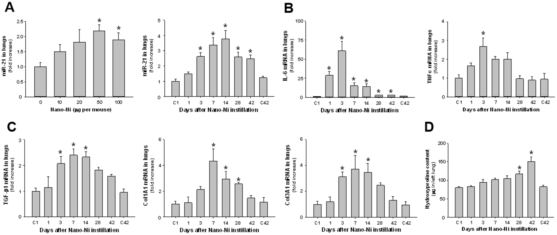
Figure 1.

Upregulation of miR-21 (A), proinflammatory cytokines (B), and fibrosis-related genes (C), and increased hydroxyproline content (D) in mouse lungs after Nano-Ni exposure. In the dose-response study, C57BL/6J mice were instilled intratracheally with 0, 10, 20, 50, or 100 μg per mouse of Nano-Ni, and lung tissues were collected at day 3 after exposure. In the time-response study, C57BL/6J mice were instilled intratracheally with 50 μg per mouse of Nano-Ni, and lung tissues were collected at multiple times after exposure. Control mice were instilled with physiological saline and sacrificed at day 1 (C1) and day 42 (C42) after exposure. The expression of miR-21 (A), proinflammatory cytokines (B), and fibrosis-related genes (C) was analyzed by real-time PCR, normalized to the endogenous control U6 snRNA (A) or β-actin (B-C), and reported as fold increase as compared to the control. The hydroxyproline content was measured in the left lung (D). Data are shown as mean ± SEM (n=4~5). * p<0.05 vs. control.
