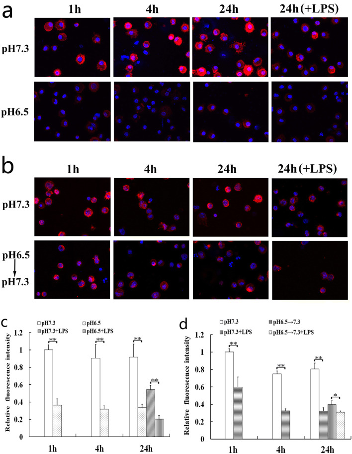
Fig. 3.
Effects of extracellular acidosis on F-actin organization of DCs. Confocal microscopy analysis was performed on DCs to analyze the effects of extracellular acidosis on F-actin organization in the acidic microenvironment (a, c) or after removing it (b, d). a, b The 3-dimensional images from a representative experiment are shown. The magnification used is × 600. c, d Results are expressed as mean fluorescence intensity values and represent the arithmetic mean ± SD of three experiments. The value of the pH 7.3 group at the 1 h after acidosis treatment (c) and removing acidosis (d) was set to 1.0. Statistically significant difference was indicated by bars with asterisks (*p < 0.05; **p < 0.01).

