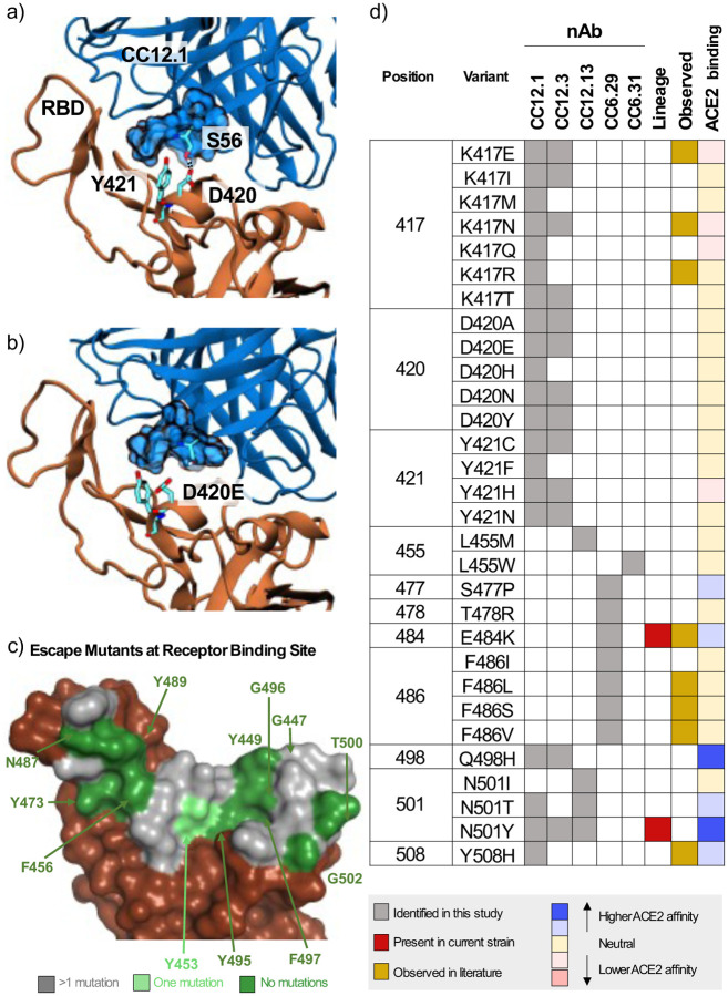
Figure 3.
Mechanistic, structural, and sequence analysis of SARS-CoV-2 escape mutants. a-b) Snapshots from MD trajectories showing a.) key interactions in the control simulation of S RBD in complex with CC12.1, and b.) mechanism of escape of S RBD from CC12.1 due to the D420E mutation. Images were rendered with Visual Molecular Dynamics (VMD (25)), and black dotted lines indicate persistent hydrogen bonds. c.) S RBS positions are colored by the number of escape mutants identified to date. RBS residues involving the S RBD-ACE2 structural complex (PDB ID: 6M0J) are colored by number of escape mutants identified to date. d.) Summary of 1-nt escape mutants identified in the present study. Lineage column indicates presence of the given mutation amongst currently circulating SARS-CoV-2 strains, while the observed column refers to an escape mutant previously identified in literature (11, 19–22). ACE2 binding indicates affinity to ACE2 based on the measurements by Starr et al. (10).

