

A 57-year-old woman with a remote history of jejunoileal bypass surgery, 37 years prior, presented with worsening abdominal bloating and increasing bowel frequency over the past several years. A computed tomography scan of the abdomen was performed, which revealed innumerable fat-containing lesions throughout the colon, consistent with colonic lipomatosis (Figures A and B). Findings suggestive of a nonobstructing intussusception were appreciated in the distal transverse colon (Figure C). A barium enema fluoroscopy study was performed, demonstrating multiple filling defects throughout the colon without evidence of intussusception or obstruction.
Figure.
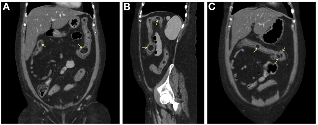
Gastrointestinal lipomas are common and most frequently found in the colon in patients greater than 50 years of age. However, very few reports of such extensive colonic lipomatosis exist in current literature. Although most are found incidentally, lipomas may present with nonspecific symptoms, such as bloating, abdominal pain, or nausea. In rare circumstances, patients may present with acute complications, such as bowel obstruction or intussusception. The mainstay of treatment for symptomatic cases involves local excision; however, most can be managed without surgical intervention. Our patient was managed conservatively, because of extensive colonic involvement, with dietary counseling and had a reduction in symptoms at her 8-week follow-up appointment.
Footnotes
Conflicts of interest
The authors disclose no conflicts.