Abstract
Alzheimer’s disease is associated with the formation of toxic aggregates of amyloid beta (Aβ) peptides. Despite tremendous efforts, our understanding of the molecular mechanisms of aggregation, as well as cofactors that might influence it, remains incomplete. The small cyclic neuropeptide somatostatin-14 (SST14) was recently found to be the most selectively enriched protein in human frontal lobe extracts that binds Aβ42 aggregates. Furthermore, SST14’s presence was also found to promote the formation of toxic Aβ42 oligomers in vitro. In order to elucidate how SST14 influences the onset of Aβ oligomerization, we performed all-atom molecular dynamics simulations of model mixtures of Aβ42 or Aβ40 peptides with SST14 molecules and analyzed the structure and dynamics of early-stage aggregates. For comparison we also analyzed the aggregation of Aβ42 in the presence of arginine vasopressin (AVP), a different cyclic neuropeptide. We observed the formation of self-assembled aggregates containing the Aβ chains and small cyclic peptides in all mixtures of Aβ42–SST14, Aβ42–AVP, and Aβ40–SST14. The Aβ42–SST14 mixtures were found to develop compact, dynamically stable, but small aggregates with the highest exposure of hydrophobic residues to the solvent. Differences in the morphology and dynamics of aggregates that comprise SST14 or AVP appear to reflect distinct (1) regions of the Aβ chains they interact with; (2) propensities to engage in hydrogen bonds with Aβ peptides; and (3) solvent exposures of hydrophilic and hydrophobic groups. The presence of SST14 was found to impede aggregation in the Aβ42–SST14 system despite a high hydrophobicity, producing a stronger “sticky surface” effect in the aggregates at the onset of Aβ42–SST14 oligomerization.
Author summary
Improper folding of proteins causes disorders known as protein misfolding diseases. Under normal conditions most proteins adopt particular folds, which allow them functioning properly. However, for reasons that are not yet fully understood, proteins may misfold and aggregate, forming deposits known as amyloid fibrils, which accumulate in the brain or other tissues. This process affects functioning of the nervous system, gradually causing loss of cognitive abilities. Alzheimer’s disease is one of the most common diseases from this group. A better understanding of the aggregation of peptides implicated in Alzheimer’s disease, known as amyloid beta (Aβ) peptides, may facilitate the development of treatments that ameliorate or prevent the disease. We use detailed molecular dynamics simulations to investigate the influence of somatostatin-14 (SST14), a cyclic neuropeptide that might be involved in the amyloidogenic aggregation of Aβ, on molecular processes occurring at the onset of Aβ aggregation. Results of these simulations explain how the presence of SST14 might alter pathways of aggregation of Aβ, shedding light upon the possible role of extrinsic factors in the aggregation at a molecular level.
Introduction
Alzheimer’s disease (AD) is one of the most devastating neurodegenerative disorders of our time due to its high prevalence in the aging population, the challenges of early diagnosis and the lack of efficient therapeutics. AD is associated with the misfolding and formation of toxic aggregates of the amyloid β (Aβ) peptide [1,2]. It is believed that misfolding in spontaneously formed aggregates of Aβ chains eventually leads to a cascade of self-replicating misfolding events producing β-sheet rich amyloid deposits in the brain. However, mounting evidence suggests that relatively small prefibrillary oligomers rather than mature amyloid fibrils may be the primary toxic assemblies underlying the pathogenesis of the disease [2,3]. Insights into the molecular mechanisms of misfolding and aggregation, as well as co-factors that might influence these processes, remain incomplete.
Contributing to this status quo is a high level of heterogeneity in regards to both the building blocks and the architecture of early oligomeric assemblies. First, the Aβ peptide itself exists in multiple alloforms. Aβ42 comprises two C-terminal residues, I41 and A42, not present in Aβ40. Although the Aβ40 variant is more abundant than Aβ42, the latter is believed to be more pathogenic, primarily on the grounds of a relative increase in Aβ42 in the brain of AD patients, and higher rates of fibrillization in-vitro [4–6]. Second, Aβ oligomers are an ensemble of highly dynamic assemblies [7,8]. Experiments indicate that the initial small aggregates tend to adopt a largely unstructured morphology [9–11]. Quaternary structures without pronounced alignments of peptide chains were also predicted by molecular dynamics (MD) simulations for dimers of Aβ40 and Aβ42 [12–14] and larger multimeric Aβ aggregates [15,16]. Characterizations by a variety of biophysical methods in vitro suggest that initially unstructured oligomers may either undergo a transition into more ordered β-sheet-rich conformations, or mediate formation of β-sheet-rich assemblies [9–12]. However, the majority of early non-fibrillary aggregates dissociate into monomers rather than convert into fibrils [7,8], albeit they are potentially toxic [8,11]. Neither the detailed molecular mechanisms of aggregation, nor the accompanying misfolding or the specific structures and morphologies of the various non-fibrillary and pre-fibrillary assemblies are sufficiently understood. On theoretical grounds, it has been inferred that the aggregation process is driven by a competition of hydrophobic collapse and hydrogen bonding, with the former favoring unstructured conformations, and the latter giving rise to β-sheets [17–19]. The toxicity of non-fibrillary and pre-fibrillary oligomeric assemblies has been hypothetically linked to the exposure of hydrophobic groups and unpaired β-strands at their surfaces [9,20], often referred to as a “sticky surface” effect. Recent modeling studies [21,22] indicate that accumulation of subtle structural perturbations of early aggregates at the onset of the oligomerization process may result in distinct aggregation pathways at later, more advanced stages of Aβ oligomerization. Altogether, both experimental and theoretical evidence suggests that molecular events occurring early in the process of aggregation play a key role in determining both the structure and toxicity of Aβ oligomers [14].
The potential role of extrinsic factors in the aggregation and amyloidogenic conversion is a relatively under-explored aspect of immense importance for both the basic understanding of the aggregation process and the rational design of therapeutic strategies [23,24]. In particular, toxic Aβ*56 complexes have been hypothesized to require unknown cofactors for their assembly [25]. The cyclic neuropeptide somatostatin-14 (SST14) was reported to promote the proteolytic degradation of Aβ through neprilysin induction [26]. The level of SST14 in the brain decreases with ageing [26] and an accelerated decline is found in AD patients [27], potentially leading to an increase in steady-state Aβ levels. Recent experiments indicate that the cyclic neuropeptide somatostatin-14 (SST14) is the most selectively enriched peptide in human frontal lobe extracts that binds oligomeric Aβ42 aggregates [28]. Moreover, SST14’s presence was found to inhibit fibrillization of Aβ42 in vitro, while promoting formation of smaller oligomers, reminiscent of toxic Aβ*56 complexes [28,29]. Interestingly, Aβ42 but not Aβ40 peptides were prone to delayed fibrillization under the influence of SST14. This effect was also not observed upon replacement of SST14 with other cyclic neuropeptides, such as arginine vasopressin (AVP) [29]. Another recent biochemical study [30] investigated binding of SST14 to a specific membrane-associated Aβ42 tetramer, termed βPFOAβ(1–42). This report validated the ability of SST14 to selectively interact only with oligomeric assemblies of Aβ42. Consistent with previous data gathered with soluble Aβ42 oligomers [29], the authors reported that the binding interface between SST14 with βPFOAβ(1–42) may involve a central tryptophan (W8) within SST14. Whereas prior experiments [29] with soluble Aβ42 oligomers had pointed toward a contribution of the N-terminal half to the SST14 binding, the interactions with the membrane associated βPFOAβ(1–42) oligomer seems to primarily rely on residues 18–20 within the C-terminal half of Aβ42. Although at first glance these results may seem contradictory, the N-terminal half of Aβ42 is required for the formation of βPFOAβ(1–42), an observation that would have precluded the ability to detect binding of SST14 in assays that relied on truncated Aβ18–42 in prior binding studies. Both studies agreed that binding occurs predominantly to Aβ42 oligomers and is not observed with oligomers formed from Aβ40. Because analyses of [30] were restricted to a highly purified membrane-associated βPFOAβ(1–42), it remains unclear whether an alternative binding pose or binding stoichiometry would be available in soluble Aβ42 oligomers. Consistent with such a scenario, the binding constant for binding of SST14 to βPFOAβ(1–42) was approximately threefold higher than the previously observed Kd for binding of SST14 to soluble Aβ42 oligomers.
In order to elucidate how SST14 might influence the onset of Aβ oligomerization, we have performed all-atom molecular dynamics (MD) simulations of model systems containing mixtures of Aβ42 or Aβ40 monomeric peptides and SST14 molecules in explicit water. We also performed similar simulations for mixtures of Aβ42 peptides and AVP molecules as negative controls. We investigated early stages of aggregation in each system, and analyzed the structure and dynamics of the early self-assembled aggregates. For this purpose, we combined well-established structural analysis tools with a novel essential collective dynamics (ECD) method developed in our group [14,16], which allows to accurately identify persistent correlations of motion (dynamics) in a molecule or supramolecular system without exhaustive conformational sampling, based on an original fundamental concept [31,32]. The method allows analysing dynamics correlations between selected pairs of atoms; characterising main-chain flexibilities; and identifying domains of correlated motion within the same framework. The presence of SST14 was found to impede the aggregation in the Aβ42–SST14 mixtures despite having a high hydrophobicity. We attribute differences in the structures and dynamics of the Aβ42–SST14, Aβ42–AVP and Aβ40–SST14 systems to distinct tendencies of SST14 and AVP to (1) interact with specific regions of the Aβ chains; (2) develop hydrogen bonds with Aβ peptides; and (3) expose hydrophilic or hydrophobic groups to the solvent.
Results
Aggregation of Aβ peptides in the presence of small cyclic peptides
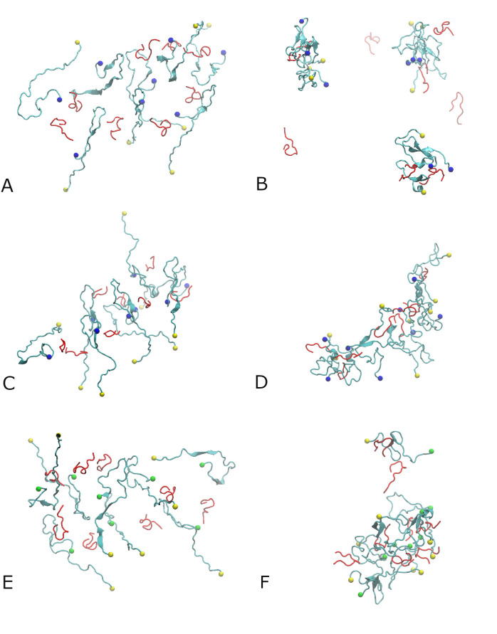
Initially, eight Aβ42 or Aβ40 chains and eight SST14 or AVP molecules were placed in the simulation box in random positions, as outlined in the Methods section. In each system, the Aβ peptide chains are labelled A, B, C,…H, and the small cyclic peptides are labelled I, J, K,…P. Three independent 500 ns long MD simulations were conducted for each of the Aβ42-SST14, Aβ42-AVP and Aβ40-SST14 systems in explicit OPC water. To distinguish between the three trajectories, each of them were numbered by I, II, and III. Unless otherwise stated, the examples displayed below originate from trajectories Aβ42-SST14-I, Aβ42-AVP-II, and Aβ40-SST14-II. These trajectories were selected as representative examples of leading trends for each of the systems. Fig 1 shows the configurations for the three representative systems after the equilibration (see Methods), and after 500 ns production MD simulations. Additional details on the structures obtained in these trajectories can be found in S1 and S2 Figs in the Supporting Information. Table 1 summarizes the aggregation status in all nine systems after 500 ns MD simulations.
Fig 1.
Eight Aβ42 monomers and eight SST14 molecules in water after equilibration (A), and the aggregates formed after 500 ns of MD simulation for system Aβ42-SST14-I (B). Eight Aβ42 monomers and eight AVP molecules in water after equilibration (C), and the octamer formed after 500 ns of MD simulation for system Aβ42-AVP-II (D). Eight Aβ40 monomers and SST14 molecules in water after equilibration (E), and the heptamer formed after 500 ns of MD simulation for system Aβ40-SST14-II (F). Aβ42 and Aβ40 chains are colored cyan, SST14 and AVP molecules are colored red. The C-termini of the Aβ42 chains are indicated by blue spheres and those of Aβ40 chains are shown as green spheres. The N-termini of Aβ42 and Aβ40 chains are indicated by yellow spheres.
Table 1. Aggregation status in all trajectories after 500 ns MD simulations.
| System | Aβ42-SST14 | Aβ42-AVP | Aβ40-SST14 |
|---|---|---|---|
| I | 3Aβ42+2SST14; 3Aβ42+1SST14; 2Aβ42+2SST14; 3 free SST14. |
6Aβ42+6AVP; 2Aβ42+2AVP. |
7Aβ40+6SST14; 1Aβ40+1SST14; 1 free SST14. |
| II | 4Aβ42+5SST14; 3Aβ42+2SST14; 1 free Aβ42; 1 free SST14. |
8Aβ42+8AVP. | 7Aβ40+6SST14; 1Aβ40+2SST14 |
| III | 5Aβ42+3SST14; 3Aβ42+3SST14; 2 free SST14. |
6Aβ42+7AVP; 2Aβ42+1AVP. |
7Aβ40+6SST14; 1 free Aβ40; 1 free SST14. |
After equilibration of the Aβ42-SST14 system, Aβ chains were still relatively sparsely positioned, as shown in Fig 1A. After the 500 ns long production MD simulations, all three trajectories for this Aβ42-SST14 system developed self-assembled aggregates. Most aggregates contained two or three Aβ42 chains and one or two SST14 molecules, as illustrated in Figs 1B and S2A for system Aβ42-SST14-I. Somewhat larger aggregates were observed occasionally along with dimers and trimers. In system Aβ42-SST14-II four Aβ42 chains and five SST14 molecules formed an aggregate, and in system Aβ42-SST14-III five Aβ42 chains and three SST14 molecules formed an aggregate (Table 1). The SST molecules tend to attach to the surface of the Aβ42 oligomers such that their N- and C- terminal arms face the solvent.
When the eight SST14 molecules were replaced by AVP molecules, the starting configurations for the Aβ42-AVP system after equilibration also exhibited sparse positioning of the chains (Fig 1C). However, the final structures from the three trajectories comprise large aggregates composed of mixed Aβ42 and AVP (Table 1). The final configuration for system Aβ42-AVP-II is shown in Figs 1D and S2B. In this case, the largest aggregate is an octamer containing all eight Aβ42 chains and eight AVP molecules. In each of the other two Aβ42-AVP trajectories Aβ42 peptides formed a hexamer and a dimer. The hexamers contained 6 or 7 AVP molecules and at least one AVP was bound to each of the Aβ42 dimers.
For the Aβ40-SST14 system containing eight Aβ40 chains and eight SST14 molecules, all three trajectories developed aggregates composed of seven Aβ peptides and six SST14 chains. One Aβ40 chain remained free in each of the trajectories (Table 1). Fig 1E shows a representative configuration in system Aβ40-SST14-II after the equilibration, and Figs 1F and S2C depict this system after 500 ns of MD simulation. In this case, seven Aβ40 and six SST14 chains formed a curved semi-hollow structured oligomer. Six SST14 molecules are inserted inside the aggregate, whereas two SST14 are attached to the remaining free Aβ40 chain preventing its attachment to the larger aggregate.
Due to the putative importance of the C-terminal residues in defining the aggregation behaviour of Aβ peptides [4,19], the locations of the Aβ42 and Aβ40 C-termini are marked, respectively, by blue and green spheres in Fig 1. From Fig 1B and 1D it appears that the C-termini of Aβ42 peptides (blue spheres) tend to remain at the surface of the self-assembled aggregates in both the Aβ42-SST14–I and Aβ42-AVP-II systems. In the Aβ40-SST14–II system, the C-termini of several Aβ40 chains were buried inside the aggregate (Fig 1F). The N-termini of both Aβ42 and Aβ40 chains tend to remain at the surface of the oligomers in all systems.
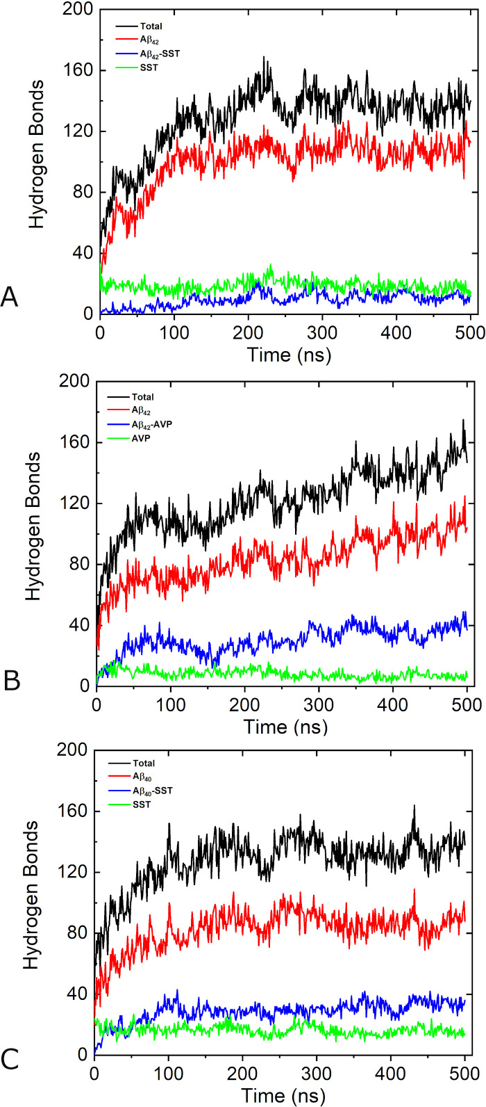
The aggregation process is accompanied by a build-up of hydrogen bonds (HBs) in each system. Fig 2 shows the numbers of hydrogen bonds between and across all chains as functions of MD simulation time in systems Aβ42-SST14-I, Aβ42-AVP-II, and Aβ40-SST14-II. Additional details can be found in S1 Table, which lists average numbers of bonds in all 9 systems during the first and the last 20 ns of the 500 ns long simulations. In Fig 2 the black lines show how the total number of HBs between all chains evolved during the MD simulations. An increase in total HB number is observed in all systems. Overall, most HBs are formed over the first 200–250 ns of production MD simulations in all systems considered. An initial stage of fast build-up of hydrogen bonding ends at approximately 150–200 ns of the production MD simulation; then the build-up slows down although the number of HBs fluctuates considerably during subsequent simulations. In the system Aβ42-AVP-II a persistent increase in the total number of HBs continued throughout the entire simulation. By 500 ns this system developed more HBs than the other two systems, presumably due to the large size of the aggregate. As can be seen in S1 Table, the average number of HBs over three Aβ42-AVP trajectories at the end of the simulations was greater than similar averages in the other two systems as well.
Fig 2.
Number of hydrogen bonds for systems Aβ42-SST14-I (A), Aβ42-AVP-II (B), and Aβ40-SST14-II (C) during 500 ns production MD simulations. Hydrogen bonds between and across all chains are shown by black lines, hydrogen bonds between Aβ chains are shown by red lines, hydrogen bonds across Aβ chains and small cyclic peptides (SST14 or AVP) are shown by blue lines, and those between small cyclic peptides are shown by green lines.
In order to provide more detailed information on the hydrogen bonding, we analyzed the number of HBs between and across Aβ chains and the small cyclic peptides separately. In Fig 2, red lines represent the evolution of the number of HBs between Aβ chains in the three representative systems. As is evident from the figure, Aβ-Aβ bonds contribute a significant portion (60–80%) of the total number of HBs, and their time dependencies resemble those of the total number of HBs. According to S1 Table, at the beginning of the simulations the average numbers of Aβ-Aβ bonds were close in Aβ42-SST14 and Aβ42-AVP systems (49 and 52, respectively), and slightly above than in the Aβ40-SST14 system (44). In the course of the simulations, the average numbers of Aβ-Aβ bonds increased approximately twice in each of the systems.
The most dramatic differences were observed for hydrogen bonding across Aβ chains and small cyclic peptides (blue lines in Fig 2), and between small cyclic peptides (green lines in Fig 2). After equilibration and early into the production runs Aβ-Aβ, SST14-SST14, and AVP-AVP bonds were almost exclusively intra-molecular, due to distances between the chains in the initial structures. Examples illustrating this can be found in S1 Fig. During the first 20 ns after equilibrations, approximately 20 SST14-SST14 bonds in average were formed for each of two somatostatin-containing systems (S1 Table). In striking contrast, only ~9 AVP-AVP bonds have developed. In the course of the simulations the number of SST14-SST14 bonds changed only slightly adopting average values of 23 (for Aβ42-SST14 system) and 17 (for Aβ40-SST14 system) over the last 20 ns. At the same time, the average number of AVP-AVP bonds decreased barely remaining above ~7 over the last 20 ns. Such a large difference cannot be explained by different numbers of HB acceptor sites in SST14 and AVP (which are 24 and 16, respectively) and rather indicates differences in bonding behaviours of SST14 and AVP. As can be seen in S1 Table, during the first 20 ns of production simulations when the small cyclic peptides just started binding to Aβ chains, the average values of cross-species hydrogen bonds were in the proportion of Aβ42-AVP > Aβ40-SST14 > Aβ42-SST14. More specifically, the greatest average number of 12 cross-species bonds was found in Aβ42-AVP system, and the smallest average number of 5 cross-species bonds was observed in Aβ42-SST14 system. During the simulations the number of cross-species bonds increased by 3–5 times in each simulation, and exhibited a significant variability across individual trajectories (S1 Table and S3A Fig). However, the average number of Aβ42-AVP bonds remained approximately 2.4 times greater than the number of Aβ42-SST14 bonds. Similarly to the other two systems, the number of Aβ40-SST14 bonds varied across trajectories. In system Aβ40-SST14-II, for instance, the number of cross-species bonds is close to that in system Aβ42-AVP-II over significant parts of the two trajectories (S3B Fig). However, the average number of Aβ40-SST14 bonds is less than that of Aβ42-AVP (S1 Table and S3A Fig). Overall, regardless of the size and morphology of these aggregates, SST and Aβ tend forming less HBs than AVP and Aβ. This explains the large total number of hydrogen bonds in AVP-containing systems.
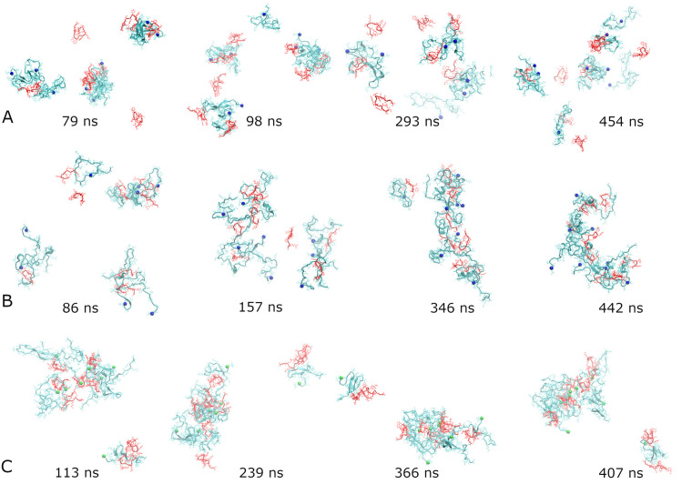
In order to better understand how fluctuations in the number of hydrogen bonds relate to the morphologies, we examined the structures adopted by the three representative systems at several local maxima and minima of the cross-species HBs’ dependencies on time. These time dependencies are shown by blue lines in Fig 2. In addition, S3B Fig depicts the three time dependencies in one plot with solid dots indicating where the snapshots were taken. The corresponding structures are presented in Fig 3. The four snapshots for system Aβ42-SST14-I in Fig 3A illustrate an overall trend towards a greater compactness of several small aggregates. This is accompanied by transient detachment-adsorption of individual Aβ42 and SST14 chains, which appears to contribute to the observed fluctuations of the hydrogen bonding. For example, the structure at local minimum of 98 ns exhibits a monomeric (“free”) Aβ42 chain, three free SST14 molecules, and one SST14 molecule semi-detached from a small aggregate. Clearly there are more free chains than for 293 ns, where a maximum of HBs is observed. However, the local maximum of hydrogen bonding at 79 ns seems to exhibit a sparser morphology than the local minimum at 454 ns. We attribute this to the dynamic nature of the small aggregates and their propensity to restructure, which emerged as another factor contributing to the fluctuations. In the Aβ42-AVP system (Fig 3B) small aggregates self-assembled early in the simulations. The local maximum of hydrogen bonding at 86 ns already exhibits four aggregates and only one free AVP molecule. This is followed by a local minimum at 157 ns, where we observe a large but sparse aggregate, two smaller ones, and one free AVP. A significant decrease in the number of HBs can be attributed to the ongoing change in the aggregation status. Two aggregates observed at 346 ns are significantly more compact, and they exhibit a pronounced increase in hydrogen bonding. By 442 ns the two aggregates have merged into one C-shaped oligomer; however, the accompanying restructuring resulted in a loss in hydrogen bonding. The system Aβ40-SST14-II (Fig 3C) quickly developed two aggregates without any free chains resulting in a maximum in HB at 113 ns. Although the gross aggregation status was retained throughout the course of the simulation, the subsequent ups and downs of the hydrogen bonding suggest continuing restructuring of the aggregates.
Fig 3.
Snapshots of representative systems Aβ42-SST14-I (A), Aβ42-AVP-II (B), and Aβ40-SST14-II (C) corresponding to maxima or minima of the number of hydrogen bonds across Aβ chains and small cyclic peptides. In (A) maxima occur at 79 ns and 293 ns, and minima occur at 98 ns and 454 ns; in (B) maxima occur at 86 ns and 346 ns, and minima occur at 157 ns and 442 ns; and in (C) maxima occur at 113 ns and 366 ns, and minima occur at 239 ns and 407 ns, as illustrated in S3B Fig. The color code for peptides and small cyclic peptides is as in Fig 1. C-termini of Aβ42/40 chains are indicated by blue/green spheres.
After the 500 ns long MD simulations, the secondary structure of the self-assembled aggregates contains predominantly random coils and turns, as depicted in Fig 1. However, stable β-sheets are also observed. S4 Fig shows the secondary structure evolution over the 500 ns of three representative trajectories, and Table 2 lists percentages of stable β-sheet content in all 9 trajectories. The corresponding time dependencies of β-sheet content are given in S5 Fig. The greatest β-sheet content is found in two SST14–containing systems with a proportion of Aβ40-SST14 > Aβ42-SST14 > Aβ42-AVP in average over each set of three simulations (Table 2). In most cases we observe anti-parallel, intramolecular β-sheets in a hairpin conformation of the chain, often involving Aβ C-terminal residues 37–40 (in Aβ42 systems) or 36–39 (in Aβ40 system) as part of the β-sheets. Regardless of the system, most inter-chain β-sheets involve Aβ residues from the central region between positions 23 and 35, although specific locations of β-strands may vary. In several simulations (Aβ42-SST14-II and III, and Aβ40-SST14-II and III) β-strands from this region formed β-sheets with N-terminal residues of a different Aβ chain. The smaller SST14 and AVP molecules appeared less prone to contribute to the stable β-sheet content (see S4 Fig), although we have observed β-sheets involving them in eight out of nine trajectories. Populations of β-bridges and α-helices in all 9 trajectories are illustrated in S5 Fig. The corresponding averages are listed in S2 and S3 Tables. During the last 20 ns of the simulations, β-bridges are observed with average populations of approximately 4%, and α-/3-π-helices are observed with average populations of 1.5–3%.
Table 2. Stable β-sheet percentages in all trajectories averaged over the last 20 ns of the 500 ns MD simulations, their averages over three trajectories for each system, and standard deviations of the data.
| System | Aβ42-SST14 | Aβ42-AVP | Aβ40-SST14 |
|---|---|---|---|
| I | 11.54 | 8.16 | 2.25 |
| II | 4.53 | 2.12 | 12.00 |
| III | 4.39 | 4.70 | 8.83 |
| Average | 6.82 | 5.00 | 7.69 |
| Standard Deviation | 4.09 | 3.03 | 4.97 |
ECD correlations of motion in self-assembled aggregates
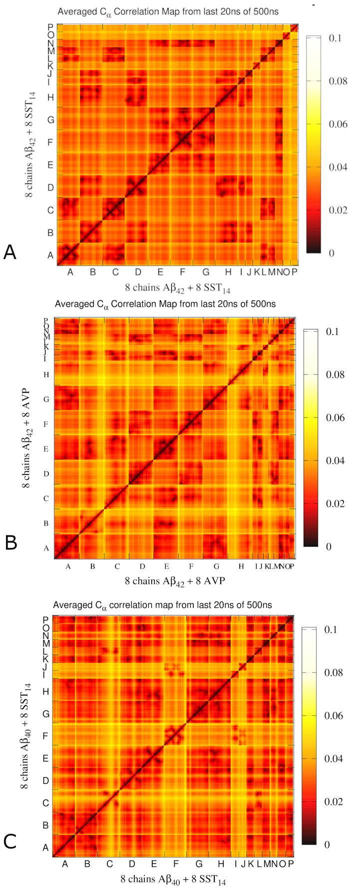
To probe collective motions of the Aβ42-SST14, Aβ42-AVP and Aβ40-SST14 systems, essential collective dynamics analyses (ECD) [16,31–35] have been performed. In the ECD method, principal eigenvectors of the covariance matrix calculated from MD trajectories are employed to characterize correlations of motion (dynamics) between atoms, as outlined in the Methods. Importantly, the method allows to reliably identify stable dynamical correlations without requiring exhaustive conformational sampling or convergence to equilibrium [31,32]. Fig 4 depicts ECD pair correlation maps [16,33,35] calculated for Cα atoms of all the chains from multiple 0.2 ns long segments of the MD trajectories and averaged over the last 20 ns of the simulations. To clarify the relation between pair correlations and morphologies, S6 Fig illustrates the three systems with all Aβ annotated similarly as in Fig 4.
Fig 4.
ECD Cα pair correlation maps for systems Aβ42-SST14-I (A), Aβ42-AVP-II (B) and Aβ40-SST14-II (C) averaged over the last 20 ns of 500 ns long MD trajectories. Stronger correlations of motion between Cα atoms are shown gradually by black, red, and orange colors, and weaker correlations are shown with white and yellow colors.
For the system Aβ42-SST14-I, Aβ chains that exhibit strong inter-chain correlations in Fig 4A are involved in three oligomers: a dimer composed of Aβ chains A and C, which also contains SST14 molecules L and M; a trimer composed of Aβ chains B, D, and H, and SST14 molecules I and J; and a trimer composed of Aβ chains E, F, and G, and SST14 molecule N. Two pairs of Aβ chains, B-H and F-G, developed anti-parallel inter-chain β-sheets, whereas chains A, C, and F each developed intra-chain anti-parallel β-sheets. In addition, extensive regions of Aβ chains E, F, and G have wrapped around SST14 chain N and developed strong inter-molecular correlations with it. However, this interaction was not accompanied by a buildup of β-sheets (S6A Fig). The pair correlation map of the Aβ42-AVP-II system is shown in Fig 4B. Consistent with the structure depicted in S6B Fig, most Aβ chains are involved in strong inter-chain correlations of motion. The exceptions are semi-detached chains B and H, large parts of which exhibit a relatively independent motion from other Aβ units. The strongest inter-chain correlations are observed across Aβ chains A-E-G and D-F, which form two sides of a dumbbell-shaped aggregate. In addition, chain E is strongly correlated with C. These two chains connect two sides of the aggregate, and form a parallel inter-chain β-sheet in the region of the connection (S6B Fig). Each AVP molecule shows pronounced correlations with several Aβ chains, inter-connecting Aβ units in the aggregate. Fig 4C shows a pair correlation map for the Aβ40-SST14-II system. In this case, strong correlations are observed across six Aβ chains A, B, D, E, G, and H which have formed a large aggregate. The seventh chain, C, is correlated with the other six only through its N-terminal part due to its location at the periphery of the aggregate (S6C Fig). The C-terminus of chain B developed an inter-chain anti-parallel β-sheet with chain A, whereas the N-terminus of chain B formed an intra-chain anti-parallel β-sheet with its central region. Six SST14 molecules K, L, M, N, O and P are strongly correlated with the entire aggregate. Remarkably, these Aβ and SST14 chains exhibit inter-molecular correlations regardless of their proximity within the tertiary structure. The other two SST14 molecules, I and J, are correlated with the single detached chain F.
The dynamics correlations identified in the ECD framework can also be visualized in the form of domains of correlated motion [16,31,32], which represent relatively rigid parts of the system consisting of atoms moving coherently. Fig 5 shows the largest domains of correlated motion color-mapped onto the secondary structure of self-assembled aggregates in the three systems. The domains are colored blue, red, green, yellow, etc. in the order of decreasing size. In the Aβ42-SST14–I system shown in Fig 5A, the three largest domains of correlated motion colored blue, red, and green are located in central parts of the three aggregates. These domains include both Aβ and SST14 chains that are strongly inter-correlated in Fig 4A, and contain most of the β-structure. Peripheral regions, which often include termini, tend to exhibit relatively independent dynamics. In the case of the Aβ42–AVP-II system shown in Fig 5B we observe three large domains as well; however, all three are included in a single dumbbell-shaped aggregate. The domain colored blue contains Aβ chains A and G and AVP chains N and O, whereas the domain colored green consists mainly of Aβ chain F and AVP chain I. These two domains are located at the core of two sides of the “dumbbell”, which are held together through the mediation of the third domain (colored red), which includes parts of Aβ chains C and E along with AVP molecule J. In the Aβ40-SST14-II system depicted in Fig 5C, the largest domain of correlated motion (colored blue) is located in the aggregate and consists primarily of Aβ chains A, B, E, G, and H; SST14 chains M, N, O, and P; and parts of several other chains. This domain contains most β-sheets of the aggregate. The second largest domain (colored red) contains the C-terminus of Aβ chain C and a part of SST14 chain L.
Fig 5.
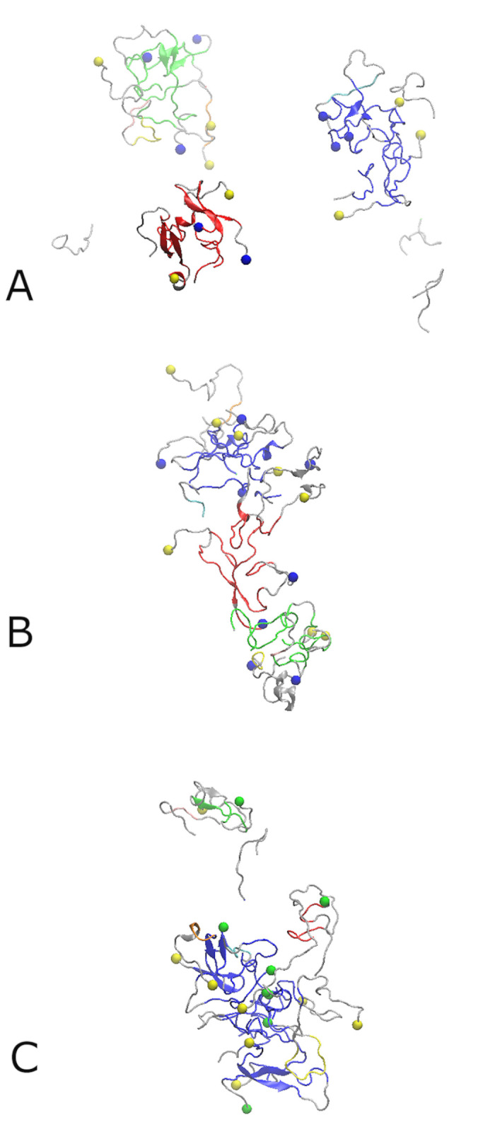
The largest ECD domains of correlated motion identified in the aggregates formed by the Aβ42-SST14–I system (A), the Aβ42–AVP-II system (B), and the Aβ40-SST14–II system (C). The dynamics domains are colored blue, red, green, yellow, cyan, orange, and pink in the order of decreasing size. Off-domain regions are colored gray. Blue and green spheres denote the C-termini of Aβ42 and Aβ40 chains, respectively. Yellow spheres denote the N-termini of the Aβ chains.
Main-chain flexibility profiles
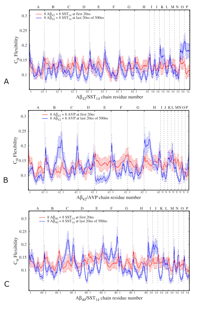
Within the same ECD framework, main-chain flexibility profiles [16,33–35] can be calculated for all the peptides in each system. High levels of the flexibility descriptor usually correspond to flexible loops and termini, whereas minima indicate more restrained regions. The main-chain flexibility profiles averaged over the first and the last 20 ns of the MD simulations for the Aβ42-SST14, Aβ42-AVP and Aβ40-SST14 systems are shown in Fig 6.
Fig 6.
ECD main chain flexibility profiles of the Aβ42-SST14–I system (A), Aβ42–AVP-II system (B) and Aβ40-SST14-II system (C). Red and blue lines indicate, respectively, the average flexibilities for the first 20 ns and the last 20 ns of the production MD simulations. The labels on top of the horizontal axis denote the chains, and those at the bottom of the horizontal axis denote the residue numbers for each chain.
In the Aβ42-SST14 system, most Aβ chains show a decreased flexibility at the last 20 ns of production MD simulation compared to the first 20 ns (Fig 6A), as one could expect since oligomerization limits mobility of the chains. The flexibility profiles of individual Aβ chains tend to adopt oscillating shapes with several maxima and minima. Although by the end of the simulations N- and C-terminal ends of the chains remain flexible, deep minima developed within 10–12 residues from one or both of the termini in most chains. This is especially the case for chains B, D, and H, which have formed a trimeric self-assembled aggregate (S6A Fig). In central regions of the Aβ chains the flexibility profiles are often M-shaped with two maxima separated by a minimum (chains B, C, and E). However in chains A, D, and G the central regions exhibit flexible loops located at the periphery of the respective aggregates. Minima of main-chain flexibility often coincide with locations of stable β-strands. In particular, this is the case for the flexibility minima in chains A, B, C, F, G and H, where stable β-strands are found (see S6A Fig). However, not all flexibility minima are necessarily associated with the presence of β-structure. For example, we do not observe stable β-sheet content in chains D and E, which exhibit pronounced minima of main-chain flexibility. A trend of adopting lower main-chain flexibility in central regions in comparison to the N- and C-termini is observed in most SST molecules in the Aβ42-SST14 system. Moreover, at the end of the simulation the central regions of the SST molecules I, J, L, and M exhibit comparable flexibilities with parts of the Aβ chains B, D, H, A, and C, with which they directly interact. Interestingly, the SST molecules K, O, and P adopted a higher flexibility during the last 20 ns in comparison to the beginning of the simulation. An analysis of the trajectory indicated that initially these molecules were surrounded by neighbouring Aβ42 chains (S1A Fig), which somewhat limited their motion. By the end of the simulations the Aβ42 chains and other SST14 molecules formed three aggregates, whereas the molecules K, O, and P remained free. Eventually, remaining in solution resulted in a less constrained motion for these SST molecules by the end of the simulation.
Fig 6B depicts the main-chain flexibility profile for the Aβ42-AVP system. After the equilibration most chains exhibit relatively uniform flexibility patterns, whereas by the end of the simulation pronounced differences emerged across the various chains. Both termini of chains B and C, as well as the N-terminus of chain F, and the C-terminus of chain G adopted higher flexibility levels in the course of the simulation, which can be explained by their location at the periphery of the self-assembled aggregate. In contrast, extensive regions of chains A, E, F, and G, which are located at the core of the three domains of correlated motion (Figs 5 and S6B), exhibited low flexibility. Flexibilities of AVP molecules tended to follow those of the Aβ chains with which they interacted over the last 20 ns of the simulation.
The main-chain flexibility profile of the Aβ40-SST14 system is shown in Fig 6C. Extensive regions of Aβ chains A, B, D, G and H, which are located in the interior regions of the aggregate, developed a decrease in flexibility during MD simulation. In contrast, chain F and most of chain C adopted relatively high flexibilities due to their less constrained positions. Central regions of SST14 molecules K, M, O, and P exhibited relatively low main-chain flexibility over the last 20 ns of the MD simulation. These chains were actively involved in the oligomerization process, which resulted in their insertion inside the self-assembled aggregate where they became a part of the largest domain of correlated motion (see also Figs 5C and S6C). SST14 molecules I and M, which attached to the free Aβ chain F, exhibited high flexibility during last 20 ns of the simulations.
Table 3 lists the main-chain flexibility of the Aβ42 or Aβ40 C-termini averaged over eight Aβ chains in each of the three MD trajectories in each system. The table also lists the results of averaging over the three MD trajectories for each system. Further details of C-terminal flexibilities in individual Aβ chains can be found in S4 Table in the Supporting Information. As Table 3 illustrates, the average flexibility of the C-termini is greater in the AVP-containing systems than in the SST-containing systems. AVP-containing systems also exhibit the greatest variation of C-terminal flexibility across individual trajectories. Interestingly, the average flexibility of the C-termini in the Aβ40-SST14 system is very close to that in the Aβ42-SST14 system (Table 3), despite the absence of two hydrophobic residues at the C-terminus of Aβ40 and the fact that the Aβ40 C-termini are often buried inside the Aβ40-SST14 aggregates.
Table 3. ECD main-chain flexibilities of the C-termini averaged over eight Aβ42 or Aβ40 chains in each trajectory over the last 20 ns of MD simulations, their averages over three trajectories for each system, and standard deviations of the data.
| System | Aβ42-SST14 | Aβ42-AVP | Aβ40-SST14 |
|---|---|---|---|
| I | 0.150 | 0.152 | 0.153 |
| II | 0.155 | 0.170 | 0.147 |
| III | 0.146 | 0.164 | 0.141 |
| Average | 0.150 | 0.162 | 0.147 |
| Standard Deviation | 0.005 | 0.009 | 0.006 |
Intermolecular interactions in self-assembled aggregates
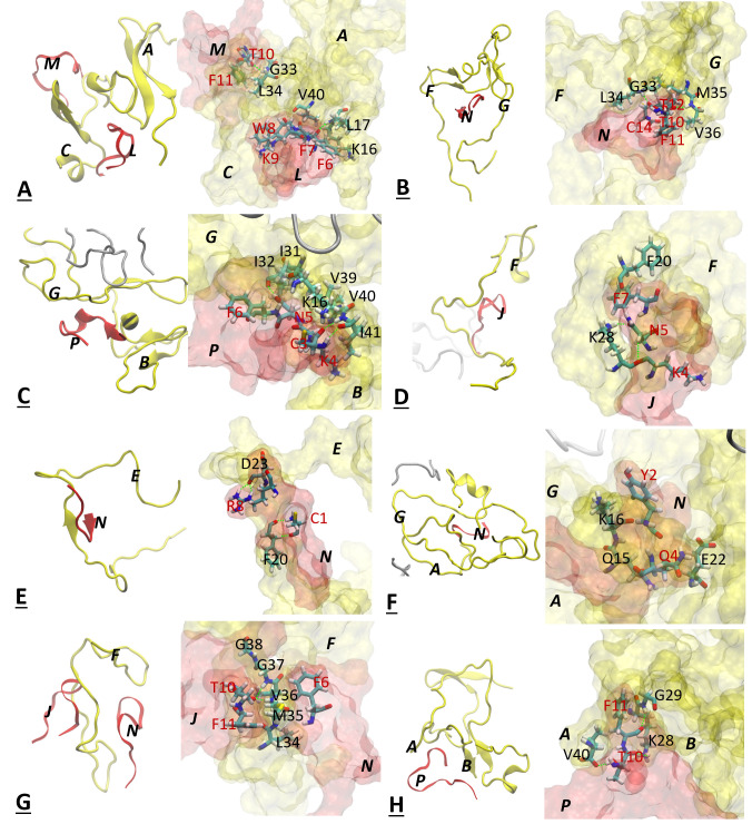
In order to elucidate the molecular mechanisms behind the influence of the small cyclic peptides onto the self-assembled aggregates, we identified the SST14 and AVP molecules that exhibited the strongest inter-molecular dynamics correlations by averaging over the last 20 ns, in each MD trajectory and for all the Aβ42-SST14, Aβ42-AVP and Aβ40-SST14 systems. Fig 7 depicts close-ups of the identified highest correlated regions involving small cyclic peptides and Aβ chains.
Fig 7.
Close-ups of interactions between Aβ chains and small cyclic peptides in systems Aβ42-SST14-I (A,B), Aβ42–SST-II (C,D), Aβ42-AVP-I (E), Aβ42-AVP-II (F), Aβ40-SST14-I (G) and Aβ40-SST14-II (H). Aβ42 or Aβ40 chains are colored yellow, and SST14 or AVP molecules are colored red. Aβ residues are annotated with black letters and small cyclic peptide residues are annotated with red letters. Hydrogen bonds are depicted by green dashed lines. The close-ups illustrate interactions resulting in the strongest ECD pair correlations averaged over the last 20 ns of 500 ns long MD trajectories (Fig 4).
Fig 7A and 7B illustrate, respectively, the interactions between an Aβ dimer (A+C) and the SST chains L and M attached to it, and between Aβ chains F+G and the SST chain N in system Aβ42-SST14-I. Fig 7C shows the interactions between Aβ chains G and B and SST chain P, and Fig 7D illustrates the interactions between Aβ chain F and SST chain J in system Aβ42-SST14-II. In these examples, the strongest inter-molecular correlations involve pairs of residues: K16-F7, G33-T10, and V40-W8, (Fig 7A), G33-T12 and M35-T10 (Fig 7B), I32-N5 and V40-K4 (Fig 7C), and K28-N5 (Fig 7D). Remarkably, the SST residues involved in strong correlations with Aβ42 are often hydrophobic (F7, W8), or carry a long hydrocarbon side chain (K4), and/or have hydrophobic neighbors such as F6 or F11. Similarly, the pairing Aβ residues often carry hydrophobic groups (M35, V40) and/or have hydrophobic neighbors (L17, I31, I32, L34, I41). These hydrophobicity-driven interactions of side chains are accompanied by backbone-backbone hydrogen bonding, for example L16-F7, G33-T10 (Fig 7A), L34-T12 and V36-T10 (Fig 7B), or I41-K4 (Fig 7C). Complementary to hydrophobic interactions and hydrogen bonding, we also detected π-π interactions between residue F20 of Aβ42 and F7 of SST14 (Fig 7D).
Next, Fig 7E and 7F depict the strongest interactions between Aβ42 and AVP molecules identified in systems Aβ42-AVP-I and Aβ42-AVP-II. In the first system illustrated by Fig 7E, AVP residue R8 from chain N developed strong dynamics correlations with D23 of the Aβ42 chain E. Moreover, residue C1 developed contacts with residue F20 located nearby, although this interaction is somewhat weaker. In this case, both AVP residues have formed hydrogen bonds with their Aβ42 counterparts resulting in formation of a cross-species β-sheet (Fig 7E). Fig 7F illustrates the strongly correlated pair consisting of Aβ42 residue K16 (chain G) and AVP residue Y2 along with a weaker interaction of closely positioned pair of E22 (chain A) and Q4. Despite an involvement of hydrophobic contacts between Y2 and a hydrocarbon group of K16, electrostatic forces rather than hydrophobicity appear to play a role in this interaction.
Interactions between Aβ40 chains and SST14 molecules are illustrated with examples from system Aβ40-SST14-I (Fig 7G) and system Aβ40-SST14-II (Fig 7H). In system Aβ40-SST14-I, the strongest dynamics correlations involve residues M35, V36, and G37 from Aβ chain F and residues T10 and T11 from SST chain J. Backbones of residues M35 and G37 developed hydrogen bonds with F11 and T10, respectively, whereas residue V38 formed a hydrogen bond with T10 as well as hydrophobic interaction with F11. In system Aβ40-SST14-II, Aβ residues V40 (chain A) and K28 (chain B) exhibit strong dynamic correlations with residues T10 and F11 from SST chain P, respectively. All these interactions involve hydrogen bonding (Fig 7H). Strong correlations that involve hydrophobic contacts are less frequent in the Aβ40-SST14 system than in the Aβ42-SST14 system, evidently due to the lack of two C-terminal residues I41 and A42 in Aβ40.
Solvent accessibility of the aggregates
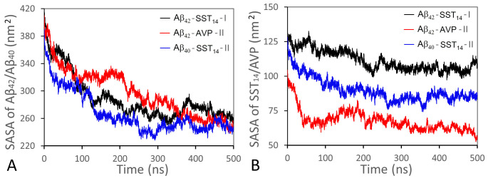
The solvent accessible surface area (SASA) is an important quantitative indicator of the oligomerization process. Fig 8 shows the evolution of total SASAs for Aβ chains and small cyclic peptides for three representative systems Aβ42-SST14-I, Aβ42-AVP-II, and Aβ40-SST14-II during 500 ns production MD simulations. Consistent with the ongoing aggregation in these systems, total SASAs of Aβ chains decrease significantly in the course of the simulations (Fig 8A). Although the greatest decrease occurs over the first 200–250 ns, a tendency of continuing collapse persists over the entire 500 ns interval. Total SASAs of small cyclic peptides also exhibit a steady decrease (Fig 8B). At the beginning of the simulations SASAs of SST14 are close across the Aβ42-SST14-I and Aβ40-SST14-II systems; however subsequently SST14 tends to exhibit a greater SASA in the Aβ42-SST14-I system than in the Aβ40-SST14-II system. There are two reasons for that. First, one or more SST14 molecules always remain free in the Aβ42-SST14-I system. Second, from our analysis of the trajectories it follows that the SST14 molecules tend to occupy peripheral regions of aggregates in Aβ42-SST14 system, whereas in Aβ40-SST14 system, they are often found in the interior of aggregates. The total SASA of AVP is less than that of SST14 in either system due to smaller size of the AVP molecules.
Fig 8.
Total SASA of Aβ chains (A) and small cyclic peptides (B) for systems Aβ42-SST14-I (black lines), Aβ42-AVP-II (red lines), and Aβ40-SST14-II (blue lines) during 500 ns production MD simulations.
Table 4 lists average hydrophobic and hydrophilic SASAs for all the peptides in each of the three systems Aβ42-SST14, Aβ42-AVP and Aβ40-SST14 during the first and the last 5 ns of 500 ns long MD simulations, and S5 Table provides similar data for SST14 and AVP molecules separately. Additional details of the SASAs in individual trajectories are given in S6 Table. As one could expect, the hydrophobic SASA is less than the hydrophilic counterpart in all systems at all stages of the production MD simulations, since the aggregation process facilitates burying of hydrophobic groups. As Table 4 indicates, exposures of both hydrophobic and hydrophilic groups to the solvent have decreased considerably in the course of the simulations. The relative decrease amounts to approximately 28–37% of the initial SASA’s values in each of the three systems. Upon averaging over three trajectories for each of the systems Aβ42-SST14, Aβ42-AVP and Aβ40-SST14, the total hydrophilic SASAs are quite uniform across all three systems during the first 5 ns of MD simulations, with a slightly increased contribution of hydrophilic residues in Aβ42-AVP (Table 4). In contrast, the initial hydrophobic SASAs of the three systems exhibit diverging trends across the three systems. At the beginning of MD simulations, total hydrophobic SASAs in all Aβ42-SST14 trajectories are higher than in any of Aβ40-SST14 trajectories (S6 Table), which can be explained by the presence of the two hydrophobic C-terminal residues in Aβ42. On the other hand, the initial hydrophobic SASAs in both SST14 containing systems are pronouncedly higher than in all Aβ42 -AVP trajectories (Tables 4 and S6), which we attribute to the larger hydrophobic SASA of SST14 molecules compared to AVP molecules (S5 Table).
Table 4. Hydrophobic and hydrophilic SASA in each system averaged over three trajectories during the first and the last 5 ns of MD simulations.
| Systems | First 5 ns (nm2) | Last 5 ns (nm2) | Difference (%) | |||
|---|---|---|---|---|---|---|
| Hydrophobic | Hydrophilic | Hydrophobic | Hydrophilic | Hydrophobic | Hydrophilic | |
| Aβ42-SST14 | 220.4 (±8.3) | 290.7 (±6.8) | 147.9 (±5.8) | 210.7 (±16.9) | 33 | 28 |
| Aβ42-AVP | 187.0 (±8.2) | 296.6 (±6.3) | 118.7 (±9.2) | 186.3 (±17.6) | 37 | 37 |
| Aβ40-SST14 | 205.6 (±5.5) | 288.1 (±4.2) | 130.5 (±2.0) | 205.1 (±13.9) | 37 | 29 |
In the course of the MD simulations, the Aβ42-SST14 system exhibited the smallest relative decrease in hydrophobic SASA (33%), consistent with a smaller size of aggregates and greater solvent exposure of Aβ chains and SST14 molecules in these systems (Table 4). The reduction in hydrophobic SASA in both Aβ42-AVP and Aβ40-SST14 systems is approximately 37% despite a relatively weak hydrophobicity of AVP. Together with a pronounced reduction in exposure of hydrophilic residues in Aβ42-AVP, this may suggest more efficient collapse in Aβ42-AVP system in comparison to Aβ40-SST14 system.
Average hydrophobic and hydrophilic SASAs calculated for SST14 alone (S5 Table) exhibit consistent trends with those in Table 4. Initially, SST14’s hydrophobic and hydrophilic SASAs are close in both systems. In the course of the subsequent simulations, the decrease in both hydrophilic and hydrophobic SASA of SST14 is less pronounced in Aβ42-SST14 system than in Aβ40-SST14 system. In the case of AVP, the relative decrease in hydrophobic and hydrophilic SASA of 49% and 43%, respectively, is the greatest of all systems. In addition, the high initial hydrophilicity in AVP results in a three times greater absolute reduction of hydrophilic SASA in comparison to the hydrophobic one.
Summarising, the greater initial hydrophobicity of the Aβ42-SST14 system might result in substantial hydrophobic collapse; however, the observed decrease in solvent exposure of hydrophobic residues in the Aβ42-SST14 system was relatively weak. Contrary to the expectations, a higher decrease of hydrophobic SASA during the MD simulations was observed in Aβ40-SST14 system instead. The Aβ42-AVP system exhibited the strongest reduction in solvent exposure of hydrophilic residues, suggesting a more important role of electrostatic interactions in the course of aggregation in these systems.
Discussion
We have conducted all-atom MD simulations of early aggregation events at the onset of oligomerization in mixtures of Aβ peptides and small cyclic peptides, Aβ42–SST14, Aβ42–AVP, and Aβ40–SST14 in water. Initially, the Aβ peptides and small cyclic peptides were sparsely positioned with respect to each other. Aggregation of the Aβ peptides, accompanied by a pronounced decrease in solvent exposure of both hydrophobic and hydrophilic groups, went on throughout the 500 ns long MD runs. In the course of the simulations, all systems developed self-assembled aggregates containing Aβ chains and small cyclic peptides. Consistent with published experiments [10,11] and MD simulations [15,16] of Aβ aggregation in the absence of small cyclic peptides, the self-assembled aggregates observed in this study exhibit largely unstructured morphologies without pronounced alignment of the chains. Although such unstructured morphologies indicate a considerable influence of hydrophobic collapse onto the oligomerization, the observed build-up of hydrogen bonding accompanied by development of stable β-sheet content also suggests a potential tendency toward β-conversion.
Overall, self-assembled Aβ42–SST14 aggregates are compact and well-correlated dynamically. However, they tend to be smaller in size than aggregates formed in the other systems; their hydrophobic groups exhibit the highest solvent accessibilities in average by the end of the 500 ns simulation; and they develop less hydrogen bonds across Aβ42 peptides and SST14 molecules in comparison to the other systems. The replacement of SST14 with AVP molecules produced larger aggregates. In addition, the Aβ42–AVP system tended to develop more hydrogen bonds than the other systems, which is especially true for cross-species hydrogen bonding. In the course of the 500 ns simulations, the AVP molecules developed almost 3 times more HBs with Aβ42 than the SST14 molecules did. Both hydrophobic and hydrophilic SASA are the lowest in the Aβ42–AVP systems in average after 500 ns simulations. The two Aβ42-containing systems exhibited similar tendencies of Aβ’s C-termini to remain at the aggregate’s surface, although C-terminal flexibility tended to be greater in AVP-containing systems than in SST14-containing systems. The most pronounced differences between the Aβ42–SST14 and Aβ40–SST14 systems concern the morphologies of the self-assembled aggregates. Unlike the Aβ42–SST14 system, the Aβ40–SST14 system formed large aggregates that contained a domain of correlated motion surrounded by relatively weakly correlated parts. In the Aβ40–SST14 system the C-termini are buried more often than in the other two systems, and they exhibit the lowest average C-terminal flexibility. According to a recent report [21], such morphological differences may lead to different pathways at later stages of oligomerization.
Our examination of inter-molecular contacts that exhibited the strongest dynamics correlations in average over the last 20 ns of the simulations revealed differences in the interactions of SST14 and AVP molecules with Aβ peptides. In Aβ42-SST14 aggregates, highly correlated regions tended to involve residues K4, N5, F7, W8, and T10 of SST14, in agreement with biochemical observations [28]. These residues were found to interact primarily with the residues 28–35 of Aβ42, although contacts with its C-terminus were also detected (K4-V40 and W8-V40). The interactions tended to involve hydrophobic contacts, although backbone-backbone hydrogen bonds were also present. In the Aβ42-AVP aggregates, strong correlations involved predominantly contacts between N-terminal AVP resides C1, Y2, and Q4, and residues 20–23 of Aβ42. In the Aβ40-SST14 aggregates, T10 and F11 of SST14 were found to interact with C-terminal residues 35–40 of Aβ40. Hydrogen bonds are found frequently between strongly correlated residues in systems Aβ42-AVP and Aβ40-SST14.
At a coarser level, we observe a tendency of SST14 molecules to develop hydrophobic contacts with surfaces of Aβ42 aggregates such that both N- and C- terminal arms of SST14 face the solvent. This may prevent hydrophobicity-driven coalescence the Aβ42 aggregates, explaining the relatively small size of aggregates in the Aβ42-SST14 system. Distinct from SST14, AVP molecules tend to penetrate inside the Aβ42 aggregates, where they develop extensive hydrogen bonding with central regions of Aβ42 peptides. When SST14 interacts with small Aβ40 aggregates, the morphologies resemble those of Aβ42-SST14, whereas large Aβ40 aggregates tend to incorporate SST14 molecules resembling the interaction of AVP with Aβ42 aggregates.
Based on the results obtained in this study, the experimentally observed differences in the aggregation behaviours of Aβ42–SST14, Aβ42–AVP, and Aβ40–SST14 mixtures [28,29] may be attributed to the different interaction of the small cyclic peptides with each of the Aβ peptides and the solvent.
First, the tendency of SST14 to develop predominantly hydrophobic contacts with residues 28–35 and the C-terminus of Aβ42 favors morphologies where several Aβ42 chains form small aggregates compacted by Aβ-Aβ hydrogen bonding, and carrying SST14 molecules attached to their surfaces. This surface attachment prevents the coalescence of small Aβ42 aggregates into bigger structures. Within the time regimes of the present MD study, this results in a prevalence of multiple small Aβ42 aggregates. The resulting delay in hydrophobicity-driven coalescence may explain the extended lag phase of Aβ42 aggregation observed experimentally [28]. In contrast, AVP molecules preferentially bind to central regions of Aβ42 peptides, and are prone to develop extensive inter-species hydrogen bonding. As a result, large Aβ42 aggregates self-assemble quickly and contain inserted AVP molecules that strengthen connections within the aggregates. The binding of SST14 to Aβ40 tended to rely on strong connections with the C-terminal region of the Aβ40 chains. Combined with the propensity of the Aβ40-SST14 system to develop inter-species hydrogen bonding, this facilitated insertion of SST14 into the interior of Aβ40 aggregates.
Second, differences in hydrophobicity of the three systems result in different surface reactivities of the aggregates. Clearly, the presence of SST14 imparts the aggregates with a greater hydrophobicity. The resulting “sticky surface” effect is enhanced even more due to the location of both SST14 molecules and hydrophobic C-termini, preferentially at the surface of the aggregates. In the Aβ42–AVP system hydrophobic C-termini are also exposed and flexible. However, the “sticky surface” effect is lessened by a lower hydrophobicity of AVP and a lesser surface area due to the larger size of the aggregates. In the Aβ40-SST14 system, the insertion of SST14 molecules into the interior of the aggregates combined with the reduced number of exposed hydrophobic C-terminal residues plays a role.
Summarising, our observations suggest that mixtures of Aβ42 and SST14, although pronouncedly more hydrophobic than their Aβ42-AVP and Aβ40-SST14 counterparts, tend to aggregate slower than the other two mixtures retaining extensive “sticky surface” areas exposed for longer times. This may explain the increased pathogenicity of Aβ42, but not Aβ40 peptides, in the presence of SST14, but not AVP molecules.
Methods
Modeling structures
Each system investigated in this study contained eight randomly positioned monomeric Aβ42 or Aβ40 peptides and eight randomly positioned small cyclic peptides, somatostatin-14 (SST14) or arginine vasopressin (AVP), as listed in Table 5. For Aβ42 peptides, the sequence 1DAEFRHDSGYEVHHQKLVFFAEDVGSNKGAIIGLMVGGVVIA42 was used, and for Aβ40 peptides the two C-terminal residues were excluded. The sequences 1AGCKNFFWKTFTSC14 and 1CYFENCPRG9-NH2 were used for SST14 and AVP, respectively, to match the conditions of the experiments [28,29]. As starting coordinates for these monomeric units, we used the coordinates of chains A–F of the NMR-derived Aβ1–42 hexamer from PDB ID 2NAO [36] and the coordinates of chains A-H of the Aβ1–40 nanomer from PDB ID 2M4J [37], which were equilibrated before modeling. For small cyclic peptides, we used the coordinates of chain A of somatostatin-14 with S-S bond from PDB ID 2MI1 [38], and coordinates of chain B of vasopressin from PDB ID 1YF4 [39]. Using the Accelrys VS [40] and VMD [41] packages, we built the starting systems to contain eight monomeric Aβ1–40/42 units from 2M4J/2NAO.pdb, and eight SST14/AVP molecules by randomly rotating and shifting the units against each other. All peptides were randomly placed in the simulation box such that distances between their surfaces were longer than 4 Å. This resulted in sparsely positioned units with a concentration of approximately 4 mM for Aβ, SST14, and AVP each.
Table 5. List of the systems studied.
| System | Details of preparation | Number of MD trajectories |
|---|---|---|
| Aβ42-SST14-I/III | Eight Aβ1–42 chains 2NAO [36], pre-equilibrated and mixed with eight SST14 chains 2MI1 [38]; then equilibrated again. | 3 |
| Aβ42-AVP-I/III control | Eight Aβ1–42 chains 2NAO [36], pre-equilibrated and mixed with eight AVP chains 1YF4 [39]; then equilibrated again. | 3 |
| Aβ40-SST14-I/III | Eight Aβ1–40 chains 2M4J [37], pre-equilibrated and mixed with eight SST14 chains 2MI1 [38]; then equilibrated again. | 3 |
Minimizations, equilibrations, and production MD simulations were carried out using the GROMACS v5.0.7 package [42]. The AMBER99SB-ILDN forcefield [43] was used for protein atoms. Then, we minimized each set of 16 randomly positioned units using a steepest descent algorithm for energy in vacuum, and added the solvent using the optimized general-purpose 4-point OPC water model [44], as well as counterions Na+/Cl− to electro-neutralize the systems. Subsequent solvent minimizations involved decreasing position restraints on non-hydrogen protein atoms, as well as heating with the Berendsen thermostats. NVT-equilibrations were performed on all the systems. To collect sufficient statistics on the systems’ dynamics, we ran independent MD trajectories for each of the systems, as listed in Table 5. The same sets of starting coordinates of atoms were used in each of the trajectories, whereas the seed numbers were varied when generating the initial Maxwell distributions of atom velocities. The three MD trajectories were labeled as “I”, “II” and “III”.
Molecular dynamics simulations
The production MD simulations, as well as the last equilibration step, were conducted at a temperature of 310 K and a pressure of 1 atm using isotropic pressure coupling (NPT ensemble), the Verlet cut-off scheme for neighbour searching, the Particle-Mesh Ewald treatment of electrostatics, a twin-range cut-off for van der Waals interactions, and bond lengths restrained through the linear constant solver (LINCS) algorithm with a fourth order of expansion. A V-rescale scheme was used for temperature coupling, and the Parrinello-Rahman algorithm was employed for pressure coupling applying the scaling to the center of mass of reference coordinates. Production MD simulations were performed for 100 ns for 9 systems containing 16 chains each (see Table 5). 2 fs time steps were used, and snapshots were saved every 20 fs in order to analyze the essential collective dynamics of the systems.
Structural analysis of the trajectories, including assessment of the secondary structure, the number of hydrogen bonds, and solvent accessible areas, has been done using the GROMACS scripts [42] and the VMD package [41]. For graphical representation, the VMD and Accelrys VS packages were utilized.
Essential collective dynamics analysis
To analyze the dynamics of the aggregates in greater depth we employed the novel essential collective dynamics (ECD) method [16,31–35], which stemmed from a recently developed statistical-mechanical framework [31,32]. In this framework, a macromolecule or supramolecular complex is described by generalized Langevin equations with a set of essential collective coordinates identified as principal eigenvectors of a covariance matrix, which in turn is calculated by applying the principal component analysis (PCA) on MD trajectories. It has been demonstrated [31,32] that persistent correlations between atoms’ motion (dynamics) can be calculated from a projected all-atom image of the system in a multi-dimensional space of the essential collective coordinates, as described in details elsewhere [16,31–33]. A suite of dynamics descriptors has been derived within this framework, including pair correlation maps [16,33,35], dynamics domains of correlated motion [16,31,33,34], and main-chain flexibilities [16,33–35]. The method has been validated extensively against NMR [31–34,45] and X-ray [33,46] structural data, and the corresponding descriptors were demonstrated to predict accurately the persistent dynamics trends from short fragments of MD trajectories without exhaustive conformational sampling [31,32]. In this study, we applied the ECD method with a set of 10 principal eigenvectors, which were sufficient to sample more than 95% of the total displacement in the systems considered. The ECD descriptors were calculated from multiple 0.2 ns long segments of the MD trajectories, and averaged over pertinent segments of production MD simulations, as specified in Results and Discussion. The pair correlation descriptors displayed in Fig 4 have been derived from the projected images of Cα atoms in the respective aggregates as specified earlier [16,33,34,45]. The dynamics domains of correlation motion, which represent relatively rigid parts of the aggregate composed of atoms moving coherently (Fig 5) were identified by applying the clustering technique described in earlier reports [31,33,34,45] with a threshold of 0.008 to the projected all-atom images of all systems. The main-chain flexibility profiles (Fig 6) illustrate the level of dynamic coupling of main-chain atoms with the entire aggregate, which in turn is represented by the centroid of the projected all-atom images of the aggregate.
Supporting information
Close-ups after equilibration at the beginning of production MD simulations illustrated by examples from representative trajectories Aβ42-SST14-I (A), Aβ42-AVP-II (B), and Aβ40-SST14 II (C). All main-chains are shown as ribbons. The C-terminal residues 36-40/42 are colored blue. SST14 molecules (A,C) and AVP molecules (B) are depicted in atomic details and colored red. Hydrophobicity of all chains is color-mapped onto solvent accessible surfaces, where blue indicates hydrophobic residues and red indicates hydrophilic residues.
(TIF)
Self-assembled aggregates in trajectories Aβ42-SST14- I (A); Aβ42-AVP-II (B); and Aβ40-SST14-II (C) after 500 ns simulations. In Aβ42 and Aβ40 chains, random coils are colored white, β-strands are colored yellow, turns are colored cyan, and α helices are colored purple; the C-terminal residues 36-40/42 of Aβ peptides are colored blue. The SST14 molecules (A,C) and AVP molecules (B) are depicted in atomic detail and colored red.
(TIF)
The number of hydrogen bonds across Aβ chains and small cyclic peptides during 500 ns production simulations. (A)–the numbers of hetero-molecular bonds as functions of time in all nine trajectories. Aβ42-SST14 systems are shown with shades of red, Aβ42-AVP–with shades of blue, and Aβ40-SST14 –with shades of green. (B)–the dependencies for Aβ42-SST14-I (red line), Aβ42-AVP-II (blue line), and Aβ40-SST14-II (green line) with solid dots indicating local maxima and minima where the snapshots presented in Fig 3 were taken.
(TIF)
Secondary structure evolution for trajectories Aβ42-SST14-I (A), Aβ42-AVP-II (B) and Aβ40-SST14-II (C) during the 500 ns long production MD simulations. β-sheets/bridges are shown with yellow/dark yellow color, α/3-π-helices–with purple/blue, turns–with green, and random coils–with white. The Y-axis represents residues in chains A-P.
(TIF)
Percentages of β-sheet (top row), β-bridge (middle row), and α/3-π-helix (bottom row) content in each of the three MD trajectories for systems Aβ42-SST14, Aβ42-AVP, and Aβ40-SST14 as functions of time.
(TIF)
Aggregates observed in systems Aβ42-SST14-I (A), Aβ42-AVP-II (B) and Aβ40-SST14-II (C) after 500 ns of simulations. In (A), chains A, B and E are colored yellow; chains C, D and F are colored cyan; and chains G and H are colored purple. In (B) and (C), chains A to H are colored yellow, cyan, purple, lime, mauve, ochre, iceblue and black, respectively. All SST14 and AVP molecules are colored red.
(TIF)
(XLSX)
The data are given in % of occupied Aβ residues.
(XLSX)
The data are given in % of occupied Aβ residues.
(XLSX)
(XLSX)
(XLSX)
(XLSX)
Acknowledgments
Molecular images were created using the VMD package [41]. The simulations were carried out using the NINT-NRC computational cluster and Compute Canada resources.
Data Availability
All relevant data are within the manuscript and its Supporting Information files.
Funding Statement
The work was supported by the Alberta Prion Research Institute (APRI), Projects 201600028 (to HW and GSU) and 201700016 (MS). Philanthropic financial support from the Borden Rosiak family is gratefully acknowledged (to GSU). The funders had no role in study design, data collection and analysis, decision to publish, or preparation of the manuscript.
References
- 1.Haass C, Selkoe DJ. Soluble protein oligomers in neurodegeneration: lessons from the Alzheimer’s amyloid beta-peptide. Nat Rev Mol Cell Biol. 2007; 8(2):101–12. 10.1038/nrm2101 [DOI] [PubMed] [Google Scholar]
- 2.Chiti F, Dobson CM. Protein misfolding, amyloid formation, and human disease: A summary of progress over the last decade. Ann Rev Biochem. 2017; 86(1): 27–68. [DOI] [PubMed] [Google Scholar]
- 3.Soto C, Pritzkow S. Protein misfolding, aggregation, and conformational strains in neurodegenerative diseases. Nat Neurosci. 2018; 21(10): 1332–40. 10.1038/s41593-018-0235-9 [DOI] [PMC free article] [PubMed] [Google Scholar]
- 4.Nasica-Labouze J, Nguyen PH, Sterpone F, Berthoumieu O, Buchete NV, Coté S, et al. Amyloid β protein and Alzheimer’s disease: When computer simulations complement experimental studies. Chem Rev. 2015; 115(9): 3518–63. 10.1021/cr500638n [DOI] [PMC free article] [PubMed] [Google Scholar]
- 5.Bitan G, Kirkitadze MD, Lomakin A, Vollers SS, Benedek GB, Teplow DB. Amyloid β-protein (Aβ) assembly: Aβ40 and Aβ42 oligomerize through distinct pathways. Proc Natl Acad Sci USA. 2003; 100(1):330–5. 10.1073/pnas.222681699 [DOI] [PMC free article] [PubMed] [Google Scholar]
- 6.Meisl G, Yang X, Hellstrand E, Frohm B, Kirkegaard JB, Cohen SI, Dobson CM, Linse S, Knowles TP. Differences in nucleation behavior underlie the contrasting aggregation kinetics of the Aβ40 and Aβ42 peptides. Proc Natl Acad Sci USA. 2014; 111(26): 9384–9. 10.1073/pnas.1401564111 [DOI] [PMC free article] [PubMed] [Google Scholar]
- 7.Michaels TCT, Šarić A, Curk S, Bernfur K, Arosio P, Meisl G, Dear AJ, Cohen SIA, Dobson CM, Vendruscolo M, Linse S, Knowles TPJ. Dynamics of oligomer populations formed during the aggregation of Alzheimer’s Aβ42 peptide. Nat Chem. 2020; 12(5):445–1. 10.1038/s41557-020-0452-1 [DOI] [PMC free article] [PubMed] [Google Scholar]
- 8.Dear AJ, Michaels TCT, Meisl G, Klenerman D, Wu S, Perrett S, Linse S, Dobson CM, Knowles TPJ. Kinetic diversity of amyloid oligomers. Proc Natl Acad Sci USA. 2020; 111(22): 12087–94. 10.1073/pnas.1922267117 [DOI] [PMC free article] [PubMed] [Google Scholar]
- 9.Breydo L, Uversky VN. Structural, morphological, and functional diversity of amyloid oligomers. FEBS Lett, 2015; 589(19A): 2640–48. [DOI] [PubMed] [Google Scholar]
- 10.Luo J, Wärmländer SKTS, Gräslund A, Abrahams JP. Alzheimer peptides aggregate into transient nanoglobules that nucleate fibrils. Biochemistry, 2014; 53(40): 6302–8. 10.1021/bi5003579 [DOI] [PubMed] [Google Scholar]
- 11.Morel B, Carrasco MP, Jurado S, Marco C, Conejero-Lara F. Dynamic micellar oligomers of amyloid beta peptides play a crucial role in their aggregation mechanisms. Phys Chem Chem Phys. 2018; 20(31), 20597–14. 10.1039/c8cp02685h [DOI] [PubMed] [Google Scholar]
- 12.Tarus B, Tran TT, Nasica-Labouze J, Sterpone F, Nguyen PH, Derreumaux P. Structures of the Alzheimer’s wild-type Aβ1–40 dimer from atomistic simulations. J Phys Chem B, 2015; 119(33): 10478–87. 10.1021/acs.jpcb.5b05593 [DOI] [PubMed] [Google Scholar]
- 13.Man VH, Nguyen PH, Derreumaux P. High-resolution structures of the amyloid-β 1–42 dimers from the comparison of four atomistic force fields. J Phys Chem B, 2017; 121(24:5977–87. 10.1021/acs.jpcb.7b04689 [DOI] [PMC free article] [PubMed] [Google Scholar]
- 14.Wille H, Dorosh L, Amidian S, Schmitt-Ulms G, Stepanova M. Combining molecular dynamics simulations and experimental analyses in protein misfolding. Adv Protein Chem Struct Biol, 2019;118: 33–110. 10.1016/bs.apcsb.2019.10.001 [DOI] [PubMed] [Google Scholar]
- 15.Barz B, Olubiyi OO, Strodel B. Early amyloid β-protein aggregation precedes conformational change. Chem Commun. 2014; 50(40): 5373–5. 10.1039/c3cc48704k [DOI] [PubMed] [Google Scholar]
- 16.Dorosh L, Stepanova M. Probing oligomerization of amyloid β peptide in silico. Mol BioSyst, 2016; 13(1), 165–82. 10.1039/c6mb00441e [DOI] [PubMed] [Google Scholar]
- 17.Cheon M, Chang I, Mohanty S, Luheshi LM, Dobson CM, Vendruscolo M, Favrin G. Structural reorganisation and potential toxicity of oligomeric species formed during the assembly of amyloid fibrils. PLoS Comput Biol, 2007; 3(9):1727–38. 10.1371/journal.pcbi.0030173 [DOI] [PMC free article] [PubMed] [Google Scholar]
- 18.Matthes D, Gapsys V, Brennecke JT, de Groot BL. An atomistic view of amyloidogenic self-assembly: Structure and dynamics of heterogeneous conformational states in the pre-nucleation phase. Sci Rep, 2016; 6:33156. 10.1038/srep33156 [DOI] [PMC free article] [PubMed] [Google Scholar]
- 19.Nagel-Steger L, Owen MC, Strodel B. An account of amyloid oligomers: Facts and figures obtained from experiments and simulations. Chem Bio Chem, 2016; 17(8): 657–76. 10.1002/cbic.201500623 [DOI] [PubMed] [Google Scholar]
- 20.Balchin D, Hayer-Hartl M, Hartl FU. In vivo aspects of protein folding and quality control. Science, 2016; 353(6294): aac4354. 10.1126/science.aac4354 [DOI] [PubMed] [Google Scholar]
- 21.Barz B, Liao Q, Strodel B. Pathways of amyloid-β aggregation depend on oligomer shape. J Am Chem Soc. 2017; 140(1): 319–27. 10.1021/jacs.7b10343 [DOI] [PubMed] [Google Scholar]
- 22.Jia Z, Schmit JD, Chen J, Amyloid assembly is dominated by misregistered kinetic traps on an unbiased energy landscape. Proc Natl Acad Sci USA. 2020; 117(19): 10322–8. 10.1073/pnas.1911153117 [DOI] [PMC free article] [PubMed] [Google Scholar]
- 23.Heller GT, Bonomi M, Vendruscolo M. Structural ensemble modulation upon small-molecule binding to disordered proteins. J Mol Biol 2018; 430(16): 2288–92. 10.1016/j.jmb.2018.03.015 [DOI] [PubMed] [Google Scholar]
- 24.Breydo L, Redington JM, Uversky VN. Effects of intrinsic and extrinsic factors on aggregation of physiologically important intrinsically disordered proteins. Int Rev Cel Mol Bio. 2017; 329: 145–85. 10.1016/bs.ircmb.2016.08.011 [DOI] [PubMed] [Google Scholar]
- 25.Larson ME, Lesné SE. Soluble Aβ oligomer production and toxicity. J Neurochem. 2012; 120 (Suppl 1): 125–39. 10.1111/j.1471-4159.2011.07478.x [DOI] [PMC free article] [PubMed] [Google Scholar]
- 26.Saito T, Iwata N, Tsubuki S, Takaki Y, Takano J, Huang S-M, Suemoto T, Higuchi M, Saido TC. Somatostatin regulates brain amyloid beta peptide Aβ42 through modulation of proteolytic degradation. Nat Med 2005; 11(4): 434–9. 10.1038/nm1206 [DOI] [PubMed] [Google Scholar]
- 27.Gahete MD, Rubio A, Duran-Prado M, Avila J, Luque RM, Castano JP. Expression of somatostatin, cortistatin, and their receptors, as well as dopamine receptors, but not of neprilysin, are reduced in the temporal lobe of Alzheimer’s disease patients. J Alzheimer’s Dis. 2010; 20(2):465–75. [DOI] [PubMed] [Google Scholar]
- 28.Wang H, Muiznieks LD, Ghosh P, Williams D, Solarski M, Fang A, Ruiz-Riquelme A, Pomès R, Watts JC, Chakrabartty A, et al. Somatostatin binds to the human amyloid β peptide and favors the formation of distinct oligomers. ELife. 2017; 6: e28401. 10.7554/eLife.28401 [DOI] [PMC free article] [PubMed] [Google Scholar]
- 29.Solarski M, Wang H, Wille H, and Schmitt-Ulms G. Somatostatin in Alzheimer’s disease: A new role for an old player. Prion, 2018; 12(1): 1–8. 10.1080/19336896.2017.1405207 [DOI] [PMC free article] [PubMed] [Google Scholar]
- 30.Puig E, Tolchard J, Riera A, Carulla N. Somatostatin, an in vivo binder to Aβ oligomers, binds to βPFOAβ(1–42) tetramers. ACS Chem Neurosci. 2020; 11(20), 3358–65. 10.1021/acschemneuro.0c00470 [DOI] [PubMed] [Google Scholar]
- 31.Stepanova M. Dynamics of essential collective motions in proteins: theory. Phys Rev. E, 2007; 76(5 Pt 1): 051918. 10.1103/PhysRevE.76.051918 [DOI] [PubMed] [Google Scholar]
- 32.Potapov A, Stepanova M. Conformational modes in biomolecules: Dynamics and approximate invariance. Phys Rev. E, 2012; 85(2): 020901. 10.1103/PhysRevE.85.020901 [DOI] [PubMed] [Google Scholar]
- 33.Issack BB, Berjanskii M, Wishart DS, Stepanova M. Exploring the essential collective dynamics of interacting proteins: Application to prion protein dimers. Proteins, 2012; 80(7): 1847–65. 10.1002/prot.24082 [DOI] [PubMed] [Google Scholar]
- 34.Blinov N, Berjanskii M, Wishart DS, and Stepanova M. Structural domains and main-chain flexibility in prion proteins, Biochemistry, 2009; 48(7): 1488–97. 10.1021/bi802043h [DOI] [PubMed] [Google Scholar]
- 35.Mane JY, Stepanova M. Understanding the dynamics of monomeric, dimeric, and tetrameric α-synuclein structures in water. FEBS Open Bio, 2016; 6(7): 666–86. 10.1002/2211-5463.12069 [DOI] [PMC free article] [PubMed] [Google Scholar]
- 36.Wälti MA, Ravotti F, Arai H, Glabe CG, Wall JS, Böckmann A, Güntert P, Meier BH, Riek R. Atomic-resolution structure of a disease-relevant Aβ(1–42) amyloid fibril. Proc Natl Acad Sci USA. 2016; 113(34): E4976–84. 10.1073/pnas.1600749113 [DOI] [PMC free article] [PubMed] [Google Scholar]
- 37.Lu J-X, Qiang W, Yau W-M Schwieters CD, Meredith SC, Tycko R. Molecular structure of β-amyloid fibrils in Alzheimer’s disease brain tissue. Cell 2013; 154(6): 1257–68. 10.1016/j.cell.2013.08.035 [DOI] [PMC free article] [PubMed] [Google Scholar]
- 38.Anoop A, Ranganathan S, Das Dhaked B Jha NN, Pratihar S, Ghosh S, Sahay S, Kumar S, Das S, Kombrabail M, et al. Elucidating the role of disulfide bond on amyloid formation and fibril reversibility of somatostatin-14: relevance to its storage and secretion. J Biol Chem. 2014; 289(24): 16884–903. 10.1074/jbc.M114.548354 [DOI] [PMC free article] [PubMed] [Google Scholar]
- 39.Syed Ibrahim B, Pattabhi V. Trypsin inhibition by a peptide hormone: crystal structure of trypsin-vasopressin complex. J Mol Biol. 2005; 348(5): 1191–8. 10.1016/j.jmb.2005.03.034 [DOI] [PubMed] [Google Scholar]
- 40.Dassault Systèmes BIOVIA Accelrys Discovery Studio Visualiser (BIOVIA), 2005.
- 41.Humphrey W, Dalke A, and Schulten K. VMD: Visual molecular dynamics. J Mol Graph. 1996; 14(1): 33–8. 10.1016/0263-7855(96)00018-5 [DOI] [PubMed] [Google Scholar]
- 42.Berendsen HJC, van der Spoel D, van Drunen R. GROMACS: A message-passing parallel molecular dynamics implementation. Comput Phys Commun. 1995; 91(1–3): 43–56. [Google Scholar]
- 43.Lindorff-Larsen K, Piana S, Palmo K, Maragakis P, Klepeis JL, Dror RO, Shaw DE. Improved side-chain torsion potentials for the Amber ff99SB protein force field. Proteins 2010; 78(8): 1950–8. 10.1002/prot.22711 [DOI] [PMC free article] [PubMed] [Google Scholar]
- 44.Shabane PS, Izadi S, Onufriev AV. General purpose water model can improve atomistic simulations of intrinsically disordered proteins. J Chem Theory Comput. 2019;15(4):2620–34. 10.1021/acs.jctc.8b01123 [DOI] [PubMed] [Google Scholar]
- 45.Santo KP, Berjanskii M, Wishart DS, & Stepanova M. Comparative analysis of essential collective dynamics and NMR-derived flexibility profiles in evolutionarily diverse prion proteins. Prion. 2001; 5(3): 188–200. [DOI] [PMC free article] [PubMed] [Google Scholar]
- 46.Dorosh L, Kharenko OA, Rajagopalan N, Loewen MC, Stepanova M. Molecular mechanisms in the activation of abscisic acid receptor PYR1. PLoS Comp Biol. 2013; 9(6): e1003114. 10.1371/journal.pcbi.1003114 [DOI] [PMC free article] [PubMed] [Google Scholar]
Associated Data
This section collects any data citations, data availability statements, or supplementary materials included in this article.
Supplementary Materials
Close-ups after equilibration at the beginning of production MD simulations illustrated by examples from representative trajectories Aβ42-SST14-I (A), Aβ42-AVP-II (B), and Aβ40-SST14 II (C). All main-chains are shown as ribbons. The C-terminal residues 36-40/42 are colored blue. SST14 molecules (A,C) and AVP molecules (B) are depicted in atomic details and colored red. Hydrophobicity of all chains is color-mapped onto solvent accessible surfaces, where blue indicates hydrophobic residues and red indicates hydrophilic residues.
(TIF)
Self-assembled aggregates in trajectories Aβ42-SST14- I (A); Aβ42-AVP-II (B); and Aβ40-SST14-II (C) after 500 ns simulations. In Aβ42 and Aβ40 chains, random coils are colored white, β-strands are colored yellow, turns are colored cyan, and α helices are colored purple; the C-terminal residues 36-40/42 of Aβ peptides are colored blue. The SST14 molecules (A,C) and AVP molecules (B) are depicted in atomic detail and colored red.
(TIF)
The number of hydrogen bonds across Aβ chains and small cyclic peptides during 500 ns production simulations. (A)–the numbers of hetero-molecular bonds as functions of time in all nine trajectories. Aβ42-SST14 systems are shown with shades of red, Aβ42-AVP–with shades of blue, and Aβ40-SST14 –with shades of green. (B)–the dependencies for Aβ42-SST14-I (red line), Aβ42-AVP-II (blue line), and Aβ40-SST14-II (green line) with solid dots indicating local maxima and minima where the snapshots presented in Fig 3 were taken.
(TIF)
Secondary structure evolution for trajectories Aβ42-SST14-I (A), Aβ42-AVP-II (B) and Aβ40-SST14-II (C) during the 500 ns long production MD simulations. β-sheets/bridges are shown with yellow/dark yellow color, α/3-π-helices–with purple/blue, turns–with green, and random coils–with white. The Y-axis represents residues in chains A-P.
(TIF)
Percentages of β-sheet (top row), β-bridge (middle row), and α/3-π-helix (bottom row) content in each of the three MD trajectories for systems Aβ42-SST14, Aβ42-AVP, and Aβ40-SST14 as functions of time.
(TIF)
Aggregates observed in systems Aβ42-SST14-I (A), Aβ42-AVP-II (B) and Aβ40-SST14-II (C) after 500 ns of simulations. In (A), chains A, B and E are colored yellow; chains C, D and F are colored cyan; and chains G and H are colored purple. In (B) and (C), chains A to H are colored yellow, cyan, purple, lime, mauve, ochre, iceblue and black, respectively. All SST14 and AVP molecules are colored red.
(TIF)
(XLSX)
The data are given in % of occupied Aβ residues.
(XLSX)
The data are given in % of occupied Aβ residues.
(XLSX)
(XLSX)
(XLSX)
(XLSX)
Data Availability Statement
All relevant data are within the manuscript and its Supporting Information files.


