Abstract
Oligonucleotides are small molecules 8–50 nucleotides in length that bind via Watson-Crick base pairing to enhance or repress the expression of target RNA. The use of oligonucleotides to manipulate gene expression in the kidney could be a valuable tool to further understand kidney pathophysiology and can serve as an important complement to genetic studies. This chapter serves as a primer on the use of oligonucleotides in the kidney. We provide an overview of the various ways that oligonucleotides can manipulate gene expression. In addition, we describe the advancements in the development of oligonucleotides for laboratory and clinical use. Finally, instruction is provided on the design and implementation of oligonucleotides for in vitro and in vivo laboratory studies.
1. Introduction
The concept of oligonucleotide-based therapeutics was first introduced by Paul Zamecnik in 1978. Zamecnik identified a terminally redundant sequence in the Rous Sarcoma Virus (RSV) 35S RNA and designed a tridecamer oligonucleotide that bound to this 13 nucleotide sequence via classic Watson-Crick base pairing. The binding of the oligonucleotide inhibited replication of the RSV virus (Stephenson & Zamecnik, 1978). At the time, the concept of “antisense” therapeutics was readily accepted due to the widespread understanding of Watson-Crick hybridization. However, it has taken several decades to develop the biochemical advancements to bring this concept to clinical practice. There are now at least four oligonucleotide-based drugs approved by the FDA and many more in clinical trials (Crooke, Witztum, Bennett, & Baker, 2018). Thus, the utilization of oligonucleotides in mouse models can provide insight into the pathogenesis of a disease and also serve as a screening tool for potential therapeutic applications in humans.
1.1. How can oligonucleotides be used to manipulate gene expression? Mechanism of action of oligonucleotides
Oligonucleotides can be defined as molecules 8–50 nucleotides in length that can bind to RNA, DNA, protein, or function on their own. For the purposes of this chapter we will mostly focus on antisense oligonucleotides (ASO). ASOs directly bind their cognate RNA via Watson-Crick base pairing to modulate the function of that RNA.
ASOs can manipulate gene expression either by triggering degradation of RNA or occupying a region on the transcript to affect its function. Degradation of RNA can be mediated through endogenous function of RNASE H RNA interference mechanisms. Rnase H is an enzyme that cleaves RNA when it is found to interact with DNA (Cerritelli & Crouch, 2009). Endogenously its function is essential in DNA replication and proof reading. ASOs designed to mimic DNA fragments can bind to RNA and also trigger RNASE H mediated degradation. Classical RNAi mechanisms have also been well characterized. ASOs can also function like small RNAs in cells to manipulate the expression of genes by utilizing Arogonaute 2-mediated cleavage of RNA (Elbashir et al., 2001; Liu et al., 2004; Meister et al., 2004). Alternatively, ASOs can alter the function of a gene simply by occupying a region on the transcript. Utilizing either of these strategies, oligonucleotides can target any RNA to manipulate gene expression. The various ways ASOs can be utilized are as follows.
Translational repression of mRNA—This is the classical function of ASOs. By binding to a mRNA transcript, the ASO triggers RNASE H-dependent degradation. Use of ASOs in this mechanism will led to the reduction in gene expression.
Splicing Modulation—Most mRNAs undergo several processing steps which include splicing and polyadenylation (Sharp, 2009). ASOs can be designed to bind to splicing enhancer or repressor regions within pre-mRNAs and affect the intermediary metabolism of mRNAs. This method can be utilized to correct gene mutations that affect pre-mRNA splicing such as in cystic fibrosis.
Anti-miRs—AntimiRs are ASOs designed to inhibit microRNAs. By binding to the microRNA, the anti-miRs prevent miRNA-mediated repression of direct mRNA targets. This leads to the de-repression of potentially hundreds of direct microRNA target mRNAs.
Pro-miR—AnoppositeofAnti-miRs,Pro-miRsareessentiallyartificialmicroRNAs. They can be designed to mimic a microRNA. Once in the cell, they function as a microRNA by binding to and repressing hundreds of direct mRNA targets.
Target Site Blocker (TSB)—TSBs, unlike anti-miRs, do not inhibit endogenous microRNA. Instead, they cover-up microRNA binding sites on target mRNAs making them inaccessible to microRNAs. As a result, the TSB-protected mRNA is now stabilized. TSBs are designed to prevent microRNAs from binding to specific target mRNAs while leaving its inhibitory activity on other target mRNAs intact.
1.2. Oligonucleotide structures/forms/modifications
It is important to appreciate that nucleic acids alone remain unprotected from the ubiquitously found nucleases, which easily destroy the phosphodiester linkages between each nucleotide. Thus, modifications must be made that protect the oligonucleotide from nucleases but also increase the affinity of the oligo to its target RNA. A multitude of modifications have been made to nucleic acid analogs to enhance their stability, delivery, and efficacy. These modifications include the manipulation of the phosphodiester backbone, the heterocycle, and the sugar moiety. We will describe some of the most important advancements below.
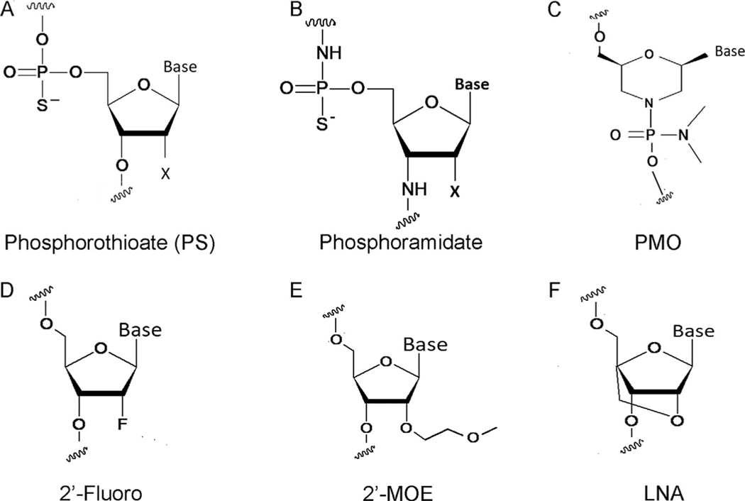
The most important advancement in the stabilization of the backbone is the development of the phosphorothioate (PS) containing oligonucleotide. PS containing oligonucleotides have a sulfur atom in place of one of the non-bridging oxygen atoms (Fig. 1A). This single modification enhances the ability of oligos to evade nuclease mediated digestion and renders them stable in tissue and plasma (Eckstein, 2000; Vortler & Eckstein, 2000). PS oligonucleotides also retain the ability to utilize RNASE H to exert their actions on their targets (Stein, Subasinghe, Shinozuka, & Cohen, 1988). The vast majority of oligonucleotides that have advanced to clinical trials possess PS modifications. Another backbone modification is the phosphoroamidate. As noted in Fig. 1B the 3′-oxygen in the deoxyribose ring is substituted for a 3′amino atom. Phophoramidates are highly stable and have a high affinity for their target RNA however they do not activate RNASE H (Gryaznov et al., 1995, 1996). This modification is only useful for situations when only RNA occupancy but not RNA degradation is required, and therefore not widely used.
FIG. 1.
The various chemical modifications that can be used to enhance oligonucleotide function.
The sugar phosphate backbone can also been replaced. The most successful modification has been the phosphorodiamidate morpholino oligonucleotide (PMO). In a PMO, the furanose has is replaced by a morpholine ring(Fig. 1C). The morpholine nitrogen atom is connected to the hydroxyl group of the 3′-side residue via a phosphorodiamidate linkage. PMOs are resistant to nucleases but again they do not activate RNASE H and thus are primarily used as blocking mechanisms or in situations where the goal is to modulate splicing (Summerton, 1999).
Several modifications to the sugar moiety are important to be aware of. First, modification to the 2′ position of the sugar moiety with a fluoro greatly increases binding affinity but does not affect stability (Fig. 1D). Moreover, the metabolites of 2-fluoro modified oligonucleotides can incorporate into the host DNA or RNA which presumably can lead to deleterious effects (Crooke et al., 2018). Therefore, ASOs which incorporate this modification need to be rigorously and thoroughly evaluated in safety and toxicity studies. Multiple drugs have entered into clinical trials with a 2′-O-methoxyethyl modification (Fig. 1E). 2-MOE modification enables resistance to endonucleases and lowers nonspecific protein binding which leads to reduced toxicity (Teplova et al., 1999). The sugar modification with the one of the largest gain in binding affinity is the locked nucleic acid (LNA). LNAs possess an analog of 2′-O-methyl RNA, where a 2′-substituent is bridged to the 4′-C atom (Fig. 1F). This modification enables nuclease resistance and enhanced binding affinity (Kumar et al., 1998). LNAs can also be used for both RNSASE H based and occupancy only targets. Multiple LNA based oligonucleotides have entered clinical trials. Finally, oligonucleotides can be conjugated to lipids, fatty acids, n-acetylgalactosamine or even packaged into lipid particles to modulate delivery.
Oligonucleotides can be single or double-stranded. Single-stranded oligos are amphipathic, readily bind to proteins, and are easily distributed to tissues. Double stranded oligos hide their hydrophobic groups and thus are hydrophilic and poorly distribute to tissues. For the purposes of pharmacokinetic discussion, we will focus on single-stranded PS-ASO which could have any 2′ modification. PS-ASOs have been effectively administered subcutaneously, intravenously, intrathecal, intravitreal, and even orally. The uptake of PS-ASOs is facilitated in part by interaction with various cell surface proteins and receptors which promote clathrin- or caveolin endocytosis (Crooke, Wang, Vickers, Shen, & Liang, 2017). The biophysical properties of PS-ASOs are such that special packaging is not necessarily required. However, the use of lipid particles to enhance delivery of ASOs to different organs has been reported (Wang et al., 2019). The bio-distribution of oligonucleotides has been characterized for various types of ASOs. In general the kidney and liver demonstrate the highest amount of accumulation (Altmann et al., 1996; Fluiter et al., 2003). Within the kidney, it has been reported that the proximal tubule has the largest degree of ASO uptake (Oberbauer, Schreiner, & Meyer, 1995). However, studies in diseased mouse models have demonstrated effective delivery to collecting duct cells and interstitial cells (Hajarnis et al., 2017). The elimination half life depends on the 2′ modification. Simple PS-ASOs have a reported half-life of 48h, whereas LNAs the half-life has been shown to be 5–7 days.
1.3. Why are oligonucleotides a good way to manipulate gene expression in the kidney?
Multiple properties of oligonucleotides make them excellent for use in the kidney. First delivery to the kidney is excellent due to the physical properties of ASOs described above. Second, depending on the modification of an ASO, the half-life in the tissue ranges from 48h to 1–2 weeks. Thus, sustained inhibition with weekly dosing is possible. Finally, ASOs are generally very well tolerated in mice without significant systemic side effects.
1.4. Examples of successful ASO usage in the kidney and other organs
Since Zamenick’s first report of ASO to inhibit RSV replication, scientists have worked to turn this concept into a new platform of therapeutic agents. The first ASO approved by the FDA was Fomivirsen in the 1990s for intravitreal use in the treatment of cytomegalovirus induced retinitis (Roehr, 1998). Since then at least four other ASOs have been approved by the FDA and dozens more are in clinical trials. Importantly, two ASOs are currently in clinical trials for the treatment of kidney diseases. RGLS4326 is an ASO targeted against miR-17 for the treatment of ADPKD. Initial studies demonstrated reduced cyst burden in mouse models of ADPKD with genetic deletion of the miR-17~92 cluster (Hajarnis et al., 2017; Patel et al., 2013). These studies led to the development of an ASO against miR-17. Pre-clinical studies have demonstrated the use of ASO against miR-17 in various mouse models of PKD reduces cyst growth (Hajarnis et al., 2017; Yheskel, Lakhia, Cobo-Stark, Flaten, & Patel, 2019). Currently RGLS4326 is in Phase 1 clinical trials in the United States. Second, RG-012 is an oligonucleotide designed to inhibit miR-21 for the treatment of Alport’s syndrome. Pre-clinical studies showed reduced kidney fibrosis with the use of ASO in a mouse model of Alport’s syndrome (Gomez et al., 2015). RG-012 is now in Phase 1 and 2 clinical trials.
2. Design of oligonucleotides
Determining the precise nucleic acid sequence can be challenging. The various modifications that are made to the ASO backbone and the nucleotide sequence both affect the melting temperature (Tm) of the molecule. A precise Tm is required to obtain high affinity binding and minimize off target effects. Most laboratories do not have the capabilities to produce their own oligonucleotides and thus must rely on commercial vendors. It is important to have clear communication with a proprietary vendor about the goals you wish to achieve with your oligonucleotide and the target sequence of interest. The vendor will then work with you on the targeted sequence to determine the optimal length and exact sequence of the oligonucleotide to reach the proper Tm to ensure optimal binding to the target RNA and minimize off target effects.
3. In vitro validation of oligonucleotides
In vitro validation is suggested prior to moving to in vivo studies for several reasons. First, in vitro studies allow for a quick assessment to determine whether an oligonucleotide is efficacious. Second, since in vitro studies require substantially less oligonucleotide, it is possible to test multiple configurations of an oligonucleotide in a cost-effective manner to determine which one has the potential to perform the best in in vivo studies. This is particularly important when designing target site blockers and ASOs designed to degrade a RNA transcript. In addition to efficacy, in vitro studies will provide the opportunity to find off-target effects. Oligonucleotides can be tested by measuring endogenous transcript levels. Alternatively, consideration can be given to introducing a luciferase vector with your transcript of choice into a cell line and then assessing luciferase activity in the presence or absence of an oligonucleotide. For the purposes of this chapter we will utilize an easily-transfectable cell line. However, these methods can be generalized to any other appropriate cell line.
3.1. Reagents
mIMCD3 cells (mouse internal medullary collecting duct cells) (ATCC: CRL-2123).
Lipofectamine 2000 (Invitrogen: Cat no. 11668019).
DMEM media (Invitrogen: Cat no. 11320082).
Fetal Bovine Serum (FBS).
Oligonucleotide.
Trypsin-EDTA 0.25% (Invitrogen: Cat no. 25200056).
1×Phosphate Buffered Saline (Invitrogen Cat no. 10010023). Centrifuge.
3.2. Protocol
Day 1
Start with a confluent 10cm plate of mIMCD3 cells maintained in DMEM media with 10% FBS serum.
Aspirate media from plate and wash adherent cells with PBS ×2.
Add 1.5mL of warm Trypsin. Place plate back in incubator for 3–5min.
Once cells are no longer adhered from plate, neutralize trypsin with 5mL of media.
Using pipet-aide move cell suspension into a 15mL falcon tube and pellet cells using centrifuge.
Aspirate media. Add 7mL of fresh media and pipet up and down 20 times to properly re-suspend cells.
Use a hemocytometer to determine volume required to plate 2×10⁵ cells per well in a six well plate. Seed cells in a six well plate and add 1.5mL of media to each well.
Day 2
Aspirate media from cells. Add exactly 1.5mL of fresh media to each well.
Prepare transfection reagent −100μL of transfection reagent is required for each well. In each 100μL combine 9μL of lipofectamine with 91μL of serum free media. Calculate how much total lipofectamine and serum free media will be needed for all wells (e.g., six wells will need 100μL×6=600μL) and make a master supply. Allow the suspension to sit for 5min at room temperature.
Prepare oligonucleotide mixture. The final concentration of oligonucleotide will be 20nM. To achieve this from a stock of 25mM of oligonucleotide, 1.2μL of oligonucleotide from at 25mM stock will be required for each well. Prepare a mixture of oligonucleotide and serum free media that reaches a total volume of 100μL for each well (e.g., 1.2μL of oligonucleotide +98.8μL of serum free media).
Combine oligonucleotide mix and transfection reagent mix. Briefly vortex. Allow suspension to sit at room temperature for 20min.
Add 200μL of the combined mixture to each well.
Change media after 6–24h.
Day 4
Harvest cells with trizol/protein lysis buffer/passive lysis buffer and proceed to validation studies.
3.2.1. In vitro validation studies
Validation will vary based on the type of oligonucleotide. Below is a list of suggested initial studies to validate efficacy of oligonucleotides based on class.
Repression of mRNA—oligonucleotide-treated cells should have reduced expression by qRT-PCR and Western blot of the targeted gene. Further studies can include assessment of the activation or inhibition of the pathway downstream of gene of interest.
Anti-miR—Measure expression by qRT-pCR of the targeted microRNA. Most importantly, the expression of mRNA targets of the microRNA of interest should be increased.
Pro-miR—First, measure expression by qRT-PCR of microRNA to ensure that transfection was successful. Second, the transcript levels of genes targeted by the microRNA should be reduced.
Target Site Blocker—The transcript level of the gene of interest should be increased by qRT-PCR. Furthermore, the protein levels of the gene of interest should also be increased.
Special considerations regarding target site blockers. Individual use of target site blocker should lead to de-repression of transcript due to endogenous microRNA regulation. However, this effect may be small and thus the use of mimics in combination with target site blockers can be considered.
4. In vivo use of oligonucleotides
Once an oligonucleotide has passed all in vitro validation tests, in vivo studies can began. Design of in vivo studies requires consideration of multiple factors. General dosing guidelines suggest 25mg/kg as a starting dose. The timing of ASO administration depends on the goals of the experiment. To demonstrate the considerations that must be given for an in vivo study design, we will use a polycystic kidney disease mouse model as an example. The principles delineated below can then be extrapolated to any kidney disease model and any other type of oligonucleotide.
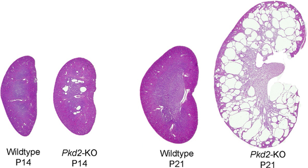
Autosomal dominant polycystic kidney disease (ADPKD) is caused by mutations in the PKD1 or PKD2 gene (Igarashi & Somlo, 2007). The disease is characterized by the formation of kidney cysts that arise from the nephron and compress the surrounding normal renal parenchyma. This leads to kidney failure. Several orthologous mouse models of ADPKD exist. For our example here we will utilize a mouse model where the Pkd2 gene has been deleted from the collecting duct using the Pkhd1;Cre transgene. In this mouse model at 14 days of age there are few cysts. However, at 21 days of age there is significant cyst burden (Lakhia et al., 2016) (Fig. 2). Several microRNAs demonstrate aberrant expression in concordance with cyst expansion. Thus, to target these microRNAs, we will first inject Anti-miRs on postnatal days 10, 11, and 12 as a loading dose (pre-cystic time points). Then will administer anti-miR on postnatal day 19 and sacrifice mice at postnatal day 28 for assessment.
FIG. 2.
Cyst progression in Pkhd1/Cre; Pkd2F/F mice is shown.
4.1. Reagents
Oligonucleotide (5mg)—commercial vendor.
Insulin syringe.
Scale.
Re-suspend oligonucleotide to a stock concentration of 20μg/μL and aliqot to avoid repeat freeze/thaw cycles.
Dilute one vial of stock oligonucleotide to 2μg/μL with isotonic buffer recommended by vendor, vortex, and keep on ice.
Weigh mouse to determine volume of oligonucleotide to administer (using a weight-based dose of 25mg/kg, a 5g mouse would receive 50μL of drug as injection).
Draw up appropriate amount of oligonucleotide into an insulin syringe.
Perform intraperitoneal/Subcutaneous injection in abdomen of mouse.
Unused diluted oligonucleotide can be stored at −20 for short periods of time.
Repeat injections on scheduled days.
Sacrifice mouse on 21 days of age. Collect blood, urine, flash freeze right kidney, perfuse and fix left kidney with PFA.
Perform molecular analysis for delivery and efficacy as delineated below.
4.2. Delivery assessment
Delivery can be assessed in several ways. First qRT-PCR will confirm delivery similar to in vitro methods. Second, a probe specific to the back-bone of the oligo can be designed such that in situ hybridization can locate the exact location of the oligo within the kidney. Finally, mass spectroscopy can be used to detect the oligonucleotide.
4.3. Efficacy assessment
The aim of oligonucleotide therapy may be to improve disease burden in a mouse model. For example, in ADPKD, we assess kidney weight/body weight ratio as a marker for cyst burden as well as serum creatinine, and cyst proliferation. Second a thorough understanding of the downstream effects of manipulating the transcript of interest is required. This will dictate the series of studies required to determine efficacy. For example, several miR-17 targets genes have been validated in ADPKD mouse models. Thus, injection of anti-miR against miR-17 should de-repress miR-17 targets genes.
4.4. Trouble-shooting/special considerations
Dosage Adjustments—As mentioned above, the recommend starting dose is 25mg/kg. If the desired molecular effect is only partially observed at this dosage, consider increasing the dosage or the frequency of administration. Although oligonucleotides have few systemic effects, higher dosages may induce unwarranted a systemic inflammatory response.
Study Design—In addition to adequately designing a study based on the expected course of disease, it is important to power the study to achieve the desired effect. Often the use of genetic studies which parallel pharmacologic studies can aide in predicting the effect and guide a power analysis. Alternatively, a preliminary trial to determine dose and efficacy can be performed and used to determine how to adequately power a study.
5. Conclusions
The use of oligonucleotides to manipulate gene expression is a valuable tool to better understand the pathophysiology of the kidney. Multiple advancements have made the use of oligonucleotides in the laboratory a relatively simple and accessible tool for gene manipulation. In conjunction with genetic studies, proper use of ASOs in disease models can serve as an excellent platform for pre-clinical drug development.
Acknowledgments
The work from the author’s laboratory is supported by National Institute of Health (R01DK102572) and the Department of Defense (D01 W81XWH1810673) to V.P. R.L. is supported by National Institute of Health (K08DK117049). Vishal Patel has applied for a patent related to the treatment of polycystic kidney disease using miR-17 inhibitors. The Patel lab has a sponsored research agreement with Regulus Therapeutics.
Footnotes
The remaining authors declare no conflict of interests.
References
- Altmann KH, Fabbro D, Dean NM, Geiger T, Monia BP, Muller M, et al. (1996). Second-generation antisense oligonucleotides: Structure-activity relationships and the design of improved signal-transduction inhibitors. Biochemical Society Transactions, 24(3), 630–637. [DOI] [PubMed] [Google Scholar]
- Cerritelli SM, & Crouch RJ(2009). Ribonuclease H: The enzymes in eukaryotes. The FEBS Journal, 276(6), 1494–1505. 10.1111/j.1742-4658.2009.06908.x. [DOI] [PMC free article] [PubMed] [Google Scholar]
- Crooke ST, Wang S, Vickers TA, Shen W, & Liang XH (2017). Cellular uptake and trafficking of antisense oligonucleotides. Nature Biotechnology, 35(3), 230–237. 10.1038/nbt.3779. [DOI] [PubMed] [Google Scholar]
- Crooke ST, Witztum JL, Bennett CF, & Baker BF (2018). RNA-targeted therapeutics. Cell Metabolism, 27(4), 714–739. 10.1016/j.cmet.2018.03.004. [DOI] [PubMed] [Google Scholar]
- Eckstein F. (2000). Phosphorothioate oligodeoxynucleotides: What is their origin and what is unique about them? Antisense & Nucleic Acid Drug Development, 10(2), 117–121. 10.1089/oli.1.2000.10.117. [DOI] [PubMed] [Google Scholar]
- Elbashir SM, Harborth J, Lendeckel W, Yalcin A, Weber K, & Tuschl T. (2001). Duplexes of 21-nucleotide RNAs mediate RNA interference in cultured mammalian cells. Nature, 411(6836), 494–498. 10.1038/35078107. [DOI] [PubMed] [Google Scholar]
- Fluiter K, ten Asbroek AL, de Wissel MB, Jakobs ME, Wissenbach M, Olsson H, et al. (2003). In vivo tumor growth inhibition and biodistribution studies of locked nucleic acid (LNA) antisense oligonucleotides. Nucleic Acids Research, 31(3), 953–962. [DOI] [PMC free article] [PubMed] [Google Scholar]
- Gomez IG, MacKenna DA, Johnson BG, Kaimal V, Roach AM, Ren S, et al. (2015). Anti-microRNA-21 oligonucleotides prevent Alport nephropathy progression by stimulating metabolic pathways. The Journal of Clinical Investigation, 125(1), 141–156. 10.1172/JCI75852. [DOI] [PMC free article] [PubMed] [Google Scholar]
- Gryaznov SM, Lloyd DH, Chen JK, Schultz RG, DeDionisio LA, Ratmeyer L, et al. (1995). Oligonucleotide N3′–>P5′ phosphoramidates. Proceedings of the National Academy of Sciences of the United States of America, 92(13), 5798–5802. [DOI] [PMC free article] [PubMed] [Google Scholar]
- Gryaznov S, Skorski T, Cucco C, Nieborowska-Skorska M, Chiu CY, Lloyd D, et al. (1996). Oligonucleotide N3′–>P5′ phosphoramidates as antisense agents. Nucleic Acids Research, 24(8), 1508–1514. [DOI] [PMC free article] [PubMed] [Google Scholar]
- Hajarnis S, Lakhia R, Yheskel M, Williams D, Sorourian M, Liu X, et al. (2017). microRNA-17 family promotes polycystic kidney disease progression through modulation of mitochondrial metabolism. Nature Communications, 8, 14395. 10.1038/ncomms14395. [DOI] [PMC free article] [PubMed] [Google Scholar]
- Igarashi P, & Somlo S. (2007). Polycystic kidney disease. Journal of the American Society of Nephrology:JASN,18(5),1371–1373.doi:ASN.2007030299[pii] 10.1681/ASN.2007030299. [DOI] [PubMed] [Google Scholar]
- Kumar R, Singh SK, Koshkin AA, Rajwanshi VK, Meldgaard M, & Wengel J. (1998). The first analogues of LNA (locked nucleic acids): Phosphorothioate-LNA and 2′-thio-LNA. Bioorganic & Medicinal Chemistry Letters, 8(16), 2219–2222. [DOI] [PubMed] [Google Scholar]
- Lakhia R, Hajarnis S, Williams D, Aboudehen K, Yheskel M, Xing C, et al. (2016). MicroRNA-21 aggravates cyst growth in a model of polycystic kidney disease. Journal of the American Society of Nephrology: JASN, 27(8), 2319–2330. 10.1681/ASN.2015060634. [DOI] [PMC free article] [PubMed] [Google Scholar]
- Liu J, Carmell MA, Rivas FV, Marsden CG, Thomson JM, Song JJ, et al. (2004). Argonaute2 is the catalytic engine of mammalian RNAi. Science, 305(5689), 1437–1441. 10.1126/science.1102513. [DOI] [PubMed] [Google Scholar]
- Meister G, Landthaler M, Patkaniowska A, Dorsett Y, Teng G, & Tuschl T. (2004). Human Argonaute2 mediates RNA cleavage targeted by miRNAs and siRNAs. Molecular Cell, 15(2), 185–197. 10.1016/j.molcel.2004.07.007. [DOI] [PubMed] [Google Scholar]
- Oberbauer R, Schreiner GF, & Meyer TW (1995). Renal uptake of an 18-mer phosphorothioate oligonucleotide. Kidney International, 48, 1226–1232. [DOI] [PubMed] [Google Scholar]
- Patel V, Williams D, Hajarnis S, Hunter R, Pontoglio M, Somlo S, et al. (2013). miR-17~92 miRNA cluster promotes kidney cyst growth in polycystic kidney disease. Proceedings of the National Academy of Sciences of the United States of America, 110(26), 10765–10770. 10.1073/pnas.1301693110. [DOI] [PMC free article] [PubMed] [Google Scholar]
- Roehr B. (1998). Fomivirsen approved for CMV retinitis. Journal of the International Association of Physicians in AIDS Care, 4(10), 14–16. [PubMed] [Google Scholar]
- Sharp PA (2009). The centrality of RNA. Cell, 136(4), 577–580. 10.1016/j.cell.2009.02.007. [DOI] [PubMed] [Google Scholar]
- Stein CA, Subasinghe C, Shinozuka K, & Cohen JS (1988). Physicochemical properties of phosphorothioate oligodeoxynucleotides. Nucleic Acids Research, 16(8), 3209–3221. [DOI] [PMC free article] [PubMed] [Google Scholar]
- Stephenson ML, & Zamecnik PC (1978). Inhibition of Rous sarcoma viral RNA translation by a specific oligodeoxyribonucleotide. Proceedings of the National Academy of Sciences of the United States of America, 75(1), 285–288. [DOI] [PMC free article] [PubMed] [Google Scholar]
- Summerton J. (1999). Morpholino antisense oligomers: The case for an RNase H-independent structural type. Biochimica et Biophysica Acta, 1489(1), 141–158. [DOI] [PubMed] [Google Scholar]
- Teplova M, Minasov G, Tereshko V, Inamati GB, Cook PD, Manoharan M, et al. (1999). Crystal structure and improved antisense properties of 2′-O-(2-methoxyethyl)-RNA. Nature Structural Biology, 6(6), 535–539. 10.1038/9304. [DOI] [PubMed] [Google Scholar]
- Vortler LC, & Eckstein F. (2000). Phosphorothioate modification of RNA for stereochemical and interference analyses. Methods in Enzymology, 317, 74–91. [DOI] [PubMed] [Google Scholar]
- Wang Y, Li Y, Zhang P, Baker ST, Wolfson MR, Weiser JN, et al. (2019). Regenerative therapy based on miRNA-302 mimics for enhancing host recovery from pneumonia caused by Streptococcus pneumoniae. Proceedings of the National Academy of Sciences of the United States of America, 116(17), 8493–8498. 10.1073/pnas.1818522116. [DOI] [PMC free article] [PubMed] [Google Scholar]
- Yheskel M, Lakhia R, Cobo-Stark P, Flaten A, & Patel V. (2019). Anti-microRNA screen uncovers miR-17 family within miR-17~92 cluster as the primary driver of kidney cyst growth. Scientific Reports, 9(1), 1920. 10.1038/s41598-019-38566-y. [DOI] [PMC free article] [PubMed] [Google Scholar]


