. 2021 Jan 5;299(1):E204–E213. doi: 10.1148/radiol.2021203957

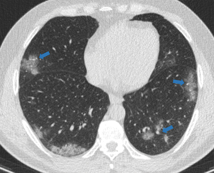
Figure 5d:

Examples of examination-level annotations on axial CT images. (a) Ground-glass opacities surrounding a nodular opacity (arrow) in the left lower lobe (halo sign). (b) Bilateral ground-glass opacities (arrows) with central clearing (reversed halo sign). (c) Reticular pattern without parenchymal opacity in the left upper lobe (arrows). (d) Perilesional vessel enlargement associated with bilateral ground-glass opacities (arrows). (e) Bronchial wall thickening most evident in the right lung (arrows). (f) Bronchiectasis in the left upper lobe (arrows). (g) Bilateral subpleural curvilinear lines (arrows). (h) Small bilateral pleural effusions (arrows). (i) Right pleural thickening (arrows). (j) Right pneumothorax (arrows). (k) Pericardial effusion (arrow). (l) Mediastinal lymphadenopathy (arrows) in the prevascular and bilateral lower paratracheal stations. (m) Pulmonary emboli (arrows) in the right lower and middle lobar pulmonary arteries.

Examples of examination-level annotations on axial CT images. (a) Ground-glass opacities surrounding a nodular opacity (arrow) in the left lower lobe (halo sign). (b) Bilateral ground-glass opacities (arrows) with central clearing (reversed halo sign). (c) Reticular pattern without parenchymal opacity in the left upper lobe (arrows). (d) Perilesional vessel enlargement associated with bilateral ground-glass opacities (arrows). (e) Bronchial wall thickening most evident in the right lung (arrows). (f) Bronchiectasis in the left upper lobe (arrows). (g) Bilateral subpleural curvilinear lines (arrows). (h) Small bilateral pleural effusions (arrows). (i) Right pleural thickening (arrows). (j) Right pneumothorax (arrows). (k) Pericardial effusion (arrow). (l) Mediastinal lymphadenopathy (arrows) in the prevascular and bilateral lower paratracheal stations. (m) Pulmonary emboli (arrows) in the right lower and middle lobar pulmonary arteries.