

Abstract
Colonoscopy is the gold-standard tool to investigate the colon which also allows to biopsy or treat intraluminal pathologies. About 900 000 colonoscopies are performed annually in UK. It is considered a relatively safe procedure; however, some serious complications might take place. The common complications of colonoscopy are bleeding and perforation. Splenic injury is a rare complication of colonoscopy which can be fatal. Our case report describes a grade two subscapular splenic haematoma after routine colonoscopy that has been managed conservatively.
Keywords: Surgery, General surgery, Ultrasonography
Background
Colonoscopy is the gold-standard tool to investigate the colon which also allows to biopsy or treat intraluminal pathologies. About 900 000 colonoscopies are performed annually in UK.1 2 It is considered a relatively safe procedure; however, some serious complications might take place. The common complications of colonoscopy are bleeding and perforation.1 2 Splenic injury is a rare complication of colonoscopy which can be fatal if not detected.3 Our case report describes a grade 2 subscapular splenic haematoma after routine colonoscopy that has been managed conservatively (table 1).
Table 1.
| Grade | Type of injury | Description of injury |
| I | Haematoma | Subcapsular,<10% |
| Laceration | Capsular tear,<1 cm in depth | |
| II | Haematoma | Subcapsular, 10%–50%; intraparenchymal,<5 cm in diameter |
| Laceration | Capsular tear, 1–3 cm in parenchymal depth, not involving trabecular vessel | |
| III | Haematoma | Subcapsular, >50% surface area or expanding, ruptured subcapsular or parenchymal haematoma; intraparenchymal haematoma, ≥5 cm or expanding |
| Laceration | >3 cm in parenchymal depth or involving trabecular vessel | |
| IV | Laceration | Segmental or hilar vessels, major devascularisation (>25%) |
| V | Laceration | Completely shattered spleen |
| Vascular | Hilar vascular injury that devascularises the spleen |
*Advance one grade for multiple injuries, up to grade III.
Case presentation
Our case was a 65-year-old woman who had a history of ischaemic heart disease, hypertension, COPD and Arnold Chiari malformation. Her medications list included aspirin, but she was not on any anticoagulants. Her surgical history includes open appendectomy, two caesarean sections, total abdominal hysterectomy and bilateral salpingo-oophrectomy.
She underwent a diagnostic colonoscopy, for altered bowel habits, which was normal and she went home on the same day. Three weeks later, she presented to the emergency department having persistent progressive dull aching pain in the left upper quadrant and radiating to her left shoulder which started 3 days after she had the colonoscopy. The pain was aggravated by movement and was hindering her breathing. She denied having fever, vomiting, chest or bowel symptoms. On examination, she was haemodynamically stable. There was no skin discoloration, but there was tenderness over the lateral side of the left lower ribs and the left upper quadrant and Kehr’s sign was positive. There were no signs of peritonitis.
Investigations
HGB level was normal. C reactive protein level was mildly elevated, but other inflammatory makers, liver and kidney functions and urinalysis were otherwise normal. A contrast-enhanced CT scan showed two subscapular splenic haematomas, the largest superior measured 11.5×6.1×8.4 cm and the smaller inferior one was 3.3×1.5×2.5 cm. Splenic parenchyma was normal and there was no free intraperitoneal fluid (figures 1 and 2). This was diagnosed as a grade 2 splenic injury according to the American Association for the Surgery of Trauma (AAST) injury scoring scale (table 1). The findings were discussed with endoscopist who performed the colonoscopy. He explained that it was a very challenging procedure which required various manoeuvring techniques, external manipulation and changing of the patient’s position.
Figure 1.

Coronal CT showing the haematoma on initial presentation
Figure 2.
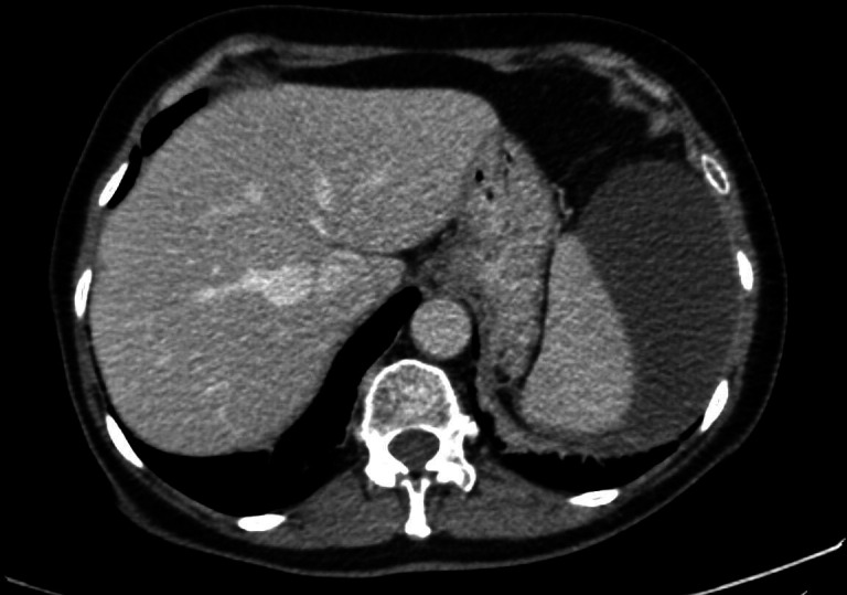
Axial CT showing the haematoma on initial presentation.
Treatment
The CT images were reviewed by the interventional radiology consultant on call who advised that there was no role for IR intervention. The patient was admitted for observation and pain control. She stayed in the hospital for 4 days during which two ultrasound scans were done and her HGB level was checked daily. The size of the haematoma as well as the HGB remained stable throughout the admission (figure 3). The patient was discharged home then and was advised on rest and strong pain killers were prescribed at home.
Figure 3.

USS showing the haematoma on day 2 after admission.
Outcome and follow-up
A CT with intravenous contrast scan was repeated a weak after discharge which showed decrease in the size of both haematomas. An ultrasound scan was done after 6 weeks which showed a residual collection measuring 4×2×1 cm. The patient was reviewed 3 months after discharge. She reported that the pain has completely settled. A follow-up ultrasound scan demonstrated resolving haematoma measuring 2.9×0.8×1 cm (figure 4). The patient has been reassured no further complications occurred so far.
Figure 4.

Showing resolving haematoma.
Discussion
Colonoscopy is an invasive procedure which carries a small risk of complications. Although the risk is low with diagnostic colonoscopy, it increases significantly when therapeutic procedures are performed. Iatrogenic injury of the spleen during colonoscopy is an extremely rare complication that has been described 174 times in medical literature with the reported incidence of 1:100 000 colonoscopies.2 It can be ranging from a minor subscapular haematoma that might not be detected to splenic rupture.
Postcolonoscopy splenic injury is more common in females, male: female ratio is 1: 3. The average age of incidence in the reported cases is 63 years.3 It is difficult to predict the precipitating factors of this condition, but the risk may be higher in patients who have adhesions from previous abdominal surgeries, splenomegaly, complex intraluminal lesions and anticoagulant medications.4
The most commonly proposed mechanism of injury is traction on the splenic ligaments or adhesions while performing manoeuvres to pass through the splenic flexure. Other mechanisms might be direct injury or blunt trauma from outside while manipulating or straightening the colonoscope.3
Splenic injury is usually presented acutely after the procedure and manifest as left upper quadrant pain with or without haemodynamic instability.4 FAST scan can be a quick tool to detect free intraperitoneal fluid. Contrast-enhanced CT is the gold standard to diagnose the grade of splenic injury and it also helps to rule out other possible complications, for example, perforation or bleeding. Management of splenic injury depends on the severity according to the AAST classification system and the haemodynamic state of the patient.5
Management options range from conservative, non-intervention approach to splenic angiography and embolisation or even splenectomy in high grade injuries or unstable patients.
Learning points.
Endoscopists and surgical trainees must be aware of this serious complication of colonoscopy despite its rarity.
Postcolonoscopy splenic injury often presents early after the procedure and a high index of suspicion is required to be identified and managed accordingly.
This condition might also run a more benign course as discussed in our manuscript.
Footnotes
Contributors: AA has initially assessed the patient and diagnosed her condition and followed her case. AK was the responsible consultant for the care of the patient throughout her admission and follow up. OA participated in writing the manuscript.
Funding: The authors have not declared a specific grant for this research from any funding agency in the public, commercial or not-for-profit sectors.
Competing interests: None declared.
Patient consent for publication: Obtained.
Provenance and peer review: Not commissioned; externally peer reviewed.
References
- 1.Shenbagaraj L, et al. Endoscopy in 2017: a national survey of practice in the UK. BMJ:2018–100970. [DOI] [PMC free article] [PubMed] [Google Scholar]
- 2.Kamath AS, Iqbal CW, Sarr MG, et al. Colonoscopic splenic injuries: incidence and management. J Gastrointest Surg 2009;13:2136–40. 10.1007/s11605-009-1064-7 [DOI] [PubMed] [Google Scholar]
- 3.Li S, Gupta N, Kumar Y, et al. Splenic laceration after routine colonoscopy, a case report of a rare iatrogenic complication. Transl Gastroenterol Hepatol 2017;2:49. 10.21037/tgh.2017.04.11 [DOI] [PMC free article] [PubMed] [Google Scholar]
- 4.Rees CJ, et al. UK Key Performance Indicators & Quality Assurance Standards for Colonoscopy. BMJ:2016–312044. [DOI] [PMC free article] [PubMed] [Google Scholar]
- 5.Moore EE, et al. Scaling system for organ specific injuries. J Trauma Inj Infect Crit Care 2010;69:1600–1. [Google Scholar]