Abstract
Objectives
The impact of Pseudomonas aeruginosa on the prognosis of bronchiectasis remains controversial. This study aimed to explore the prognostic value of P. aeruginosa in adult patients with bronchiectasis in central-southern China.
Patients and methods
This prospective cohort study enrolled 1,234 patients with bronchiectasis between 2013 and 2019. The independent impact of P. aeruginosa on all-cause mortality, annual exacerbations, and hospitalizations was assessed.
Results
P. aeruginosa was isolated from 244 patients (19.8%). A total of 188 patients died over a follow-up period of 16 (1–36) months. Patients with P. aeruginosa had a longer disease course, poorer lung function, more lung lobe involvement, and more severe Bronchiectasis Severity Index (BSI) stage than those without P. aeruginosa. The independent impact of P. aeruginosa was observed on frequent hospitalizations but not on mortality and frequent exacerbations. Moderate- or high-risk comorbidities increased the risk of mortality (hazard ratio [HR]: 1.93, 95% confidence interval [CI]: 1.26–2.95), and this effect was magnified by the presence of P. aeruginosa (HR: 2.11, 95% CI: 1.28–3.48).
Conclusions
P. aeruginosa infection acts as a marker of disease severity as well as predictor of frequent hospitalizations. P. aeruginosa had no independent effect on all-cause mortality. P. aeruginosa combined with moderate- or high-risk comorbidities posed an increased risk of mortality. The management of comorbidities may be a critical target during the treatment of P. aeruginosa infection in bronchiectasis.
KEY MESSAGE:
P. aeruginosa increased the risk of frequent hospitalizations; however, it had no independent impact on all-cause mortality.
P. aeruginosa combined with moderate- or high-risk comorbidities posed an increased risk of mortality.
The management of comorbidities may be a critical target during the treatment of P. aeruginosa infection in bronchiectasis.
Keywords: Bronchiectasis, Pseudomonas aeruginosa, mortality, hospitalizationsexacerbations
Introduction
Non-cystic fibrosis bronchiectasis (hereafter referred to as bronchiectasis) is a chronic progressive bronchus or bronchiole dilation due to complex interactions between recurrent infection, unbalanced immune regulation, impaired mucociliary clearance, and progressive airway structure damage or obstruction [1]. The estimated prevalence of bronchiectasis is 52.3 cases per 100,000 in the United States [2] and 67 cases per 100,000 in Germany [3], and the prevalence increases each year by 8.7% [4]. The crude annual mortality rate of bronchiectasis varies between 2% and 10% [5–7], which continues to pose a heavy disease burden on both developing and developed countries.
Pseudomonas aeruginosa (P. aeruginosa) tops the list of organisms isolated from sputum in patients with bronchiectasis, either during stable state or exacerbation [8,9]. It is usually considered to be highly responsible for the poor clinical outcomes of bronchiectasis. As bronchiectasis is a heterogeneous disease, the aetiology, microbial spectrum, and disease severity vary under different geographic and economic background [10]. Some studies have suggested that chronic infection with P. aeruginosa independently affects clinical outcomes, including hospitalizations, exacerbations [11,12] and mortality [12,13], in bronchiectasis, whereas other studies have drawn contrasting conclusions [5,14,15]. It remains controversial whether P. aeruginosa has an independent effect on clinical outcomes or is simply a marker of disease severity.
In China, bronchiectasis has long been neglected, and studies regarding clinical profiles and the impact of P. aeruginosa infection on its prognosis are scanty, mainly concentrated in two developed metropolises [12,16]. Therefore, we aimed to explore the clinical profiles of P. aeruginosa infection in bronchiectasis exacerbation and its effect on all-cause mortality, future hospitalizations, and exacerbations in a large tertiary hospital in central-southern China.
Patients and methods
Study design and participants
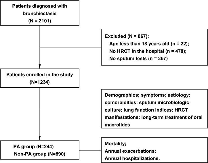
This prospective cohort study collected data on patients with bronchiectasis exacerbation between 2013 and 2019 at the Second Xiangya Hospital of Central South University. A flowchart of patient enrolment is displayed in Figure 1. Consecutive adult patients with a diagnosis of bronchiectasis based on high-resolution computed tomography (HRCT) scans and presenting corresponding respiratory symptoms attributable to bronchiectasis exacerbation were enrolled according to the guidelines [17]. Patients were excluded if they lacked HRCT scan results or records of sputum culture in the hospital. Only the first medical records of patients with multiple admissions were reviewed for our analysis. This study was approved by the institutional review board of the Second Xiangya Hospital in China (luoh202006), and informed consent was obtained from all participants.
Figure 1.
Flow chart of patient enrolment and analysis. HRCT: high-resolution computed tomography; PA: Pseudomonas aeruginosa.
Data collection
Data on demographics (age, sex, occupation, height, and weight), disease course, symptoms, aetiology, comorbidities, forced expiratory volume in the first second (FEV1% predicted), radiologic findings, long-term treatment of macrolides, and Medical Research Council (MRC) dyspnoea score were collected. The aetiology and diseases associated with bronchiectasis were identified according to Spanish guidelines [18].
The Bronchiectasis Aetiology Comorbidity Index (BACI) [19], which is a sum score (range, 0–55) of 13 weighted diseases, was used to assess comorbidities, where higher scores denote an increasing burden of comorbidities specified in bronchiectasis. Patients were classified into the following tertiles: low-risk comorbidities (for patients with a score of 0), moderate-risk comorbidities (for patients with scores ≥1 and <6), and high-risk comorbidities (for patients with a score ≥6).
The radiological severity of bronchiectasis was assessed using a modified Reiff score (range,1–18), which rates the number of involved lobes (of a total of six, with the lingula considered separate) and the degree of dilatation (tubular = 1, varicose = 2, and cystic = 3), as in previous bronchiectasis studies [11,20].
The Bronchiectasis Severity Index (BSI) (age, body mass index [BMI], dyspnoea, exacerbation, FEV1% predicted, microbial colonization, radiological extension, and range of 0–26) was used to assess bronchiectasis severity as mild (score range, 0–4), moderate (score range, 5–8), and severe (score range, 9–26). As microbial colonization status was unavailable in our study, we used data on baseline sputum microbial culture in the hospital as an alternative to microbial colonization.
Sputum microbiology and group assignment
All microbiological tests were performed on spontaneous sputum samples. Sputum samples were considered eligible if they contained more than 25 leukocytes and less than 10 squamous cells per low-powered field (10 × 10). Specific microorganism infection was considered if sputum cultures were positive on one or more occasions and accorded with clinical physician judgement.
We assigned patients into the following two groups: (1) the PA group for those with P. aeruginosa isolation from sputum and (2) the non-PA group for those without P. aeruginosa isolation from sputum.
Follow-up and clinical outcomes
Patients were followed up after discharge via specifically designed telephone interviews every 1–3 months. Follow-up was completed on 30 June 2020. There was a median follow-up duration of 16 (1–36) months, in which the overall number of exacerbations and hospitalizations due to exacerbations during the follow-up period were recorded, and mortality was assessed.
The clinical outcomes in this study were all-cause mortality, frequency of annual exacerbations, and frequency of annual hospitalizations due to exacerbations.
An exacerbation of bronchiectasis was defined as at least three of the following symptoms: cough frequency, sputum volume and/or consistency, sputum purulence, dyspnoea and/or exercise tolerance, fatigue and/or malaise, and haemoptysis; where the condition deteriorated for at least 48 h, and beyond the daily variation range, a change in treatment was required [21]. Patients were considered to have frequent exacerbations if there were two or more annual exacerbations (total number of exacerbations divided by follow-up years) and frequent hospitalizations if there were one or more annual hospitalizations (total number of hospitalizations divided by follow-up years).
Statistical analysis
Descriptive statistics of demographic and clinical variables are presented as median (interquartile range) or counts (n) and proportions (%), unless otherwise stated. Data comparisons were applied between the PA and non-PA groups. The chi-squared test was used for categorical data. The student’s t-test was used for continuous variables showing normal distributions and the Kruskal–Wallis test for those with non-normal distributions. Unless stated otherwise, the significance level was set at p < .05.
Kaplan–Meier curves were constructed to illustrate survival data, and Cox proportional hazard regression analysis was used for univariate and multivariable mortality analysis to estimate hazard ratios (HRs) and their 95% confidence intervals (CIs). Similarly, univariate and multivariate logistic regression analyses were performed to identify variables associated with bronchiectasis hospitalizations and exacerbations, and odds ratios (ORs) and their 95% CIs were calculated. We included the following baseline variables when studying potential in the univariate analyses: sex; age; BMI; smoking habit; symptoms of haemoptysis; exacerbations in the previous year; MRC dyspnoea score; Reiff score; FEV1% predicted; BACI score; aetiology and associated diseases (unknown aetiology, chronic respiratory disease; connected tissue disease, post-infectious); long-term oral macrolides; sputum microbiologic culture (P. aeruginosa, Acinetobacter baumannii, Klebsiella pneumoniae, Escherichia coli, Haemophilus influenzae, Staphylococcus aureus, Candida, and Aspergillus fumigatus); BSI stage; status of PA; and comorbidities (only for survival analysis). Each variable was initially tested individually before we added all variables that showed a univariate association (p < .10) to the multivariable model, except for sex and age, which had to appear in both models. Backward stepwise selection (likelihood ratio) (pin < 0.05 and pout > 0.10) was used to determine the factors associated with mortality, hospitalizations, and exacerbations. The Hosmer–Lemeshow goodness-of-fit test was performed to assess the overall fit of the final model. All data were analysed using SPSS version 26.0 (SPSS Inc., Chicago, IL, USA).
Results
Patients characteristics
We recruited 1,234 patients in this study (Table 1). Overall, there were slightly more men than women (52% versus 48%). The median age was 62.5 years. Of the patients, 94.2% were over 40 years of age and 39.5% over 65 years of age. Two fifths of them had a history of smoking. The median disease course was eight years. Regarding aetiology of bronchiectasis, 407 patients (33%) had post-infection, 355 (28.8%) chronic respiratory disease (chronic obstructive pulmonary disease [COPD], asthma, and pneumoconiosis), 73(5.9%) connective tissue disease, and 254 (20.6%) unknown aetiology (Table 1). A total of 428 patients (34.7%) had low-risk comorbidities (BACI score of 0), 495 (40.1%) moderate-risk comorbidities (BACI score of 1–6), and 311 (25.2%) high-risk comorbidities (BACI score > 6). The majority of the patients (84.7%) were in the moderate or severe stage according to BSI score.
Table 1.
General characteristics of bronchiectasis patients.
| Total | PA group | Non-PA group | p-value | |
|---|---|---|---|---|
| Patients | 1234 (100%) | 244 (19.8%) | 990 (81.2%) | – |
| Demographics | ||||
| Male | 642 (52%) | 106 (43.4%) | 536 (54.1%) | .03 |
| Age (years) | 62.5 (53.0–70.0) | 62 (53–68) | 63 (53–70) | .532 |
| Body mass index (kg*m–2) | 20.83 (18.29–23.60) | 20.03 (17.63–22.66) | 21.05 (18.43–23.88) | <.001 |
| Smokers and ex-smokers | 487 (39.5%) | 84 (34.4%) | 403 (40.7%) | .072 |
| Clinical status | ||||
| Disease course (years) | 8 (1–20) | 13 (5–30) | 5.5 (1–20) | <.001 |
| Haemoptysis | 112 (45.9%) | 132 (54.1%) | .077 | |
| MRC dyspnoea score | 2 (0–3) | 2 (1–2) | 2 (0–3) | .641 |
| Annual exacerbations | 2 (1–3) | 1 (1–2) | 1 (1–2) | .397 |
| Annual hospitalizations | 0 (0–1) | 1 (0–2) | 0.5 (0–1) | <.001 |
| Aetiology and associated diseases | ||||
| Post-infectious | 407 (33.0%) | 80 (32.8%) | 327(33.0%) | .942 |
| Associated with chronic respiratory disease | 355 (28.8%) | 76 (31.1%) | 280(28.3%) | .376 |
| Associated with connected tissue disease | 73 (5.9%) | 9 (3.7%) | 64(6.5%) | .128 |
| Unknown aetiology | 254 (20.5%) | 59 (24.2%) | 195 (19.7%) | .133 |
| Comorbidities | ||||
| Chronic obstructive pulmonary disease | 346 (28.0%) | 70 (28.7%) | 276 (27.9%) | .801 |
| Asthma | 137 (11.1%) | 29 (11.9%) | 108 (10.9%) | .733 |
| Pulmonary hypertention | 107 (8.7%) | 40 (16.4%) | 67 (6.8%) | <.001 |
| Ischaemic cardiomyopathy | 134 (10.9%) | 17 (7.0%) | 117 (11.8%) | .029 |
| Diabetes | 100 (8.1%) | 21 (8.6%) | 79 (8.0%) | .748 |
| Connective tissue disease | 110 (8.9%) | 20 (8.2%) | 90 (9.1%) | .661 |
| BACI score | 3 (0–6) | 3 (0–7) | 3 (0–6) | .186 |
| BACI score = 0 | 428 (34.7%) | 78 (32.0%) | 350 (35.4) | – |
| BACI score 1–6 | 495 (40.1%) | 98 (40.2%) | 397 (40.1%) | – |
| BACI score over 6 | 311 (25.2%) | 68 (27.9%) | 243 (24.5%) | – |
| Lung function | ||||
| FEV1(% predicted) | 53.12 (36.47–71.27) | 45.5 (31.5–65.1) | 52.9 (35.8–72.1) | <.001 |
| FEV1(% predicted) <50% | 555 (45.0%) | 74 (30.3%) | 195 (19.7%) | <.001 |
| Radiology | ||||
| Cystic bronchiectasis | 244 (19.8%) | 154 (63.1%) | 90 (36.9%) | <.001 |
| Involved lung Lobes | 3 (0–6) | 6 (3–6) | 2 (1–6) | <.001 |
| Reiff score | 6 (1–18) | 12(6–18) | 4 (2–9) | <.001 |
| BSI stage | <.001 | |||
| Mild | 189 (15.3%) | 0 (0%) | 189 (19.1%) | – |
| Moderate | 604 (59.0%) | 0 (0%) | 189 (19.1%) | – |
| Severe | 441 (35.7%) | 46 (18.9%) | 558 (56.4%) | – |
| Treatment | 3 (0–22) | 198 (81.1%) | 243 (24.5%) | – |
| Long-term oral macrolides | 54 (4.4%) | 18 (7.4%) | 36 (3.6%) | .011 |
| Follow up | ||||
| More than 2 annual exacerbations | 149 (12.1%) | 43 (17.6%) | 106 (10.7%) | .002 |
| More than 1 annual hospitalization | 226 (18.3%) | 65 (26.6%) | 161 (16.3%) | <.001 |
| Mortality rate (deaths per person*year of observation) | 188/2204 | 45/444 | 143/1760 | – |
Data are presented as n (%) or median (interquartile range) unless otherwise stated. PA: Pseudomonas aeruginosa; MRC: Medical Research Council; FEV1: forced expiratory volume in 1 s; BACI: Bronchiectasis Aetiology Comorbidity. Index; BSI: Bronchiectasis Severity Index.
Microbiology
Overall, 479 (38.8%) patients had positive sputum-test results for pathogenic microorganisms. Sixty-three patients (5.1%) were co-infected with P. aeruginosa and other microorganisms. As regards bacteria, P. aeruginosa was the most common positive pathogen in 244 (19.8%) patients, followed by Acinetobacter baumannii in 48 (3.9%), Klebsiella pneumoniae in 47 (3.8%), Escherichia coli in 21 (1.7%), and Haemophilus influenzae in 13 (1.0%). Regarding fungi, Candida was more common (n = 108, 8.8%) than Aspergillus fumigatus (n = 57, 4.6%). The details are shown in Table 2.
Table 2.
Microbiological characteristics of subjects with bronchiectasis.
| Pathogens | Numbers (N) | Percentage# | Percentage* |
|---|---|---|---|
| Total# | 1234 | – | – |
| Total* | 496 | 40.2% | – |
| Bacteriologic | |||
| Pseudomonas aeruginosa | 244 | 19.8% | 49.2% |
| Acinetobacter baumannii | 48 | 3.9% | 9.7% |
| Klebsiella pneumoniae | 47 | 3.8% | 9.5% |
| Stenotrophomonas maltophilia | 24 | 1.9% | 4.8% |
| Escherichia coli | 21 | 1.7% | 4.2% |
| Mycoplasma pneumoniae | 24 | 1.9% | 4.8% |
| Staphylococcus aureus | 17 | 1.4% | 3.4% |
| Haemophilus influenzae | 13 | 1.1% | 2.6% |
| Mycobacterium tuberculosis | 13 | 1.1% | 2.6% |
| Enterobacter cloacae | 9 | 0.7% | 1.8% |
| Legionella pneumophila | 7 | 0.6% | 1.4% |
| Serratia marcescens | 5 | 0.4% | 1.0% |
| Mycological | |||
| Candida | 108 | 8.8% | 21.8% |
| Aspergillus fumigatus | 57 | 4.6% | 11.5% |
| Other species | 27 | 2.20% | 5.4% |
#Indicates the patients included in this study. *Indicates the patients who had positive culture in sputum. Other species include Streptococcus pneumoniae, Enterococcus faecium, Proteus mirabilis, Alcaligenes, Malodorous pseudomonas, Acinetobacter roffei, Moraxella catarrhalis, Enterobacter aerogenes, Candida tropicalis and Mucor.
Differences in characteristics between PA group and non-PA group patients
Compared to those in the non-PA group (Table 1), patients in the PA group had a higher female ratio (56.6% versus 45.9%), lower BMI (20.04[17.63–22.66] kg/m2 versus 21.05 [18.43–23.88] kg/m2, p < .001), and longer disease course (13 [5–30] years versus 5.5 [1–20] years). As regards etiology, there was no significant difference between the PA and non-PA groups. In terms of comorbidities, patients in the PA group had a higher proportion of pulmonary hypertension (16.4% versus 6.8%, p < .001) and a similar BACI score to those in the non-PA group. Patients in the PA group had poorer lung function (lower FEV1% predicted), more cystic bronchiectasis, more lung lobes involved, and more severe disease stages according to BSI score. Patients in the PA group also had more frequent future annual exacerbations (17.6% versus 10.1%, p = .002) and hospitalizations (26.6% versus 16.3%, p < .001).
All-cause mortality
Overall, 188 patients (15.2%) died during the follow-up period. The all-cause mortality was 18.4% (45 of 244 patients) in the PA group and 14.4% (143 of 990 patients) in the non-PA group.
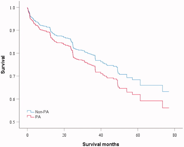
The univariate analysis of the influence of P. aeruginosa on mortality showed a higher risk trend but without statistically significant differences (HR: 1.26, 95% CI: 0.90–1.76, p = 0.176) (Figure 2). Multivariate analysis showed that age (HR: 1.03, 95% CI: 1.02–1.05, p < .001), BACI score (HR: 1.06, 95% CI: 1.03–1.10, p < .001), and Acinetobacter baumannii (HR: 1.96, 95% CI: 1.21–3.18, p = .006) contributed independently to a higher risk of mortality, whereas higher BMI (HR: 0.93, 95% CI: 0.90–0.97, p = .001) had a protective effect against mortality (Table 3). In the second multivariate analysis model of composite scores, BSI stage was associated with mortality.
Figure 2.
Kaplan–Meier log-rank test survival curve and univariate analysis for mortality. Pseudomonas aeruginosa (PA) versus non-PA. Hazard ratio for death for PA infection was 1.26 (95% CI 0.90–1.76, p = .176) in Cox proportional hazard regression analysis.
Table 3.
Univariate and multivariate Cox regression analysis of factors associated with mortality.
| Variables | Crude HR (95% CI) | p value | Adjusted HR (95% CI)a | p value | Adjusted HR (95% CI)b | p value | Adjusted HR (95% CI)c | p value |
|---|---|---|---|---|---|---|---|---|
| Pseudomonas aeruginosa | 1.26 (0.90–1.76) | .176 | – | – | – | – | – | – |
| Female vs male | 0.90 (0.67– 1.19) | .452 | 1.09 (0.74–1.60) | .663 | 1.12 (0.78–1.62) | 0.535 | 1.01 (0.69–1.48) | .948 |
| Age (years) | 1.04 (1.03–1.06) | <.001 | 1.03 (1.02–1.05) | <.001 | – | – | 1.04 (1.02–1.05) | <.001 |
| BMI (kg*m–2) | 0.92 (0.89–0.96) | <.001 | 0.93 (0.90–0.97) | <.001 | – | – | 0.93 (0.90–0.97) | <.001 |
| Smoking vs non-smoking | 1.31 (0.98–1.74) | .066 | 1.18 (0.80–1.73) | .401 | 1.21 (0.84–1.75) | 0.311 | 1.19 (0.82–1.74) | .355 |
| Haemoptysis | 0.69 (0.51–0.94) | .018 | 0.83 (0.60–1.14) | .251 | 0.77 (0.56–1.06) | 0.104 | 0.81 (0.59–1.12) | .198 |
| Exacerbations in the previous year | 1.03 (0.94–1.13) | .496 | – | – | – | – | – | – |
| MRC dyspnoea score | 0.95 (0.84–1.07) | .376 | – | – | – | – | – | – |
| Reiff score | 1.01 (0.98–1.03) | .675 | – | – | – | – | – | – |
| FEV1% predicted <50% versus ≥ 50% | 1.12 (0.84–1.50) | .427 | – | – | – | – | – | – |
| BACI score | 1.09 (1.06–1.12) | <.001 | 1.06 (1.03–1.10) | <.001 | 1.08(1.06–1.12) | <0.001 | – | – |
| Aetiology and associated diseases | – | – | ||||||
| Unknown aetiology | 1.00 (reference) | – | 1.00 (reference) | – | 1.00 (reference) | – | 1.00 (reference) | – |
| Associated with chronic respiratory disease | 1.51 (0.97–1.36) | .071 | 0.91 (0.56–1.46) | .683 | 0.88 (0.54–1.42) | 0.594 | 1.00 (0.63–1.60) | .996 |
| Associated with connected tissue disease | 1.19 (0.59–2.39) | .623 | 0.99 (0.49–2.03) | .993 | 0.83 (0.41–1.68) | 0.600 | 0.98 (0.48–2.02) | .960 |
| Post-infectious | 1.47 (0.95–2.27) | .085 | 1.34 (0.86–2.10) | .198 | 1.26 (0.81–1.96) | 0.313 | 1.50 (0.96–2.34) | .075 |
| Long-term oral macrolides | 0.74 (0.30–1.79) | .497 | – | – | – | – | – | – |
| Acinetobacter baumannii | 2.84 (1.77–4.57) | <.001 | 1.96 (1.21–3.18) | .006 | – | – | – | – |
| Klebsiella pneumoniae | 1.43 (0.76–2.70) | .271 | – | – | – | – | – | – |
| Escherichia coli | 0.84 (0.27–2.63) | .765 | – | – | – | – | – | – |
| Haemophilus influenzae | 0.713 (0.1–5.09) | .736 | – | – | – | – | – | – |
| Staphylococcus aureus | 2.55 (1.13–5.75) | .024 | – | – | – | – | – | – |
| Candida | 2.27 (1.54–3.33) | <.001 | 1.30 (0.85–1.98) | .226 | – | – | – | – |
| Aspergillus fumigatus | 1.33 (0.70–1.52) | .38 | – | – | – | – | – | – |
| BSI stage | ||||||||
| Mild | 1.00 (reference) | – | – | – | 1.00 (reference) | – | – | – |
| Moderate | 1.80 (1.07–3.03) | .026 | – | – | 1.75 (1.04–2.95) | 0.035 | – | – |
| Severe | 2.57 (1.53–4.33) | <.001 | – | – | 2.36 (1.40–3.98) | 0.001 | – | – |
| Status of PA and comorbidities | ||||||||
| Non-PA with low-risk comorbidities | 1.00 (reference) | – | – | – | – | – | 1.00 (reference) | – |
| Non-PA with moderate or high-risk comorbidities | 2.46(1.62–3.74) | <.001 | – | – | – | – | 1.93 (1.26–2.95) | .003 |
| PA with low-risk comorbidities | 1.55(0.70–3.40) | .28 | – | – | – | – | 1.56 (0.71–3.44) | .268 |
| PAwith moderate or high-risk comorbidities | 2.77(1.69–4.55) | <.001 | – | – | – | – | 2.11 (1.28–3.48) | .003 |
HR: Hazard ratio; CI: confidence interval; BMI: body mass index; PA: Pseudomonas aeruginosa; MRC: Medical Research Council; FEV1: forced expiratory volume in 1 s; BSI: Bronchiectasis Severity Index; BACI: Bronchiectasis Aetiology Comorbidity Index. Data are shown as estimated HRs (95% CIs) of the explanatory variables.
aInitial multivariable model comprised gender, age, BMI, smoking habit, haemoptysis, BACI score, aetiology and associated diseases, Acinetobacter baumanniiand Candida. Final multivariable model was adjusted for age, BMI, BACI score, Acinetobacter baumannii.
bInitial multivariable model comprised gender, smoking habit, haemoptysis, BACI score, aetiology and associated diseases and BSI stage. Final multivariable model was adjusted for BACI score and BSI stage.
cInitial multivariable model comprised gender, age, BMI, smoking habit, haemoptysis, aetiology and associated diseases and status of PA and comorbidities. Final multivariable model was adjusted for age, BMI and status of PA and comorbidities.
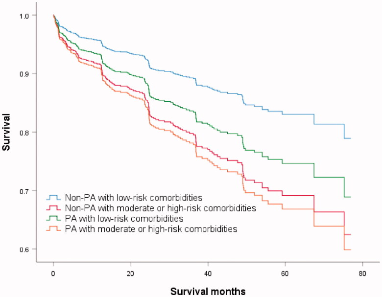
Figure 3.
Kaplan–Meier log-rank test survival curve. Comparison between four subgroups: non-Pseudomonas aeruginosa (PA) with low-risk comorbidities; non-PA with moderate or high-risk comorbidities; PA with low-risk comorbidities; PA with moderate or high-risk comorbidities.
We hypothesised that the association of P. aeruginosa with mortality may be comorbidity-dependent. Based on the condition of the P. aeruginosa infection andBACI score, we reclassified the patients into four subgroups as follows: (1) non-PA with low-risk comorbidities (n = 350); (2) non-PA with moderate- or high-risk comorbidities (n = 640); (3) PA with low-risk comorbidities (n = 78); and (4) PA with moderate- or high-risk comorbidities (n = 166). The survival curves of the four subgroups are shown in Figure 3. In the third multivariable analysis model of subgroups (Table 3), P. aeruginosa with low-risk comorbidities had no impact on mortality (HR: 1.56, 95% CI: 0.71–3.44, p = 0.268). Moderate- or high-risk comorbidities increased mortality risk (HR: 1.93, 95% CI: 1.26–2.95, p = .003), and the effect was magnified in the presence of P. aeruginosa (HR: 2.11, 95% CI: 1.28–3.48, p = .003).
Future exacerbations and hospitalizations
Of the 1,234 patients, 494 (40.0%) experienced frequent future exacerbations (two or more future annual exacerbations), and 258 (20.9%) had frequent future hospitalizations (more than one future annual hospitalization). Multivariate analysis indicated that P. aeruginosa did not increase the risk of frequent future exacerbations (odds ratio [OR]: 1.15, 95% CI: 0.83–1.59, p = .400) (Table 4); however, it indicated a higher possibility of frequent hospitalizations (OR: 1.51, 95% CI: 1.08–2.13, p = .018) (Table 5). Comorbidities (BACI score) and BSI score also increased the risk of frequent hospitalizations (OR: 1.06, 95% CI: 1.02–1.10, p = .003).
Table 4.
Univariate and multivariate logistic regression analysis of factors associated with frequent exacerbations.
| Variables | Crude OR (95% CI) | p value | Adjusted OR (95% CI)a | p value | Adjusted OR (95% CI)b | p value |
|---|---|---|---|---|---|---|
| Pseudomonas aeruginosa | 1.47 (1.11–1.95) | .008 | 1.15 (0.83–1.59) | .400 | – | – |
| Female versus male | 1.01 (0.81–1.27) | .907 | 1.14 (0.87–1.48) | .339 | 1.14 (0.89–1.45) | .299 |
| Age (years) | 1.01 (1.00–1.02) | .153 | 1.00 (0.99–1.02) | .414 | – | – |
| Body mass index (kg*m–2) | 0.95 (0.93–0.98) | .001 | 0.97 (0.94–1.01) | .099 | – | – |
| Smoking vs non-smoking | 1.19 (0.94–1.50) | .152 | – | – | – | – |
| Haemoptysis | 0.82 (0.65–1.04) | .094 | 0.96 (0.86–1.07) | .750 | 1.02 (0.80–1.32) | .865 |
| Exacerbations in the previous year | 2.01 (1.79–2.26) | <.001 | 1.93 (1.72–2.18) | <.001 | – | – |
| MRC dyspnoea score | 0.820 (0.65–1.04) | .094 | 0.96 (0.86–1.07) | .444 | – | – |
| Reiff score | 1.03 (1.01–1.04) | .009 | 1.01 (0.99–1.03) | .485 | – | – |
| FEV1% predicted <50% versus ≥ 50% | 1.14 (0.91–1.44) | .252 | – | – | – | – |
| BACI score | 1.06 (1.03–1.09) | <.001 | 1.04 (1.01–1.07) | .015 | 1.04 (1.01–1.07) | .012 |
| Aetiology and associated diseases | ||||||
| Unknown aetiology | 1.00 (reference) | – | 1.00 (reference) | – | 1.00 (reference) | – |
| Associated with chronic respiratory disease | 2.38 (1.69–3.34) | <.001 | 1.60 (1.07–2.38) | .022 | 1.89 (1.32–2.71) | .001 |
| Associated with connected tissue disease | 1.13 (0.65–1.97) | .676 | 1.13 (0.61–2.09) | .701 | 1.01 (0.57–1.78) | .982 |
| Post-infectious | 1.36 (0.97–1.90) | .075 | 1.26 (0.87–1.81) | .225 | 1.26 (0.90–1.78) | .180 |
| Long-term oral macrolides | 1.24 (0.70–2.20) | .458 | – | – | – | – |
| Acinetobacter baumannii | 1.40 (0.78–2.50) | .257 | – | – | – | – |
| Klebsiella pneumoniae | 0.93 (0.51–1.69) | .805 | – | – | – | – |
| Escherichia coli | 1.37 (0.58–3.25) | .476 | – | – | – | – |
| Haemophilus influenzae | 0.78 (0.26–2.33) | .651 | – | – | – | – |
| Staphylococcus aureus | 1.70 (0.65–4.43) | .279 | – | – | – | – |
| Candida | 1.50 (1.01–2.22) | .046 | 1.21 (0.77–1.89) | .405 | – | – |
| Aspergillus fumigatus | 1.37 (0.80–1.33) | .249 | – | – | – | – |
| BSI stage | ||||||
| Mild | 1.00 (reference) | – | – | – | 1.00 (reference) | – |
| Moderate | 1.61 (1.12–2.31) | .01 | – | – | 1.44 (1.00–2.08) | .052 |
| Severe | 2.65 (1.82–3.84) | <.001 | – | – | 2.31 (1.58–3.37) | <.001 |
OR: odds-ratio; CI: confidence interval; BMI: body mass index; MRC: Medical Research Council; FEV1: forced expiratory volume in 1 s; BACI: Bronchiectasis Aetiology Comorbidity Index; BSI: Bronchiectasis Severity Index. Data are shown as estimated ORs (95% CIs) of the explanatory variables.
aInitial multivariable model comprised gender, age, BMI, smoking habit, exacerbations in the previous years, haemoptysis, mMRC dyspnoea score, Reiff score, BACI score, aetiology and associated diseases, Pseudomonas aeruginosa and Candida. Final multivariable model was adjusted for exacerbations in the previous year, BACI score and aetiology and associated diseases.
bInitial multivariable model comprised gender, haemoptysis, BACI score and aetiology and associated diseases, BSI stage. Final multivariable model was adjusted for BACI score, aetiology and associated diseases and BSI stage.
Table 5.
Multivariate logistic regression analysis of factors associated with frequent hospitalizations.
| Variables | Crude OR (95% CI) | p value | Adjusted OR (95% CI)a | p value | Adjusted OR (95% CI)b | p value |
|---|---|---|---|---|---|---|
| Pseudomonas aeruginosa | 1.76 (1.28–2.42) | <.001 | 1.51 (1.08–2.13) | .018 | – | – |
| Female versus male | 1.17 (0.89–1.54) | .276 | 1.10 (0.75–1.59) | .63 | 1.16 (0.81–1.66) | .416 |
| Age (years) | 1.02 (1.01–1.03) | <.001 | 1.02 (1.00–1.03) | .005 | – | – |
| Body mass index (kg*m–2) | 0.97 (0.94–1.01) | .100 | 0.99 (0.96–1.03) | .716 | – | – |
| Smoking vs non-smoking | 1.53 (1.16–2.02) | .003 | 1.27 (0.87–1.85) | .216 | 1.39 (1.04–1.85) | .024 |
| Haemoptysis | 0.89 (0.67–1.18) | .427 | – | – | – | – |
| Exacerbations in the previous year | 1.60 (1.44–1.77) | <.001 | 1.58 (1.42–1.76) | <.001 | – | – |
| MRC dyspnoea score | 0.98 (0.87–1.01) | .702 | – | – | – | – |
| Reiff score | 1.01 (0.99–1.03) | .409 | – | – | – | – |
| FEV1% predicted <50% versus ≥ 50% | 1.34 (1.02–1.77) | .036 | 1.21 (0.90–1.64) | .203 | – | – |
| BACI score | 1.08 (1.05–1.11) | <.001 | 1.07 (1.03–1.10) | <.001 | 1.07 (1.04–1.10) | <.001 |
| Aetiology and associated diseases | ||||||
| Unknown aetiology | 1.00 (reference) | – | 1.00 (reference) | – | 1.00 (reference) | – |
| Associated with chronic respiratory disease | 2.36 (1.55–3.61) | <.001 | 1.41 (0.88–2.26) | .159 | 1.61 (1.02–2.52) | .040 |
| Associated with connected tissue disease | 1.57 (0.80–3.01) | .188 | 1.63 (0.80–3.32) | .181 | 1.33 (0.67–2.65) | .410 |
| Post-infectious | 1.44 (0.93–2.20) | .100 | 1.30 (0.82–2.06) | .271 | 1.28 (0.83–1.99) | .269 |
| Long-term oral macrolides | 0.75 (0.36–1.55) | .435 | – | – | – | – |
| Acinetobacter baumannii | 1.13 (0.57–2.24) | .727 | – | – | – | – |
| Klebsiella pneumoniae | 1.31 (0.67–1.56) | .428 | – | – | – | – |
| Escherichia coli | 0.89 (0.30–2.66) | .833 | – | – | – | – |
| Haemophilus influenzae | 0.69 (1.51–3.11) | .625 | – | – | – | – |
| Staphylococcus aureus | 2.69 (1.02–7.15) | .047 | 2.31 (0.75–7.07) | .143 | – | – |
| Candida | 1.76 (1.14–2.72) | .011 | 1.21 (0.74–1.98) | .446 | – | – |
| Aspergillus fumigatus | 1.25 (0.67–2.31) | .488 | – | – | – | – |
| BSI stage | ||||||
| Mild | 1.00 (reference) | – | – | – | 1.00 (reference) | – |
| Moderate | 1.51 (0.94–2.44) | .088 | – | – | 1.42 (0.88–2.30) | .154 |
| Severe | 2.72 (1.69–4.38) | <.001 | – | – | 2.52 (1.56–4.07) | <.001 |
OR: odds-ratio; CI: confidence interval; BMI: body mass index; MRC: Medical Research Council; FEV1: forced expiratory volume in 1 s; BACI: Bronchiectasis Aetiology Comorbidity Index; BSI: Bronchiectasis Severity Index. Data are shown as estimated ORs (95% CIs) of the explanatory variables.
aInitial multivariable model comprised gender, age, BMI, smoking habit, exacerbations in the previous years, FEV1% predicted, BACI score, aetiology and associated diseases, Pseudomonas aeruginosa, Staphylococcus aureus and Candida. Final multivariable model was adjusted for exacerbations in the previous year, BACI score and aetiology and associated diseases.
bInitial multivariable model comprised gender, smoking habit, BACI score, aetiology and associated diseases and BSI stage. Final multivariable model was adjusted for smoking habit, BACI score, aetiology and associated diseases and BSI stage.
Discussion
To the best of our knowledge, this is the largest Chinese single-center study on clinical profiles and outcomes of P. aeruginosa infection in bronchiectasis. Our study demonstrates that P. aeruginosa increased the risk of frequent hospitalizations in bronchiectasis but had no independent role in all-cause mortality and frequent exacerbations. Moderate- or high-risk comorbiditiesincreased the risk of all-cause mortality, and the risk could have been magnified by P. aeruginosa.
P. aeruginosa was the most common organism (19.8%) isolated from the sputum of bronchiectasis patients, which is consistent with the findings of some previous reports [5,16,22] yet different from thoseof others [10,13,14]. The reason for these disparities may be the difference in the definition of P. aeruginosa infection, geographic and economic background, and race.
In our study, patients with P. aeruginosa had many different clinical profiles from those without P. aeruginosa, such as a longer clinical course, poorer lung function, higher rate of pulmonary hypertension, more radiologic involvement, and more frequent future hospitalizations, indicating that P. aeruginosa is a marker of disease severity.
The all-cause mortality was 15.2% with a mean follow-up of 16 months in our cohort, which is higher than that reported by Araujo et al. (10.8% in 5 years) and Wang et al. (7.6% in 44 months) [12,14]. The reason for the higher mortality rate in this cohort might have been the complex comorbidities in these patients, considering that 40.1% of the patients had moderate-risk comorbidities and 25.2% high-risk comorbidities. Another reason might have been the severe disease stage of the patients, as the majority of these patients fell into the moderate or severe BSI stage.
The effect of P. aeruginosa on the risk of mortality in patients with bronchiectasis is controversial. Several studies have reported that chronic infection with P. aeruginosa increases the risk of mortality in bronchiectasis patients [5,13,23]; however, Chalmers et al. [11] and Araujo et al. [14] analysed data from a large cohort of 2,596 patients and found that P. aeruginosa had no independent impact on mortality. Previous studies have assessed the impact of certain comorbidities, such as COPD, asthma, and connective tissue disease, without a systematic review of comorbidities [5,6,11,12,14,15]. An international, multicenter cohort analysis of outpatients with bronchiectasis from Europe utilized the BACI score to assess comorbidities comprehensively and demonstrated that the BACI score could predict mortality; nevertheless, this study did not explore the compound effect of comorbidities with other clinically important factors, such as P. aeruginosa [19]. In our cohort, we found that the independent predictors of all-cause mortality were age, low BMI and BACI score, and BSI stage but not P. aeruginosa infection. In the subgroup analysis, we found that moderate- or high-risk comorbidities increased the risk of all-cause mortality. Furthermore, P. aeruginosa magnified the risk of mortality in patients with moderate- or high-risk comorbidities. These data suggest the importance of managing comorbidities in all bronchiectasis patients, especially in cases of moderate- or high-risk comorbidities combined with P. aeruginosa presence.
Our study showed that P. aeruginosa infection was independently associated with frequent hospitalizations due to exacerbations, which is consistent with the findings of a previous study [11,12,14,15,24]. On the other hand, our study found no independent association of P. aeruginosa infection with frequent exacerbations, contradicting the findings from the previous study [11,12,14,15]. As oral antibiotics and mucolytic are available in many places in China [25], bronchiectasis patients often take self-prescribed antibiotics irregularly to suppress the symptoms instead of consulting doctors, which potentially underestimates the exacerbation rates and may be the reason we failedto observe the independent effect of P. aeruginosa on frequent exacerbations. Hospitalizations signify a more severe disease condition, heavier disease burden, and high expenditure, despite different admission criteria in different areas. The eradication of P. aeruginosa colonization may reduce hospitalization and disease burden in the bronchiectasis population. Recently, the eradication of P. aeruginosa has focussed on antibiotic therapy [18,26]. Limited randomised controlled studies have shown that long-term oral macrolides can prevent bronchiectasis exacerbations [27–29]. As bronchiectasis progression is a complex interaction of the host, environment, and microorganisms, interventions for P. aeruginosa infection in bronchiectasis are not limited to antibiotics. Host immune regulation, including passive and active immunity, and pulmonary rehabilitation, especially the airway clearance technique, have been utilized in individual treatments [30]. Large randomized controlled trials are still required to clarify the management of bronchiectasis with P. aeruginosa infection.
Our study has several limitations. First, this was a prospective study in a single-center tertiary hospital, where we could not define the PA group as chronic P. aeruginosa colonization because we were unable to perform subsequent repeated sputum tests to confirm their state of bacterial colonization. It would be valuable to trace multiple cultures in future longitudinal studies to assess chronic P. aeruginosa colonization, and large prospective cohort or randomized controlled trials are warranted to further evaluate the influence of P. aeruginosa infection on mortality and exacerbations. Second, we described microbiologic features based on sputum culture on account of its availability, which may underestimate microbial diversity, especially in viruses. Metagenomics sequencing of respiratory specimens may provide more insights into microbes in the future [31]. Third, we did not analyse the impact of the P. aeruginosa resistance pattern and phenotype/strain type on clinical outcomes. The existence of multidrug-resistant P. aeruginosa (MDR-PA) isolates is an emergent health threat worldwide. However, evidence on the association between MDR-PA isolates and bronchiectasis prognosis is limited [16] and MDR-PA needs to be investigated further in future studies. Whereas a cross-sectional study of mucoid P. aeruginosa and non-mucoid P. aeruginosa in bronchiectasis presented different hospitalizations and exacerbations in the previous year [32], analyses of the prognostic effects of mucoid P. aeruginosa and non-mucoid P. aeruginosa on bronchiectasis are limited. Therefore, more well-designed prospective studies are required to explore the relationship between P. aeruginosa resistance patterns and phenotype/strain type on clinical outcomes. Additionally, we did not consider infections by nontuberculous mycobacterium (NTM) in this study because most patients did not undergo examinations for NTM. NTM infection in bronchiectasis is essential for full assessment, and it is critically important to perform NTM cultures in bronchiectasis in future clinical management.
In conclusion, our study provides a more comprehensive clinical view of the role of P. aeruginosa infection in disease severity and burden. P. aeruginosa infection in bronchiectasis indicated longer disease courses, poorer lung function, and more severe radiologic involvement. P. aeruginosa increased the risk of frequent hospitalizations but had no independent role in all-cause mortality and frequent exacerbations in bronchiectasis. Moderate- or high-risk comorbidities increased the risk of all-cause mortality, and the risk was potentially magnified by the presence of P. aeruginosa. Therefore, P. aeruginosa infection acts as a marker of disease severity as well as predictor of frequent hospitalizations. The management of comorbidities in bronchiectasis may be a critical target during the treatment of P. aeruginosa infections.
Acknowledgments
We acknowledge Dr. Xi Chen for information searching assistance and Jing-zheng Shi for advice in statistics. We would like to thank Editage (www.editage.cn) for English language editing.
Funding Statement
This study was supported by the National Natural Science Foundation of China (81470202, 81770002, 82070003, 81900002), Natural Science Foundation of Hunan Province, China (2020JJ5805), Fundamental Research Funds for Central Universities of Central South University (2020zzts292, 2020zzts281), the Hunan Provincial Innovation Foundation For Postgraduate (CX20200289), the Hunan Province Health Commission Scientific Research Project (B2019159), the Science and Technology Program of Changsha, China (kq1901120), Xiangya Clinical Big Data System Construction Project in Pulmonary Inflammatory Disease of Central South University, and the National Key Clinical Specialty Construction Projects of China.
Disclosure statements
No potential conflict of interest was reported by the author(s).
Data availability statement
The data is available on Open Science Framework (https://osf.io/qhe3t/?view_only=851a31a564b444b28e8be2e132bddcfd)
References
- 1.Chalmers JD, Chang AB, Chotirmall SH, et al. Bronchiectasis. Nat Rev Dis Primers. 2018;4(1):45. [DOI] [PubMed] [Google Scholar]
- 2.Goeminne PC, Hernandez F, Diel R, et al. The economic burden of bronchiectasis – known and unknown: a systematic review. BMC Pulm Med. 2019;19(1):54. [DOI] [PMC free article] [PubMed] [Google Scholar]
- 3.Diel R, Chalmers JD, Rabe KF, et al. Economic burden of bronchiectasis in Germany. Eur Respir J. 2019;53(2):1802033. [DOI] [PubMed] [Google Scholar]
- 4.Seitz AE, Olivier KN, Adjemian J, et al. Trends in bronchiectasis among medicare beneficiaries in the United States, 2000 to 2007. Chest. 2012;142(2):432–439. [DOI] [PMC free article] [PubMed] [Google Scholar]
- 5.Loebinger MR, Wells AU, Hansell DM, et al. Mortality in bronchiectasis: a long-term study assessing the factors influencing survival. Eur Respir J. 2009;34(4):843–849. [DOI] [PubMed] [Google Scholar]
- 6.Onen ZP, Gulbay BE, Sen E, et al. Analysis of the factors related to mortality in patients with bronchiectasis. Respir Med. 2007;101(7):1390–1397. [DOI] [PubMed] [Google Scholar]
- 7.Goeminne PC, Nawrot TS, Ruttens D, et al. Mortality in non-cystic fibrosis bronchiectasis: a prospective cohort analysis. Respir Med. 2014;108(2):287–296. [DOI] [PubMed] [Google Scholar]
- 8.Cox MJ, Turek EM, Hennessy C, et al. Longitudinal assessment of sputum microbiome by sequencing of the 16S rRNA gene in non-cystic fibrosis bronchiectasis patients. PLoS One. 2017;12(2):e0170622. [DOI] [PMC free article] [PubMed] [Google Scholar]
- 9.Purcell P, Jary H, Perry A, et al. Polymicrobial airway bacterial communities in adult bronchiectasis patients. BMC Microbiol. 2014;14:130. [DOI] [PMC free article] [PubMed] [Google Scholar]
- 10.Dhar R, Singh S, Talwar D, et al. Bronchiectasis in India: results from the European Multicentre Bronchiectasis Audit and Research Collaboration (EMBARC) and Respiratory Research Network of India Registry. Lancet Glob Health. 2019;7(9):e1269–e1279. [DOI] [PubMed] [Google Scholar]
- 11.Chalmers JD, Goeminne P, Aliberti S, et al. The bronchiectasis severity index. An international derivation and validation study. Am J Respir Crit Care Med. 2014;189(5):576–585. [DOI] [PMC free article] [PubMed] [Google Scholar]
- 12.Wang H, Ji XB, Mao B, et al. Pseudomonas aeruginosa isolation in patients with non-cystic fibrosis bronchiectasis: a retrospective study. BMJ Open. 2018;8(3):e014613. [DOI] [PMC free article] [PubMed] [Google Scholar]
- 13.Martinez-Garcia MA, de Gracia J, Vendrell Relat M, et al. Multidimensional approach to non-cystic fibrosis bronchiectasis: the FACED score. Eur Respir J. 2014;43(5):1357–1367. [DOI] [PubMed] [Google Scholar]
- 14.Araujo D, Shteinberg M, Aliberti S, et al. The independent contribution of Pseudomonas aeruginosa infection to long-term clinical outcomes in bronchiectasis. Eur Respir J. 2018;51(2):1701953. [DOI] [PubMed] [Google Scholar]
- 15.McDonnell MJ, Jary HR, Perry A, et al. Non cystic fibrosis bronchiectasis: a longitudinal retrospective observational cohort study of Pseudomonas persistence and resistance. Respir Med. 2015;109(6):716–726. [DOI] [PubMed] [Google Scholar]
- 16.Gao YH, Guan WJ, Zhu YN, et al. Antibiotic-resistant Pseudomonas aeruginosa infection in patients with bronchiectasis: prevalence, risk factors and prognostic implications. Int J Chron Obstruct Pulmon Dis. 2018;13:237–246. [DOI] [PMC free article] [PubMed] [Google Scholar]
- 17.Pasteur MC, Bilton D, Hill AT.. British Thoracic Society guideline for non-CF bronchiectasis. Thorax. 2010;65(Suppl 1):i1–58. [DOI] [PubMed] [Google Scholar]
- 18.Martinez-Garcia MA, Maiz L, Olveira C, et al. Spanish guidelines on treatment of bronchiectasis in adults. Arch Bronconeumol. 2018;54(2):88–98. [DOI] [PubMed] [Google Scholar]
- 19.McDonnell MJ, Aliberti S, Goeminne PC, et al. Comorbidities and the risk of mortality in patients with bronchiectasis: an international multicentre cohort study. Lancet Respir Med. 2016;4(12):969–979. [DOI] [PMC free article] [PubMed] [Google Scholar]
- 20.Reiff DB, Wells AU, Carr DH, et al. CT findings in bronchiectasis: limited value in distinguishing between idiopathic and specific types. AJR Am J Roentgenol. 1995;165(2):261–267. [DOI] [PubMed] [Google Scholar]
- 21.Hill AT, Haworth CS, Aliberti S, et al. Pulmonary exacerbation in adults with bronchiectasis: a consensus definition for clinical research. Eur Respir J. 2017;49(6):1700051. [DOI] [PubMed] [Google Scholar]
- 22.Kelly MG, Murphy S, Elborn JS.. Bronchiectasis in secondary care: a comprehensive profile of a neglected disease. Eur J Intern Med. 2003;14(8):488–492. [DOI] [PubMed] [Google Scholar]
- 23.Finch S, McDonnell MJ, Abo-Leyah H, et al. A comprehensive analysis of the impact of pseudomonas aeruginosa colonization on prognosis in adult bronchiectasis. Ann Am Thorac Soc. 2015;12(11):1602–1611. [DOI] [PubMed] [Google Scholar]
- 24.Alcaraz-Serrano V, Gimeno-Santos E, Scioscia G, et al. Association between physical activity and risk of hospitalisation in bronchiectasis. Eur Respir J. 2020;55(6):1902138. [DOI] [PubMed] [Google Scholar]
- 25.Chang Y, Chusri S, Sangthong R, et al. Clinical pattern of antibiotic overuse and misuse in primary healthcare hospitals in the southwest of China. PLoS One. 2019;14(6):e0214779. [DOI] [PMC free article] [PubMed] [Google Scholar]
- 26.Hill AT, Sullivan AL, Chalmers JD, et al. British Thoracic Society Guideline for bronchiectasis in adults. Thorax. 2019;74(Suppl 1):1–69. [DOI] [PubMed] [Google Scholar]
- 27.Altenburg J, de Graaff CS, Stienstra Y, et al. Effect of azithromycin maintenance treatment on infectious exacerbations among patients with non-cystic fibrosis bronchiectasis the bat randomized controlled trial. Jama-J Am Med Assoc. 2013;309(12):1251–1259. [DOI] [PubMed] [Google Scholar]
- 28.Serisier DJ, Martin ML, McGuckin MA, et al. Effect of long-term, low-dose erythromycin on pulmonary exacerbations among patients with non-cystic fibrosis bronchiectasis the BLESS randomized controlled trial. Jama-J Am Med Assoc. 2013;309(12):1260–1267. [DOI] [PubMed] [Google Scholar]
- 29.Wong C, Jayaram L, Karalus N, et al. Azithromycin for prevention of exacerbations in non-cystic fibrosis bronchiectasis (EMBRACE): a randomised, double-blind, placebo-controlled trial. Lancet. 2012;380(9842):660–667. [DOI] [PubMed] [Google Scholar]
- 30.Chalmers JD, Chotirmall SH.. Bronchiectasis: new therapies and new perspectives. Lancet Respir Med. 2018;6(9):715–726. [DOI] [PubMed] [Google Scholar]
- 31.Chiu CY, Miller SA.. Clinical metagenomics. Nat Rev Genet. 2019;20(6):341–355. [DOI] [PMC free article] [PubMed] [Google Scholar]
- 32.Alcaraz-Serrano V, Fernandez-Barat L, Scioscia G, et al. Mucoid Pseudomonas aeruginosa alters sputum viscoelasticity in patients with non-cystic fibrosis bronchiectasis. Respir Med. 2019;154:40–46. [DOI] [PubMed] [Google Scholar]
Associated Data
This section collects any data citations, data availability statements, or supplementary materials included in this article.
Data Availability Statement
The data is available on Open Science Framework (https://osf.io/qhe3t/?view_only=851a31a564b444b28e8be2e132bddcfd)


