PLOS ONE logoLink to PLOS ONE
. 2021 Mar 26;16(3):e0249184. doi: 10.1371/journal.pone.0249184

Mycobacterium leprae promotes triacylglycerol de novo synthesis through induction of GPAT3 expression in human premonocytic THP-1 cells

Kazunari Tanigawa 1, Yasuhiro Hayashi 2, Kotaro Hama 3, Atsushi Yamashita 2, Kazuaki Yokoyama 3, Yuqian Luo 4, Akira Kawashima 4, Yumi Maeda 5, Yasuhiro Nakamura 1, Ayako Harada 1, Mitsuo Kiriya 4, Ken Karasawa 1, Koichi Suzuki 4,5,*
Editor: Delphi Chatterjee6
PMCID: PMC7997041  PMID: 33770127

Abstract

Mycobacterium leprae (M. leprae) is the etiological agent of leprosy, and the skin lesions of lepromatous leprosy are filled with numerous foamy or xanthomatous histiocytes that are parasitized by M. leprae. Lipids are an important nutrient for the intracellular survival of M. leprae. In this study, we attempted to determine the intracellular lipid composition and underlying mechanisms for changes in host cell lipid metabolism induced by M. leprae infection. Using high-performance thin-layer chromatography (HPTLC), we demonstrated specific induction of triacylglycerol (TAG) production in human macrophage THP-1 cells following M. leprae infection. We then used [14C] stearic acid tracing to show incorporation of this newly synthesized host cell TAG into M. leprae. In parallel with TAG accumulation, expression of host glycerol-3-phosphate acyltransferase 3 (GPAT3), a key enzyme in de novo TAG synthesis, was significantly increased in M. leprae-infected cells. CRISPR/Cas9 genome editing of GPAT3 in THP-1 cells (GPAT3 KO) dramatically reduced accumulation of TAG following M. leprae infection, intracellular mycobacterial load, and bacteria viability. These results together suggest that M. leprae induces host GPAT3 expression to facilitate TAG accumulation within macrophages to maintain a suitable environment that is crucial for intracellular survival of these bacilli.

Introduction

Leprosy is a chronic infectious disease caused by Mycobacterium leprae (M. leprae), which mainly affects the skin and peripheral nerves. In 2018, 208,641 new cases of leprosy were reported in 127 countries worldwide [1]. Leprosy is a neglected tropical disease (NTD) that is classified into two forms based on clinical, histological and bacteriological features [2]: lepromatous leprosy and tuberculoid leprosy. Lepromatous leprosy is a progressive and disseminated disease characterized by widespread skin lesions in which bacilli undergo unrestricted multiplication inside foamy histiocytes. In these lesions, M. leprae replicates within enlarged, lipid-filled phagosomes that facilitate its infectious activity [3].

The surface of lipid droplets is decorated by proteins, particularly adipose differentiation-related protein (ADRP) and perilipin, which are involved in regulation of lipid metabolism [4, 5]. We previously reported that ADRP and perilipin are highly expressed in foamy macrophages in dermal leprosy granulomas [6]. Consistent with in vivo data, ADRP and perilipin are also strongly induced by M. leprae infection and localize within M. leprae-containing phagosomes in human macrophages in vitro [6]. Meanwhile, M. leprae suppresses degradation of lipid droplets by suppressing expression of hormone-sensitive lipase (HSL), thus contributing to a lipid-rich environment in host macrophages [7]. These results suggest that M. leprae infection profoundly modifies host cell lipid metabolism. However, the exact composition of lipid droplets in M. leprae-infected cells is not well defined and which lipids are actually utilized by M. leprae is not known.

M. leprae is reported to increase expression of cholesterol synthase (HMG-CoA reductase) in host cells, whereas inhibition of de novo cholesterol synthesis by lovastatin reduces mycobacterial viability [8]. Mycobacterium tuberculosis (M. tuberculosis), which uses host cell-derived cholesterol as a carbon source, incorporates cellular lipids into bacterial cell membranes via the Mce4 transporter [9]. However, M. leprae has lost the mce4 operon and thus cannot use cholesterol directly as a nutrient source. In order to survive, M. leprae may instead actively convert cholesterol to cholestenone via cholesterol oxidase (ML1492) [10]. In Schwann cells, M. leprae stimulates glucose uptake and activates the pentose phosphate pathway that contributes to triacylglycerol (TAG) synthesis [11].

Together, these findings demonstrate the importance of host-derived lipids for mycobacteria parasitism. Thus, a comprehensive analysis of M. leprae-induced lipid droplets in macrophages and elucidation of how M. leprae utilizes host-derived lipids can contribute to a better understanding of its parasitism mechanism. In this study, we examined changes in intracellular lipid composition induced by M. leprae infection, as well as underlying molecular mechanisms in macrophages that are involved in these changes.

Materials and methods

M. leprae preparation and cell culture

The M. leprae were grown in the footpads of nude mice and prepared at the Leprosy Research Center, National Institute of Infectious Diseases, Tokyo, Japan as described previously [1214]. The human premonocytic cell line THP-1 was obtained from the American Type Culture Collection (ATCC; Manassas, VA). Cells were cultured in 10-cm tissue culture dishes in RPMI-1640 medium supplemented with 10% charcoal-treated fetal bovine serum (FBS) and 50 mg/ml penicillin/streptomycin at 37°C and 5% CO2. THP-1 cells (3 × 10⁶) were treated with latex beads (Fluoresbrite microspheres; Technochemical, Tokyo, Japan), peptidoglycan (Sigma, St Louis, MO) or live or heat-killed (80°C, 30 min) bacilli (1.5 × 10⁸) at a multiplicity of infection (MOI) of 50. The animal experiment was reviewed and approved by the Experimental Animal Committee of the National Institute of Infectious Diseases (Permit No. 118028), and all experiments were conducted according to the recommended guidelines.

Lipid analysis by High-Performance Thin-Layer Chromatography (HPTLC)

THP-1 cells (3 × 10⁶) were treated with either latex beads (Fluoresbrite microspheres; Technochemical, Tokyo, Japan), peptidoglycan (Sigma, St Louis, MO), or 1.5 × 10⁸ bacilli, which were either live or heat-killed, at a multiplicity of infection (MOI) of 50. After treatment, lipids were extracted from the cells using the Bligh and Dyer method [15], and the solutions were evaporated to dryness. The dried lipids were dissolved in 10 μl chloroform/methanol (2:1, v/v), and applied to silica gel 60 HPTLC plates (10 × 10 cm, Merck, Darmstadt, Germany) that were developed in hexane/ethyl ether/acetic acid (60:40:1, v/v/v) until the solvent front reached the middle of the plate. The plates were then further developed in hexane/chloroform/acetic acid (80:20:1, v/v/v). After separation, the plates were stained by spraying a charring solution containing 10% CuSO4 and 8% H3PO4 and heated at 180°C for 10 min. Lipid bands were quantified by photo densitometry using ImageJ/Fiji software (https://imagej.net/Fiji).

RNA preparation and quantitative real-time PCR (qRT-PCR)

RNA from THP-1 cells was prepared using an RNeasy Plus Mini Kit (Qiagen Inc., Valencia, CA) as described previously [6, 13]. The concentrations and quality of total RNA samples were evaluated with an e-Spect spectrophotometer (BMBio, Tokyo, Japan). Total RNA from each sample was reverse-transcribed to cDNA using a High Capacity cDNA Reverse Transcription Kit (Applied Biosystems, Foster City, CA) as described previously [6, 13]. Primers used for PCR to amplify specific cDNAs are listed in S1 Table. Quantitative real-time PCR (qRT-PCR) was performed on an ABI 7500 Fast system using SYBR Select Master Mix (Applied Biosystems). Target genes were normalized to the β-actin level in each sample. Quantitative measurements were performed using the ΔΔCt method.

Protein preparation and Western blot analysis

Cellular protein was extracted and analyzed as previously described [16, 17]. Briefly, cells were washed three times with ice-cold PBS and lysed in lysis buffer containing 50 mM HEPES, 150 mM NaCl, 5 mM EDTA, 0.1% NP40, 20% glycerol, and protease inhibitor cocktail (Complete Mini, Roche, Indianapolis, IN) for 1 h. Cells were then transferred to a 1.5 ml tube and centrifuged. The supernatant was transferred into a new tube, and then the protein concentration was measured. Proteins (10 μg) were heated in SDS sample loading buffer at 70°C for 10 min and loaded onto NuPage 4–12% Bis-Tris gels (Thermo Fisher Scientific). After electrophoresis, proteins were transferred to a PVDF membrane using an iBlot 2 transfer system (Thermo Fisher Scientific). The membrane was washed with PBST (PBS with 0.1% Tween 20), blocked overnight with ImmunoBlock (KAC Co., Ltd., Kyoto, Japan), and then incubated with anti-GPAT3 (Thermo Fisher Scientific; 1:2,000 dilution) or anti-β-actin (Cell Signaling Technology, Danvers, MA; 1:2,000 dilution) antibodies. After washing with PBST, the membrane was incubated for 1 h with HRP- conjugated goat anti-rabbit IgG secondary antibody (Thermo Fisher Scientific; 1:1,500 dilution). The signal was developed using Amersham ECL Prime (GE Healthcare, Buckinghamshire, UK), and the images were scanned with an Amersham Imager 680 RGB (GE Healthcare).

Generation of CRISPR/Cas9-based GPAT3 Knockout (KO) cells

To establish GPAT3 KO THP-1 cells, guide RNAs were designed using target design software developed by Dr. Feng Zhang’s group at the Massachusetts Institute of Technology (http://crispr.mit.edu). The guide RNA sequence, 5’-ATGGAGGGCGCAGAGCTGGC-3’, was cloned into the pSpCas9(BB)-2A-GFP (PX458) vector (Addgene, Watertown, MA). The construct was then transfected into THP-1 cells using the Xfect Transfection Reagent (Clontech Laboratories, Inc., Mountain View, CA) according to the manufacturer’s instructions. The transfected cells were cultured for 48 h and GFP-positive cells were selected using a FACSAria III cell sorter (BD Biosciences, Franklin Lakes, NJ). Clonal populations of GPAT3 KO cells were isolated using limiting dilution. Disruption of the GPAT3 gene was confirmed by DNA sequencing and protein expression.

Immunofluorescence staining

THP-1 cells were infected with FITC-conjugated M. leprae and cultured on glass-bottom dishes (Matsunami Glass, Osaka, Japan) for 24 h. After discarding the supernatant, the cells were washed with PBS to remove excess extracellular M. leprae, fixed with 3% paraformaldehyde (Wako Pure Chemicals, Osaka, Japan) in PBS for 15 min, then permeabilized with 0.1% Triton X-100 (Wako Pure Chemical) in PBS for 10 min. After treating with blocking buffer (ImmunoBlock) for 10 min, the cells were incubated with HCS LipidTOX Red neutral lipid stain (Thermo Fisher Scientific; 1:1,000 dilution) with blocking buffer for 30 min at room temperature in the dark. After washing with PBS, the nuclei were counterstained with Hoechst 33258 (Thermo Fisher Scientific; 1:2,000 dilution) with blocking buffer for 10 min at room temperature. Immunofluorescence was visualized and the images were captured with a FV10i confocal laser-scanning microscope (Olympus, Tokyo, Japan). Quantification of LipidTOX staining was performed by tracing the region-of-interest (ROI) to generate fluorescence intensity values using FV10i software (Olympus).

Flow cytometry

THP-1 cells (1 × 10⁵) cultured in 6-well plates were infected with FITC-conjugated M. leprae (MOI: 0, 50, 100, 200 and 500), and incubated for 24 h. The culture medium was discarded, the cells were washed three times with 3 ml of warm PBS to remove extracellular M. leprae, and then fixed with 3% buffered formalin. Cells were suspended in PBS with 1 mM EDTA to analyze the fluorescence intensity using a FACSCanto II flow cytometer (BD Biosciences) and FlowJo software (FlowJo LLC, Ashland, OR).

Metabolic labeling of TAG

THP-1 cells (1 × 10⁶) were inoculated with ether live or heat-killed M. leprae (MOI: 10 and 50) for 24 h, then incubated with 0.2 μCi [14C] stearic acid (American Radiolabeled Chemicals, Saint Louis, MO) for 16 h at 37°C with 5% CO2. The cells were washed three times with 0.25% BSA in PBS and suspended in 0.05% Tween80/HBSS. After homogenization by 20 passages through a 22G needle, the homogenates were centrifuged at 250×g for 10 min at 4°C. The supernatant was collected in a new tube, 1/5 volume of 0.25% trypsin was added, and the mixture was incubated for 1 h at 37°C. After centrifugation, the supernatant was discarded. The pellet was vortexed with 0.05% Tween80/HBSS prior to the addition of 0.5N NaOH and incubation at 37°C for 15 min. After incubation, M. leprae was washed twice with 0.05% Tween/HBSS by centrifugation at 3,000×g for 20 min at 4°C. The presence of M. leprae was confirmed by PCR using a primer for hsp70. M. leprae-derived lipids were prepared as described above and applied to a TLC plate, which was developed with hexane/ether/acetic acid (80/30/1, v/v/v). Radioactive TAG on the TLC plate was detected with a Typhoon FLA 9500 instrument (GE Healthcare) and quantified using ImageQuant TL software (GE Healthcare).

Quantification of M. leprae RNA expression

THP-1 cells (3 × 10⁶) were infected with live M. leprae (MOI: 50) for 24 h and M. leprae was purified as described above. RNA was extracted using an RNeasy Plus Mini Kit (Qiagen) and reverse-transcribed to cDNA using a High Capacity cDNA Reverse Transcription Kit (Applied Biosystems) as described previously [18]. PCR primers to amplify cDNAs are listed in S1 Table. Touchdown PCR was performed using a Thermal Cycler Dice (Takara) as described previously [7, 19]. The PCR products were analyzed by 2% agarose gel electrophoresis.

Statistical analysis

Statistical analyses were performed with GraphPad PRISM 6 (GraphPad Software). For comparisons, Student’s t test or one-way analysis of variance (ANOVA) followed by the Dunnett test were used. The results are shown as the mean ± S.D.

Results

M. leprae promoted TAG accumulation in THP-1 cells

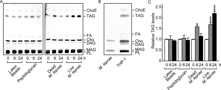
To clarify the effect of M. leprae infection on the host cell lipid composition, we first examined the lipid components of M. leprae-infected THP-1 cells using HPTLC conditions suitable for separating neutral lipids [8]. Heat-killed M. leprae, latex beads and peptidoglycan were used as controls. The positions of cholesterol ester (ChoE), triacylglycerol (TAG), fatty acid (FA), cholesterol (Cho), diacylglycerol (DAG), monoacylglycerol (MAG) and phospholipid (PL) were determined by their relative to front (RF) values based on the position of standard lipids (S1 Fig).

The results showed that the absolute amount of TAG visualized on the TLC plates increased only after a live M. leprae infection (Fig 1A). TAG accumulation was transiently induced by dead bacilli at 6 h, but the levels returned to baseline within 24 h (Fig 1A). The observed transient TAG induction corresponds with previous observations that heat-killed M. leprae changed host mRNA levels of ADRP, perilipin, and HSL [6, 7]. Cells treated with latex beads or peptidoglycan showed no significant change in the amount of TAG (Fig 1A). Meanwhile, the absolute amount of TAG visualized on TLC plates was clearly increased after live M. leprae infection (Fig 1A). Since TAG was not detected in the lipids derived from M. leprae before infection (Fig 1B), it suggests that the observed lipids were derived from host cells.

Fig 1. M. leprae infection enhanced TAG accumulation in THP-1 cells.

Fig 1

(A) THP-1 cells (3 × 10⁶) were cultured in 6-well plates with live M. leprae (MOI: 20), heat killed (80°C for 30 min) M. leprae, 2 μg/ml peptidoglycan, or latex beads for the indicated period of time. Total lipids were extracted and analyzed by HPTLC. (B) Total lipids extracted from M. leprae and THP-1 cells were analyzed by TLC. (C) The amount of TAG as measured by densitometry and expressed relative to levels at 0 h. Values represent the mean ± S.D. from three independent experiments. Significance was determined by a one-way ANOVA followed by a Dunnett test. One and three asterisks indicate p<0.05 and p<0.001, respectively. ChoE: Cholesterol ester; TAG: Triacylglycerol; FA: Fatty acid; Cho: Cholesterol; DAG: Diacylglycerol; MAG: Monoacylglycerol; PL: Phospholipid.

We also measured the density of each lipid and calculated the relative proportion of TAG among all the lipid types. The proportion of TAG among the overall lipid population was also significantly increased by M. leprae infection (Fig 1C). Other lipid components, such as PL, FA, Cho and ChoE, showed no change even after live M. leprae infection (data not shown). These results suggest that TAG is the key lipid that is specifically induced by M. leprae infection in host macrophages.

Glycerol-3-phosphate acyltransferase 3 (GPAT3) expression was induced in THP-1 cells following M. leprae infection

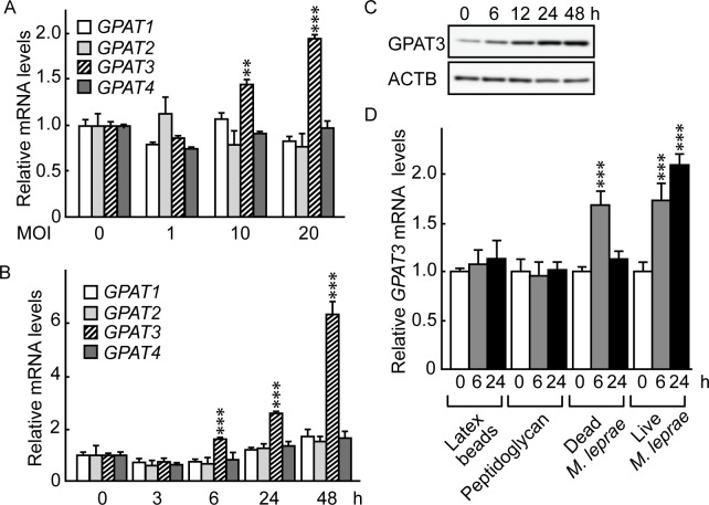
Glycerol-3-phosphate acyltransferase (GPAT) is a rate-limiting enzyme in TAG biosynthesis and is present in four isoforms: GPAT1, GPAT2, GPAT3 and GPAT4 [2023]. To investigate the potential involvement of these GPAT isoforms in M. leprae-infected host cells, we used qRT-PCR to examine the mRNA expression levels of GPAT1, GPAT2, GPAT3 and GPAT4 in THP-1 cells after M. leprae infection. Endogenous GPAT isoforms were expressed at a similar level in THP-1 cells (S2 Fig). Among the four isoforms, only GPAT3 mRNA levels increased after M. leprae infection at MOI 10 and 20 in 24 h (Fig 2A). The increase in GPAT3 mRNA was apparent as early as 6 h after M. leprae infection and increased up to 48 h post-infection (Fig 2B). In accordance with mRNA levels, Western blotting showed that the amount of GPAT3 protein expression also increased following M. leprae infection (Fig 2C).

Fig 2. M. leprae infection induced GPAT3 expression.

Fig 2

(A) THP-1 cells (3 × 10⁶) were cultured in 6-well plates and inoculated with M. leprae (MOI: 0, 1, 10 and 20). After a 24 h incubation, total RNA was purified and the expression level of GPAT isoforms was evaluated by qRT-PCR. Expression levels of mRNA were normalized relative to β-actin (ACTB) levels and are expressed relative to their levels at MOI 0. (B and C) THP-1 cells were cultured in 6-well plates and inoculated with M. leprae (MOI: 20). Total RNA and protein were purified from the cells for (B) qRT-PCR and (C) Western blotting analysis. GPAT mRNA levels were normalized against ACTB and are expressed relative to its level at 0 h. (D) THP-1 cells were cultured with live M. leprae, dead M. leprae, 2 μg/ml of peptidoglycan, or latex beads for the indicated period of time. Total RNA was purified and subjected to an qRT-PCR analysis. Statistical significance was determined by a one-way ANOVA followed by a Dunnett test. Two and three asterisks indicate p<0.01 and p<0.001, respectively. The representative results of three independent experiments are shown.

To clarify whether the observed increase in GPAT3 expression was specific for M. leprae infection, or instead was due to non-specific effects accompanying phagocytosis of particles and/or macrophage activation, we examined the effects of dead M. leprae, latex beads and peptidoglycan on GPAT3 expression. Neither latex beads nor peptidoglycan affected GPAT3 mRNA expression, but treatment of cells with heat-killed M. leprae transiently increased GPAT3 mRNA levels at 6 h before they returned to their original levels by 24 h (Fig 2D). The sustained induction of GPAT3 by live M. leprae and transient induction of GPAT3 by dead M. leprae were in accordance with changes in intracellular TAG accumulation shown in Fig 1, suggesting that GPAT3 is the key molecule that mediates increases in the amount of TAG following M. leprae parasitism.

M. leprae induces formation of lipid droplets through GPAT3

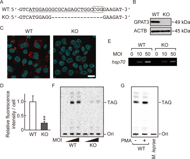
To further evaluate the effect of GPAT3 on M. leprae-induced accumulation of TAG, we generated GPAT3 KO THP-1 cells using the CRISPR/Cas9 gene-editing system. Among the three KO clones isolated, we used one clone with a 16-nucleotide deletion in exon 1 for subsequent experiments. This deletion produced a homozygous deletion of GPAT3 with a frameshift in both alleles (Fig 3A). The lack of GPAT3 protein in the KO clone was confirmed by Western blot (Fig 3B). We then compared the effect of M. leprae infection on lipid droplet formation in wild-type and GPAT3 KO cells using LipidTOX staining (Fig 3C) and quantified the LipidTOX fluorescence intensity in cells (Fig 3D). We observed a large number of lipid droplets in wild-type cells following M. leprae infection, whereas the number of lipid droplets was reduced by about 80% in GPAT3 KO cells, compared to the wild-type cells. Although both GPAT3 and GPAT4 are microsomal enzymes that share similar functions, the expression of GPAT4 in GPAT3 KO cells did not change, even after M. leprae infection (S3 Fig), confirming that GPAT3 is the main isoform that responds to M. leprae infection (Fig 2).

Fig 3. M. leprae utilizes host TAG synthesized via a GPAT3-dependent pathway.

Fig 3

(A) The 20-bp target sequence of gRNA used for the CRISPR/Cas9 gene editing system (underlined) and the PAM sequence (boxed) in wild-type (WT) GPAT3. A dashed line in the GPAT3 KO sequence indicates the frameshifting deletion as detected by DNA sequencing. (B) Western blot confirmed the absence of GPAT3 protein in KO cells. (C) LipidTOX staining (red) of WT and GPAT3 KO THP-1 cells infected or uninfected with M. leprae (MOI: 50) for 24 h with Hoechst 33258 counterstaining (blue). Scale bar: 5 μm. (D) Quantification of LipidTOX staining using fluorescence intensity values to quantify lipid droplets. Significance was determined with a Student’s t test. Two asterisks indicate p<0.01. (E and F) WT and GPAT3 KO THP-1 cells were cultured in medium containing 0.2 μCi of [14C] stearic acid for 16 h after M. leprae infection (MOI: 10 and 50). (E) M. leprae isolated from infected THP-1 cells was confirmed by PCR amplification of the M. leprae hsp70 DNA. (F) M. leprae was purified from cells, and the bacilli lipids were extracted and separated by TLC. (G) THP-1 cells were treated with PMA (20 ng/mL) for 24 h to promote lipid droplet formation, then incubated with 0.2 μCi of [14C] stearic acid for 16 h. Cell lysate was sonicated and mixed with M. leprae then incubated for 24 h. M. leprae was isolated and extracted lipids were separated by TLC to evaluate radioactivity.

To determine whether M. leprae utilizes the newly synthesized TAG that exists in lipid droplets, we performed metabolic labelling of THP-1 cells with [14C] stearic acid to follow de novo synthesis of TAG. Twenty-four hours after M. leprae inoculation of wild-type and GPAT3 KO THP-1 cells, [14C] stearic acid was added, and the cells were cultured for 16 h. Then, radioactivity of TAG in the bacilli was examined on a TLC plate. The presence of bacilli isolated from M. leprae-infected cells was confirmed by expression of M. leprae-specific hsp70 gene expression (Fig 3E). Radioactive TAG was detected only in M. leprae extracted from wild-type cells, but not from GPAT3 KO cells (Fig 3F). While strong radioactivity was detected in live M. leprae, heat-killed bacteria also showed weak signal (S4 Fig), suggesting a potential nonspecific binding of TAG on the surface of M. leprae. We therefore mixed live M. leprae with lipid-rich cell lysate prepared from PMA-treated THP-1 cells. In this case, however, the 14C signal was not detected in the TAG fraction prepared from M. leprae (Fig 3G), suggesting that the nonspecific binding of TAG on live M. leprae is insignificant. Radioactive signals from other lipids were also detected in a darker image, suggesting the utilization of [14C] stearic acid for the biosynthesis of other lipids (data not shown). Together, these results suggest that M. leprae induces de novo synthesis of TAG in host macrophages in a GPAT3-dependent manner, and that newly synthesized TAG following M. leprae infection may be utilized by M. leprae itself.

GPAT3 is essential for the intracellular survival of M. leprae

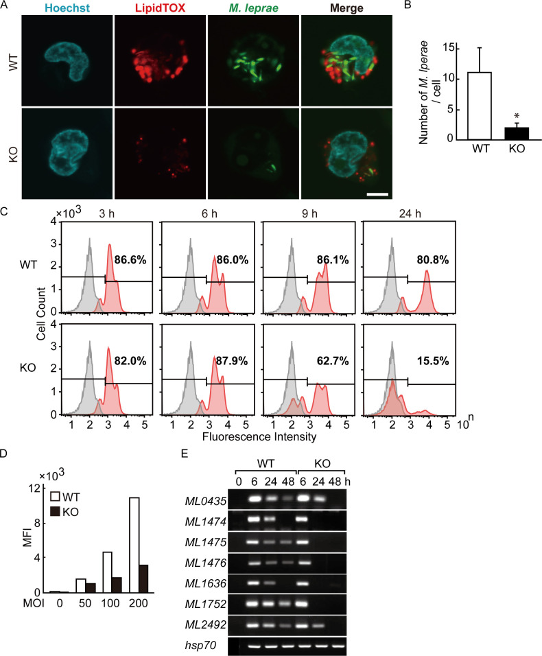
We visualized both intracellular localization of M. leprae and lipid droplets by fluorescence imaging using confocal laser scanning microscopy (Fig 4A). The bacilli were counted in 30 cells and are presented as the mean and standard deviation for wild-type and GPAT3 KO cells (Fig 4B). The MOI 200 was used to better visualize the results. After infection, M. leprae localized in lipid droplets in wild-type cells, whereas the number of both intracellular M. leprae and lipid droplets was clearly reduced in GPAT3 KO cells.

Fig 4. Intracellular M. leprae viability requires GPAT3.

Fig 4

(A) Wild-type (WT) and GPAT3 KO THP-1 cells infected with FITC-conjugated M. leprae (MOI: 200) were cultured for 24 h and confocal laser scanning microscopy was used to analyze M. leprae and lipid droplet localization. Fluorescence images of lipid droplets stained with LipidTOX (red), M. leprae (green), Hoechst 33258 (blue) and their merged images are shown. Scale bar: 5 μm. (B) Quantification of M. leprae in WT and GPAT3 KO cells (n = 30 each) shown by the mean ± S.D. Significance was determined with a Student’s t test. One asterisk indicates p<0.01. (C) WT and GPAT3 KO THP-1 cells were infected with FITC-conjugated M. leprae (MOI: 200) for 3, 6, 9, and 24 h, and the fluorescence intensity of 100,000 cells was analyzed by flow cytometry. Gray and red peaks indicate the fluorescence intensity of uninfected and infected cells, respectively. (D) Mean fluorescence intensity (MFI) of cells infected with different MOI (0, 50, 100, and 200). (E) Wild-type and GPAT3 KO cells (3 × 10⁶) were cultured in 6-well plates and inoculated with M. leprae. After incubation for the indicated period of time, total RNA was purified and an RT-PCR analysis for M. leprae pseudogenes were performed. A representative result from three independent experiments is shown in each panel.

We also performed a flowcytometric analysis of THP-1 cells to compare the mass engulfment of FITC-labeled M. leprae by wild-type and GPAT3 KO cells. More than 80% of THP-1 cells were fluorescent at 3 h post-infection and the levels of such cells were similar between wild-type and GPAT3 KO cells until 6 h (Fig 4C). The FITC-positive cells began to decline at 9 h in GPAT3 KO cells and were reduced to 15.5% by 24 h. Conversely, the fluorescence of wild-type cells remained high (>80%) at 24 h (Fig 4C). Similarly, the mean fluorescence intensity (MFI) in each cell type was higher in wild-type cells than in GPAT3 KO cells (Fig 4D), which is consistent with the confocal microscopy observations (Fig 4A and 4B).

We previously reported that pseudogenes and noncoding regions in live M. leprae are not silent but instead are strongly transcribed and can be detected as RNA [14, 18, 24, 25]. The expression levels of these RNA are variable among patients with lepromatous leprosy [18], and decrease after antibiotic treatment (unpublished data). Therefore, we used the expression levels of these RNA to estimate the viability of M. leprae. For seven M. leprae pseudogenes, similar levels of mRNA were induced by wild-type and GPAT3 KO cells 6 h after infection. However, for GPAT3 KO cells the M. leprae pseudogene expression was rapidly reduced compared to that seen for wild-type cells (Fig 4E). The expression of M. leprae hsp70 mRNA was used as a control, since its expression level is stable [26] and did not change in both cells. These results suggest that the viability of M. leprae was lower in GPAT3 KO cells. Together, these data indicate that GPAT3 expression in host cells could be important to maintain the intracellular environment required for M. leprae to successfully parasitize within host macrophages.

Discussion

Our group and others have shown that M. leprae modifies host lipid metabolism to induce lipid droplet formation in infected host macrophages [6, 7, 27]. However, changes in lipid composition in infected cells, the mechanism associated with these changes, and the role of such lipids in M. leprae are not well understood. In this study, we performed a qualitative lipid analysis using HPTLC to show that TAG is the main lipid that accumulates in lipid droplets in M. leprae-infected THP-1 cells. TAG is known to be a major component of lipid droplets and plays a central role in maintaining energy homeostasis [28]. Sustained accumulation of intracellular TAG was unlikely to be a non-specific cell response toward bacterial components or due to macrophage activation or phagocytosis, but instead is a specific event associated with infection by viable M. leprae. This phenomenon involving live bacilli is similar to our previous findings [6, 7, 29].

For lipid accumulation associated with live M. leprae, induction of ADRP and perilipin expression is needed to enhance storage of neutral lipids and reduce the expression of HSL, which is involved in lipid droplet degradation [6, 7]. In addition, tryptophan aspartate-containing coat protein (CORO1A), which inhibits phagosome-lysosome fusion, localizes in the membrane of phagosomes containing M. leprae in skin lesions from leprosy patients [13, 30]. Accumulation of CORO1A persisted on the phagosome membrane around live bacilli and there was transient accumulation in dead bacilli when M. leprae or M. bovis BCG was infected in macrophages [30, 31]. Since the induction of GPAT3 expression and TAG accumulation were not observed with the addition of PGN, a major cell wall component of mycobacteria, a hitherto unknown component(s) specific to live M. leprae could have significant effects on host cells. The effect of other mycobacterial species on GPAT3 expression and the accumulation of TAG in host cells should be a topic of future investigation.

TAG biosynthesis occurs mainly via the glycerol-3-phosphate (G3P) de novo pathway in which three fatty acid acyl chains are successively bound to a glycerol backbone (i.e., acylation) [32, 33]. In the first step of acylation catalyzed by glycerol-3-phosphate acyltransferase (GPAT), an acyl-CoA is bound to G3P to form lysophosphatidic acid (LPA). Then, 1-acylglycerol-3-phosphate acyltransferase (AGPAT) catalyzes LPA to form phosphatidic acid (PA). The slow reaction rate of GPAT suggests that it is the rate-limiting enzyme in TAG biosynthesis [34].

Four different genes encode GPAT isoforms 1–4, and isoform-specific differences in tissue expression patterns, subcellular location, fatty acyl-CoA substrate preference, and sensitivity to N-ethylmaleimide have been observed [35]. In this study we demonstrated that GPAT3 expression alone was increased after M. leprae infection, and this increase occurred in parallel with TAG accumulation in host cells. In GPAT3 KO cells, the formation of lipid droplets was significantly suppressed relative to wild-type cells after M. leprae infection. In addition, expression levels of M. leprae pseudogenes were rapidly reduced in GPAT3 KO cells compared to wild-type cells, indicating that M. leprae viability is not sustained in GPAT3 KO cells. Together, these results suggest a possibility that host GPAT3-mediated TAG synthesis could be responsible for the foamy-cell formation induced by M. leprae infection and that GPAT3 activity might be important for maintaining intracellular parasitization by M. leprae. Further studies are needed to confirm these points.

Other studies have reported that M. leprae infection promotes cholesterol accumulation rather than TAG [8]. This discrepancy may be due to the different MOI and cell systems used. The number of bacteria used in our study was 10 times higher than that in previous reports. Additionally, the cells used in the previous report were primary cultures of human macrophages, and primary cells infected with M. leprae are known to be rich in cholesterol and cholesterol esters [27]. Differences in the lipid moieties accumulated in response to M. leprae infection between primary cells and THP-1 cells need to be clarified in the future. Two additional bands can be seen above the TAG spot in wild-type cells (Fig 3F) that could be metabolites of the TAG synthesis pathway, but the nature of these moieties is not yet clear.

How M. leprae infection induces host GPAT3 expression is not clear. Activation of the peroxisome proliferator-activated receptor γ (PPARγ) signaling pathway is reported to be responsible for up-regulation of Gpat3 gene expression during adipocyte differentiation [20, 36, 37]. PPARγ is an important transcription factor that regulates expression of genes that are closely related to lipogenesis, lipid metabolism, and foam cell formation in macrophages [38]. In addition to GPAT3, PPARγ also targets ADRP [39], which has a similar induction following M. leprae infection in THP-1 cells [6]. Recently, we have reported that activation of PPARγ and PPARδ is important for lipid accumulation in M. leprae-infected THP-1 cells [40]. In Schwann cells, phenolic glicolipid-1 (PGL-1) of M. leprae promoted lipid droplet formation by activating crosstalk between CD206 and PPARγ [41]. Therefore, M. leprae might utilize the signal transduction pathway(s) mediated by PPARγ to induce GPAT3 expression in infected cells.

We showed that intracellular localization of M. leprae to lipid droplets was clearly reduced in GPAT3 KO cells compared to wild-type cells. This observation is in agreement with a recent study showing that in bone marrow-derived macrophages from Gpat3-/- mice, not only was the formation of Kdo2-lipid A (KLA)-inducible lipid droplets suppressed, intriguingly, the phagocytic capacity of the macrophages was also reduced [42]. In the present study, however, we have clearly shown that GPAT3 does not affect the ability of THP-1 cells to undergo phagocytosis. The role of GPAT3 on the phagocytosis of different cell types should be elucidated in the future.

Another interesting question is how M. leprae utilizes intracellular lipids during its parasitism. Lipids represent approximately 60% of the dry weight of mycobacteria cell walls and include lipid varieties such as phospholipids, glycolipids and mycolic acid (a long fatty acid). An intact “greasy” cell wall is considered to be important for mycobacteria viability. Medications such as isoniazid, ethionamide, isoxyl and thiacetazone that inhibit mycolic acid synthesis have shown antimycobacterial effects [43, 44]. From this perspective, M. leprae may rely on host-derived lipids to generate its own cell wall. Here, we labeled newly-synthesized intracellular TAG with [14C] stearic acid to examine incorporation of host-derived lipids in the cell wall of M. leprae following infection. The labeled TAG content of intracellular M. leprae in wild-type cells was markedly higher than that in GPAT3 KO cells. These results suggest a possibility that M. leprae utilizes host-derived lipids during intracellular parasitization.

M. leprae has substantially fewer functional genes compared to M. tuberculosis [45]. Several of the lost genes are involved in lipid metabolism and the lack of these genes could be associated with their slow growth in vivo and the inability to cultivate M. leprae in vitro. However, across evolution, M. leprae acquired the ability to use host cell resources to support its parasitism. In this study, we showed that M. leprae infection increased intracellular TAG accumulation via induction of host GPAT3 expression that in turn maintains a lipid-rich environment in host macrophages. Our finding indicates that GPAT3 also plays an important role in the intracellular survival of M. leprae, suggesting that GPAT3 may become a novel target for leprosy treatment.

Supporting information

S1 Table. List of primers used in RT-PCR.

(DOCX)

S1 Fig. Confirmation of the lipid fraction in M. leprae-infected THP-1 cells using standard controls.

THP-1 cells (3 × 10⁶) were cultured in 6-well plates with live M. leprae (MOI: 20) for 24 h. Total lipids extracted from cells with 20 nmol of control lipids (PL, Cho, FA and TAG) were spotted on an HPTLC plate. After separation, the plate was stained with a charring solution containing 10% CuSO4 and 8% H3PO4 and heated at 180°C for 10 min.

(TIF)

S2 Fig. The endogenous expression levels of GPAT isoforms in THP-1 cells.

Total RNA was extracted and the expression level of GPAT isoforms was evaluated by qRT-PCR. The results were normalized relative to ACTB levels. Each bar represents the mean ± S.D in triplicate.

(TIF)

S3 Fig. M. leprae infection does not affect GPAT4 expression in GPAT3 KO cells.

WT and GPAT3 KO cells (3 × 10⁶) were cultured in 6-well plate and infected with live M. leprae (MOI: 50). After incubating for the indicated time, total RNA was purified and RT-PCR analysis was performed.

(TIF)

S4 Fig. Analysis of nonspecific binding of TAG on the surface of M. leprae.

Wild-type THP-1 cells were inoculated with either live or heat-killed M. leprae (MOI: 10 and 50), then cultured with 0.2 μCi of [14C] stearic acid for 16 h. M. leprae was isolated and extracted lipids were separated by TLC to evaluate radioactivity.

(TIF)

S1 Raw images

(TIF)

S2 Raw images

(TIF)

Acknowledgments

We are grateful to Prof. J. Aoki and Dr. A. Inoue (Tohoku University, Sendai, Japan) for valuable technical advice regarding the CRISPR-Cas9 system.

Data Availability

All relevant data are within the manuscript and its Supporting Information files.

Funding Statement

This work was supported by AMED under Grant Numbers JP17fk0108303 (to K.S.) and JP20fk0108064 (to K.S.), MEXT KAKENHI Grant Numbers 15K190097 (to K.T.) and 18K15150 (to K.T.), and Sasakawa Scientific Research Grant Number 26-428 (to K.T.).

References

  • 1.WHO. Weekly epidemiological record. 2019; 94:389–412. [Google Scholar]
  • 2.Ridley DS, Jopling WH. Classification of leprosy according to immunity. A five-group system. Int J Lepr Other Mycobact Dis. 1966; 34(3):255–273. [PubMed] [Google Scholar]
  • 3.Chatterjee KR, Das Gupta NN, De ML. Electron microscopic observations on the morphology of Mycobacterium leprae. Exp Cell Res. 1959; 18:521–527. 10.1016/0014-4827(59)90317-9 [DOI] [PubMed] [Google Scholar]
  • 4.Gross DN, Miyoshi H, Hosaka T, Zhang HH, Pino EC, Souza S, et al. Dynamics of lipid droplet-associated proteins during hormonally stimulated lipolysis in engineered adipocytes: stabilization and lipid droplet binding of adipocyte differentiation-related protein/adipophilin. Mol Endocrinol. 2006; 20(2):459–466. 10.1210/me.2005-0323 [DOI] [PubMed] [Google Scholar]
  • 5.Sztalryd C, Kimmel AR. Perilipins: lipid droplet coat proteins adapted for tissue-specific energy storage and utilization, and lipid cytoprotection. Biochimie. 2014; 96:96–101. 10.1016/j.biochi.2013.08.026 [DOI] [PMC free article] [PubMed] [Google Scholar]
  • 6.Tanigawa K, Suzuki K, Nakamura K, Akama T, Kawashima A, Wu H, et al. Expression of adipose differentiation-related protein (ADRP) and perilipin in macrophages infected with Mycobacterium leprae. FEMS Microbiol Lett. 2008; 289(1):72–79. 10.1111/j.1574-6968.2008.01369.x [DOI] [PubMed] [Google Scholar]
  • 7.Tanigawa K, Degang Y, Kawashima A, Akama T, Yoshihara A, Ishido Y, et al. Essential role of hormone-sensitive lipase (HSL) in the maintenance of lipid storage in Mycobacterium leprae-infected macrophages. Microb Pathog. 2012; 52(5):285–291. 10.1016/j.micpath.2012.02.003 [DOI] [PubMed] [Google Scholar]
  • 8.Mattos KA, Oliveira VC, Berredo-Pinho M, Amaral JJ, Antunes LC, Melo RC, et al. Mycobacterium leprae intracellular survival relies on cholesterol accumulation in infected macrophages: a potential target for new drugs for leprosy treatment. Cell Microbiol. 2014; 16(6):797–815. 10.1111/cmi.12279 [DOI] [PMC free article] [PubMed] [Google Scholar]
  • 9.Pandey AK, Sassetti CM. Mycobacterial persistence requires the utilization of host cholesterol. Proc Natl Acad Sci U S A. 2008; 105(11):4376–4380. 10.1073/pnas.0711159105 [DOI] [PMC free article] [PubMed] [Google Scholar]
  • 10.Marques MA, Berredo-Pinho M, Rosa TL, Pujari V, Lemes RM, Lery LM, et al. The essential role of cholesterol metabolism in the intracellular survival of Mycobacterium leprae is not coupled to central carbon metabolism and energy production. J Bacteriol. 2015; 197(23):3698–3707. 10.1128/JB.00625-15 [DOI] [PMC free article] [PubMed] [Google Scholar]
  • 11.Medeiros RC, Girardi KD, Cardoso FK, Mietto BS, Pinto TG, Gomez LS, et al. Subversion of schwann cell glucose metabolism by Mycobacterium leprae. J Biol Chem. 2016; 291(41):21375–21387. 10.1074/jbc.M116.725283 [DOI] [PMC free article] [PubMed] [Google Scholar]
  • 12.Matsuoka M. The history of Mycobacterium leprae Thai-53 strain. Lepr Rev. 2010; 81(2):137. [PubMed] [Google Scholar]
  • 13.Suzuki K, Takeshita F, Nakata N, Ishii N, Makino M. Localization of CORO1A in the macrophages containing Mycobacterium leprae. Acta Histochem Cytochem. 2006; 39(4):107–112. 10.1267/ahc.06010 [DOI] [PMC free article] [PubMed] [Google Scholar]
  • 14.Suzuki K, Nakata N, Bang PD, Ishii N, Makino M. High-level expression of pseudogenes in Mycobacterium leprae. FEMS Microbiol Lett. 2006; 259(2):208–214. 10.1111/j.1574-6968.2006.00276.x [DOI] [PubMed] [Google Scholar]
  • 15.Bligh EG, Dyer WJ. A rapid method of total lipid extraction and purification. Can J Biochem Physiol. 1959; 37(8):911–917. 10.1139/o59-099 [DOI] [PubMed] [Google Scholar]
  • 16.Suzuki K, Yanagi M, Mori-Aoki A, Moriyama E, Ishii KJ, Kohn LD. Transfection of single-stranded hepatitis A virus RNA activates MHC class I pathway. Clin Exp Immunol. 2002; 127(2):234–242. 10.1046/j.1365-2249.2002.01767.x [DOI] [PMC free article] [PubMed] [Google Scholar]
  • 17.Luo Y, Akama T, Okayama A, Yoshihara A, Sue M, Oda K, et al. A novel role for flotillin-containing lipid rafts in negative-feedback regulation of thyroid-specific gene expression by thyroglobulin. Thyroid. 2016; 26(11):1630–1639. 10.1089/thy.2016.0187 [DOI] [PubMed] [Google Scholar]
  • 18.Nakamura K, Akama T, Bang PD, Sekimura S, Tanigawa K, Wu H, et al. Detection of RNA expression from pseudogenes and non-coding genomic regions of Mycobacterium leprae. Microb Pathog. 2009; 47(3):183–187. 10.1016/j.micpath.2009.06.006 [DOI] [PubMed] [Google Scholar]
  • 19.Suzuki K, Lavaroni S, Mori A, Okajima F, Kimura S, Katoh R, et al. Thyroid transcription factor 1 is calcium modulated and coordinately regulates genes involved in calcium homeostasis in C cells. Mol Cell Biol. 1998; 18(12):7410–7422. 10.1128/mcb.18.12.7410 [DOI] [PMC free article] [PubMed] [Google Scholar]
  • 20.Cao J, Li JL, Li D, Tobin JF, Gimeno RE. Molecular identification of microsomal acyl-CoA:glycerol-3-phosphate acyltransferase, a key enzyme in de novo triacylglycerol synthesis. Proc Natl Acad Sci U S A. 2006; 103(52):19695–19700. 10.1073/pnas.0609140103 [DOI] [PMC free article] [PubMed] [Google Scholar]
  • 21.Nagle CA, Vergnes L, Dejong H, Wang S, Lewin TM, Reue K, et al. Identification of a novel sn-glycerol-3-phosphate acyltransferase isoform, GPAT4, as the enzyme deficient in Agpat6-/- mice. J Lipid Res. 2008; 49(4):823–831. 10.1194/jlr.M700592-JLR200 [DOI] [PMC free article] [PubMed] [Google Scholar]
  • 22.Linden D, William-Olsson L, Rhedin M, Asztely AK, Clapham JC, Schreyer S. Overexpression of mitochondrial GPAT in rat hepatocytes leads to decreased fatty acid oxidation and increased glycerolipid biosynthesis. J Lipid Res. 2004; 45(7):1279–1288. 10.1194/jlr.M400010-JLR200 [DOI] [PubMed] [Google Scholar]
  • 23.Wang S, Lee DP, Gong N, Schwerbrock NM, Mashek DG, Gonzalez-Baro MR, et al. Cloning and functional characterization of a novel mitochondrial N-ethylmaleimide-sensitive glycerol-3-phosphate acyltransferase (GPAT2). Arch Biochem Biophys. 2007; 465(2):347–358. 10.1016/j.abb.2007.06.033 [DOI] [PMC free article] [PubMed] [Google Scholar]
  • 24.Akama T, Tanigawa K, Kawashima A, Wu H, Ishii N, Suzuki K. Analysis of Mycobacterium leprae gene expression using DNA microarray. Microb Pathog. 2010; 49(4):181–185. 10.1016/j.micpath.2010.05.010 [DOI] [PubMed] [Google Scholar]
  • 25.Akama T, Suzuki K, Tanigawa K, Kawashima A, Wu H, Nakata N, et al. Whole-genome tiling array analysis of Mycobacterium leprae RNA reveals high expression of pseudogenes and noncoding regions. J Bacteriol. 2009; 191(10):3321–3327. 10.1128/JB.00120-09 [DOI] [PMC free article] [PubMed] [Google Scholar]
  • 26.Sugita Y, Suga C, Ishii N, Nakajima H. A case of relapsed leprosy successfully treated with sparfloxacin. Arch Dermatol. 1996; 132(11):1397–1398. [PubMed] [Google Scholar]
  • 27.Mattos KA, D’Avila H, Rodrigues LS, Oliveira VG, Sarno EN, Atella GC, et al. Lipid droplet formation in leprosy: Toll-like receptor-regulated organelles involved in eicosanoid formation and Mycobacterium leprae pathogenesis. J Leukoc Biol. 2010; 87(3):371–384. 10.1189/jlb.0609433 [DOI] [PubMed] [Google Scholar]
  • 28.Birsoy K, Festuccia WT, Laplante M. A comparative perspective on lipid storage in animals. J Cell Sci. 2013; 126(Pt 7):1541–1552. 10.1242/jcs.104992 [DOI] [PubMed] [Google Scholar]
  • 29.Degang Y, Akama T, Hara T, Tanigawa K, Ishido Y, Gidoh M, et al. Clofazimine modulates the expression of lipid metabolism proteins in Mycobacterium leprae-infected macrophages. PLoS Negl Trop Dis. 2012; 6(12):e1936. 10.1371/journal.pntd.0001936 [DOI] [PMC free article] [PubMed] [Google Scholar]
  • 30.Tanigawa K, Suzuki K, Kimura H, Takeshita F, Wu H, Akama T, et al. Tryptophan aspartate-containing coat protein (CORO1A) suppresses Toll-like receptor signalling in Mycobacterium leprae infection. Clin Exp Immunol. 2009; 156(3):495–501. 10.1111/j.1365-2249.2009.03930.x [DOI] [PMC free article] [PubMed] [Google Scholar]
  • 31.Ferrari G, Langen H, Naito M, Pieters J. A coat protein on phagosomes involved in the intracellular survival of mycobacteria. Cell. 1999; 97(4):435–447. 10.1016/s0092-8674(00)80754-0 [DOI] [PubMed] [Google Scholar]
  • 32.Kennedy EP, Smith SW, Weiss SB. New synthesis of lecithin in an isolated enzyme system. Nature. 1956; 178(4533):594–595. 10.1038/178594a0 [DOI] [PubMed] [Google Scholar]
  • 33.Coleman RA, Mashek DG. Mammalian triacylglycerol metabolism: synthesis, lipolysis, and signaling. Chem Rev. 2011; 111(10):6359–6386. 10.1021/cr100404w [DOI] [PMC free article] [PubMed] [Google Scholar]
  • 34.Bell RM, Coleman RA. Enzymes of glycerolipid synthesis in eukaryotes. Annu Rev Biochem. 1980; 49:459–487. 10.1146/annurev.bi.49.070180.002331 [DOI] [PubMed] [Google Scholar]
  • 35.Wang H, Airola MV, Reue K. How lipid droplets "TAG" along: Glycerolipid synthetic enzymes and lipid storage. Biochim Biophys Acta Mol Cell Biol Lipids. 2017; 1862(10 Pt B):1131–1145. 10.1016/j.bbalip.2017.06.010 [DOI] [PMC free article] [PubMed] [Google Scholar]
  • 36.Gorga A, Rindone GM, Regueira M, Pellizzari EH, Camberos MC, Cigorraga SB, et al. PPARgamma activation regulates lipid droplet formation and lactate production in rat Sertoli cells. Cell Tissue Res. 2017; 369(3):611–624. 10.1007/s00441-017-2615-y [DOI] [PubMed] [Google Scholar]
  • 37.Lee MJ, Jash S, Jones JEC, Puri V, Fried SK. Rosiglitazone remodels the lipid droplet and britens human visceral and subcutaneous adipocytes ex vivo. J Lipid Res. 2019; 60(4):856–868. 10.1194/jlr.M091173 [DOI] [PMC free article] [PubMed] [Google Scholar]
  • 38.Yessoufou A, Wahli W. Multifaceted roles of peroxisome proliferator-activated receptors (PPARs) at the cellular and whole organism levels. Swiss Med Wkly. 2010; 140:w13071. 10.4414/smw.2010.13071 [DOI] [PubMed] [Google Scholar]
  • 39.Vosper H, Patel L, Graham TL, Khoudoli GA, Hill A, Macphee CH, et al. The peroxisome proliferator-activated receptor delta promotes lipid accumulation in human macrophages. J Biol Chem. 2001; 276(47):44258–44265. 10.1074/jbc.M108482200 [DOI] [PubMed] [Google Scholar]
  • 40.Luo Y, Tanigawa K, Kawashima A, Ishido Y, Ishii N, Suzuki K. The function of peroxisome proliferator-activated receptors PPAR-gamma and PPAR-delta in Mycobacterium leprae-induced foam cell formation in host macrophages. PLoS Negl Trop Dis. 2020; 14(10):e0008850. 10.1371/journal.pntd.0008850 [DOI] [PMC free article] [PubMed] [Google Scholar]
  • 41.Diaz Acosta CC, Dias AA, Rosa T, Batista-Silva LR, Rosa PS, Toledo-Pinto TG, et al. PGL I expression in live bacteria allows activation of a CD206/PPARgamma cross-talk that may contribute to successful Mycobacterium leprae colonization of peripheral nerves. PLoS Pathog. 2018; 14(7):e1007151. 10.1371/journal.ppat.1007151 [DOI] [PMC free article] [PubMed] [Google Scholar]
  • 42.Quiroga IY, Pellon-Maison M, Suchanek AL, Coleman RA, Gonzalez-Baro MR. Glycerol-3-phosphate acyltransferases 3 and 4 direct glycerolipid synthesis and affect functionality in activated macrophages. Biochem J. 2019; 476(1):85–99. 10.1042/BCJ20180381 [DOI] [PubMed] [Google Scholar]
  • 43.Banerjee A, Dubnau E, Quemard A, Balasubramanian V, Um KS, Wilson T, et al. inhA, a gene encoding a target for isoniazid and ethionamide in Mycobacterium tuberculosis. Science. 1994; 263(5144):227–230. 10.1126/science.8284673 [DOI] [PubMed] [Google Scholar]
  • 44.Grzegorzewicz AE, Kordulakova J, Jones V, Born SE, Belardinelli JM, Vaquie A, et al. A common mechanism of inhibition of the Mycobacterium tuberculosis mycolic acid biosynthetic pathway by isoxyl and thiacetazone. J Biol Chem. 2012; 287(46):38434–38441. 10.1074/jbc.M112.400994 [DOI] [PMC free article] [PubMed] [Google Scholar]
  • 45.Cole ST, Eiglmeier K, Parkhill J, James KD, Thomson NR, Wheeler PR, et al. Massive gene decay in the leprosy bacillus. Nature. 2001; 409(6823):1007–1011. 10.1038/35059006 [DOI] [PubMed] [Google Scholar]

Decision Letter 0

Delphi Chatterjee

Transfer Alert

This paper was transferred from another journal. As a result, its full editorial history (including decision letters, peer reviews and author responses) may not be present.

10 Sep 2020

PONE-D-20-21795

Mycobacterium leprae promotes triacylglycerol de novo synthesis through induction of GPAT3 expression in host macrophages

PLOS ONE

Dear Dr. Suzuki,

Thank you for submitting your manuscript to PLOS ONE. After careful consideration, we feel that it has merit but does not fully meet PLOS ONE’s publication criteria as it currently stands. Therefore, we invite you to submit a revised version of the manuscript that addresses the points raised during the review proce

Both  reviewers  did not agree with your conclusions and  felt there were serious flaws that need to be resurrected.  This also entails new experiments.  

Please submit your revised manuscript within 90days. If you will need more time than this to complete your revisions, please reply to this message or contact the journal office at plosone@plos.org. When you're ready to submit your revision, log on to https://www.editorialmanager.com/pone/ and select the 'Submissions Needing Revision' folder to locate your manuscript file.

Please include the following items when submitting your revised manuscript:

  • A rebuttal letter that responds to each point raised by the academic editor and reviewer(s). You should upload this letter as a separate file labeled 'Response to Reviewers'.

  • A marked-up copy of your manuscript that highlights changes made to the original version. You should upload this as a separate file labeled 'Revised Manuscript with Track Changes'.

  • An unmarked version of your revised paper without tracked changes. You should upload this as a separate file labeled 'Manuscript'.

If you would like to make changes to your financial disclosure, please include your updated statement in your cover letter. Guidelines for resubmitting your figure files are available below the reviewer comments at the end of this letter.

If applicable, we recommend that you deposit your laboratory protocols in protocols.io to enhance the reproducibility of your results. Protocols.io assigns your protocol its own identifier (DOI) so that it can be cited independently in the future. For instructions see: http://journals.plos.org/plosone/s/submission-guidelines#loc-laboratory-protocols

We look forward to receiving your revised manuscript.

Kind regards,

Delphi Chatterjee

Academic Editor

PLOS ONE

Journal Requirements:

When submitting your revision, we need you to address these additional requirements.

1. Please ensure that your manuscript meets PLOS ONE's style requirements, including those for file naming. The PLOS ONE style templates can be found at

https://journals.plos.org/plosone/s/file?id=wjVg/PLOSOne_formatting_sample_main_body.pdf and

https://journals.plos.org/plosone/s/file?id=ba62/PLOSOne_formatting_sample_title_authors_affiliations.pdf

2.PLOS ONE now requires that authors provide the original uncropped and unadjusted images underlying all blot or gel results reported in a submission’s figures or Supporting Information files. This policy and the journal’s other requirements for blot/gel reporting and figure preparation are described in detail at https://journals.plos.org/plosone/s/figures#loc-blot-and-gel-reporting-requirements and https://journals.plos.org/plosone/s/figures#loc-preparing-figures-from-image-files. When you submit your revised manuscript, please ensure that your figures adhere fully to these guidelines and provide the original underlying images for all blot or gel data reported in your submission. See the following link for instructions on providing the original image data: https://journals.plos.org/plosone/s/figures#loc-original-images-for-blots-and-gels.

In your cover letter, please note whether your blot/gel image data are in Supporting Information or posted at a public data repository, provide the repository URL if relevant, and provide specific details as to which raw blot/gel images, if any, are not available. Email us at plosone@plos.org if you have any questions.

[Note: HTML markup is below. Please do not edit.]

Reviewers' comments:

Reviewer's Responses to Questions

Comments to the Author

1. Is the manuscript technically sound, and do the data support the conclusions?

The manuscript must describe a technically sound piece of scientific research with data that supports the conclusions. Experiments must have been conducted rigorously, with appropriate controls, replication, and sample sizes. The conclusions must be drawn appropriately based on the data presented.

Reviewer #1: Partly

Reviewer #2: Partly

**********

2. Has the statistical analysis been performed appropriately and rigorously?

Reviewer #1: Yes

Reviewer #2: Yes

**********

3. Have the authors made all data underlying the findings in their manuscript fully available?

The PLOS Data policy requires authors to make all data underlying the findings described in their manuscript fully available without restriction, with rare exception (please refer to the Data Availability Statement in the manuscript PDF file). The data should be provided as part of the manuscript or its supporting information, or deposited to a public repository. For example, in addition to summary statistics, the data points behind means, medians and variance measures should be available. If there are restrictions on publicly sharing data—e.g. participant privacy or use of data from a third party—those must be specified.

Reviewer #1: Yes

Reviewer #2: Yes

**********

4. Is the manuscript presented in an intelligible fashion and written in standard English?

PLOS ONE does not copyedit accepted manuscripts, so the language in submitted articles must be clear, correct, and unambiguous. Any typographical or grammatical errors should be corrected at revision, so please note any specific errors here.

Reviewer #1: Yes

Reviewer #2: Yes

**********

5. Review Comments to the Author

Please use the space provided to explain your answers to the questions above. You may also include additional comments for the author, including concerns about dual publication, research ethics, or publication ethics. (Please upload your review as an attachment if it exceeds 20,000 characters)

Reviewer #1: The manuscript entitled ‘Mycobacterium leprae promotes triacylglycerol de novo synsthesis through induction of GPAT3 expression in host macrophages’ by Tanigawa et al; claims to demonstrate that M. leprae infection induces the production of triacylglycerols (TAGs) in a macrophage cell line. Authors linked this host production of TAGs to the function of glycerol-3-phosphagte acyltransferase 3 (GPAT3), which may be essential for intracellular survival of M. leprae within host cells. This is an interesting study working with a difficult pathogen, defining how M. leprae could stimulate the host cell physiology to create an environment that favors its intracellular survival. This study is well conducted and sounded; however, authors have some bold claims that are not well supported by their data. Major concerns are:

1/ Analysis of host lipids performing HPTLC. Although this is a valid technique, a LC/MS analysis will provide a clear picture (and quantification) of all the host lipids that are being altered after M. leprae infection of TH-1 cells.

As depicted, authors focused on DAGs/TAGs and the phospholipid fractions (a major fraction in host lipids) is not clearly studied in the solvent system presented (PLs practically did not migrated on the HPTLC from the origin to state that these are not being modified). Moreover, it will be important to add reference standard controls on the HPTLCs to be certain of the nature of the lipids being identified. THP-1 cells lipids and M. leprae lipids alone controls are also missing. Provide also the TLCs in color, so this can be better appreciate it.

2/ In the production of TAGs by host cells it also seems to be a dose dependent effect (different MOIs induced different expression levels at 24 h), but this is not being follow up. Authors, did also not explain why there is a spike of TAG production in THP-1 cells infected with dead M. leprae.

3/ Related to the induction of GPAT3, it will be important to assess if this is induction is M. leprae specific or if other bacteria/mycobacteria induces GPAT3 in Th-1 cell line. For example, M. bovis BCG, M. avium, M. smegmatis, or even M. tuberculosis.

4/ Fig 3C needs better images and with the same background contrast. GPAT3 KO background seems lighter than the WT background making difficult to interpret. A quantification will be also required demonstrating the author’s claim of decrease in lipid formation in GPAT3 cells infected with live M. leprae.

5/ Line 291-292: ‘newly synthesized TAG following M. leprae infection is in turn utilized by M. leprae itself’ There is not conclusive data supporting this statement. Could authors rationalize why other M. leprae lipids are not radioactive and thus not being seeing on the autoradiogram provided? One will be expecting seeing radioactive free fatty acids, DAG, etc. and not only DAG.

6/ Fig. 4A: Quantification by Confocal microscopy counting bacteria per cell is necessary. It will be important to show the flow cytometry plots describing the strategy used removing extracellular FITC-M. leprae bacilli and focusing only in infected cells. Also to show graphs at % of infected vs. non-infected cells. How many times this study was done?

7/ A good control will be to have one of the others GPAT KOs, and further determine that their absence does not affect M. leprae uptake/survival in THP-1 cells.

8/ Lines 379-381: “Together, these results suggest that host GPAT-3-mediated TAG synthesis is responsible for foamy cell formation induced by M. leprae infection, and that GPAT3 activity is necessary for maintaining intracellular parasitization by M. leprae.”This is not proven in this manuscript. The infection went in vitro for 24 h. It may participate in the establishment of the infection in THP-1 cells.

9/ Line 415-416: It is not show that the produced host TAG can be used as a source for mycobacterial lipids and that GPAT3 is essential (would say plays an important role) for intracellular survival of M. leprae.

10/ Discussion: Is the absence of GPAT3 affecting M. leprae replication, survival or both?

11/ Fig 2A does not show what is discussed in the text.

12/ In Fig 2B: It is not clear if the statistical analyses are relative to ACTB, to others GPAT measured, or if these are comparing GPAT3 RNA transcription levels across time. In figure legends, it is not clear how many times these analyses were performed.

13/ Some figure legends have a Scale bar that do not apply.

14/ Animals studies are missing but an ethics statement is provided.

Reviewer #2: General comments

This article explores the mechanisms by which M. leprae modulates host cell lipid metabolism. The capacity of M. leprae to induce lipid accumulation in infected cells, an aspect that seems to be essential for bacterial pathogenesis, has been demonstrated by several reports during the last two decades. In this study, the authors infected cells of the monocytic cell line THP-1 and based on this model of infection, they concluded that TAG are the major class of lipids accumulated during infection. They also show that M. leprae induces the expression of GPAT3, one of the four isoforms of the enzyme that catalyzes the rate-limiting step in the pathway of TAG biosynthesis. Moreover, by Knocking down the GPAT3 gene in THP-1 cells, they conclude that host TAG is used by M. leprae and that this nutrient source is important for bacterial intracellular survival. Although the subject is relevant and the data generated are original, experiments are incomplete, controls are missing, and alternative methods are needed to validate the conclusions of the study. Importantly, the authors should consider the limitations imposed by their in vitro model based on THP-1 cells and discuss the discrepancies observed between their results and those generated by others along recent years.

Specific comments

Title

In the title it is said that the observations are in infected macrophages, however, this is not thru since no protocol for differentiating THP-1 monocytes into macrophages is mentioned in the methods. This needs to be corrected.

Results

Figure 1- Results differ from Mattos et al. 2014, in which in human primary monocytes/ macrophages infected with M. leprae in a MOI of 5:1 cholesterol and cholesterol esters are the most abundant lipids and TAG actually decreases with the infection. The cell model is different, and MOI is higher in this work (50:1). This needs to be addressed in the discussion section. Also, tumor cells frequently express a lipogenic phenotype and how this may influence the results observed in THP-1 monocytes should be addressed at some point.

Figure 2- From the literature it seems that GPAT1 and 2 are located in the mitochondria, while GPAT3 and 4 are related to LDs. It would be interesting to show the relative basal level of expression of each isoform in THP-1 monocytes. Is GPAT3 the more abundant isoform in this cell line?

Figure 3- It would be interesting to show that KO cells of the other GPAT isoforms, specially GPAT4, are not related to TAG biosynthesis in the context of M. leprae infection. This would strengthen the data from the previous figure that only showed modulation of GPAT3 expression. 3C- The quality of the microscopy images is not good. I would also suggest increasing the size. 3E- The conclusion drawn from this experiment that M. leprae utilizes host TAG is not convincing. During cell disruption and M. leprae isolation, TAG molecules could nonspecifically bind to the hydrophobic bacterial cell envelope. This would be more likely to occur in WT cells where the levels of labeled TAG are higher. To rule out this possibility, the inclusion of a control in which heat-killed M. leprae is incubated with cell lysates prepared from monocytes pre-labeled for 24 h with stearic C14 is strongly recommended. Also, based on the results shown in Figure 4, it would be expected a smaller number of bacilli being recovered from KO cells. So, why the signal of the hsp70 gene is similar in WT and KO cells? Finally, a second TLC showing the levels of labeled TAG in the host cell fraction will nicely complement the data. There are two slight bands above TAG in the WT infected cells that are also not seen in the GPAT3 KO. Would the authors believe that these are products derived of TAG utilization by M. leprae or other possible products related to GPAT3?

Figure 4- Figures A-C show that GPAT3 KO is apparently affecting M. leprae internalization, since less bacteria is seen inside the cells at 24 h of infection. The reduced number of bacteria in KO cells could also be related to a decrease in mycobacterial viability. So, the inclusion of earlier time points of infection in the analysis shown in figures A-C, such as 4-6 h, is imperative and will definitively discriminate between these possibilities. If KO cells show less phagocytic capacity, as described in an earlier study referred in the discussion, this will absolutely compromise the conclusions drawn from figures 3 and 4. The reduction in LDs accumulation in the KO cells could be because less bacteria is infecting the cell and therefore less modulation of lipid metabolism is seen and not so much because of the absence of GPAT3. Concerning figure D, a detailed explanation of how bacterial viability is determined is missing in the methods section. In this case, different from figure 3, they mention that mRNA instead of DNA of the hsp70 gene was measured. Why? In these experiments they are also using a MOI of 200. Why? I couldn’t follow whether hsp70 was used to normalize potential differences in bacterial loads between WT and KO cells. I would advise to do a qRT-PCR, which is indeed more quantitative to evaluate viability percentages. Actually, in several other studies M. leprae viability has been determined by the 16S rRNA/16S rDNA ratio as described by Martinez AN et al, J Clin Microbiol. 2009; 47(7):2124–30.

Discussion

This section will need an extensive revision after the inclusion of new experiments as pointed out before. So far, the conclusions taken based on the assays with the GPAT-3 KO cells are not correct. The authors should also take into account the limitations imposed by their in vitro model based on THP-1 cells and discuss the discrepancies observed between their results and those generated by others along recent years. Previous studies have shown that in context of human primary monocytes, both live and dead M. leprae induces LDs accumulation. Also, infected primary monocytes were found enriched in cholesterol and cholesterol ester LDs, finding not confirmed in THP-1 cells.

**********

6. PLOS authors have the option to publish the peer review history of their article (what does this mean?). If published, this will include your full peer review and any attached files.

If you choose “no”, your identity will remain anonymous but your review may still be made public.

Do you want your identity to be public for this peer review? For information about this choice, including consent withdrawal, please see our Privacy Policy.

Reviewer #1: No

Reviewer #2: No

[NOTE: If reviewer comments were submitted as an attachment file, they will be attached to this email and accessible via the submission site. Please log into your account, locate the manuscript record, and check for the action link "View Attachments". If this link does not appear, there are no attachment files.]

While revising your submission, please upload your figure files to the Preflight Analysis and Conversion Engine (PACE) digital diagnostic tool, https://pacev2.apexcovantage.com/. PACE helps ensure that figures meet PLOS requirements. To use PACE, you must first register as a user. Registration is free. Then, login and navigate to the UPLOAD tab, where you will find detailed instructions on how to use the tool. If you encounter any issues or have any questions when using PACE, please email PLOS at figures@plos.org. Please note that Supporting Information files do not need this step.

PLoS One. 2021 Mar 26;16(3):e0249184. doi: 10.1371/journal.pone.0249184.r002

Author response to Decision Letter 0


13 Jan 2021

Reviewer I

The manuscript entitled ‘Mycobacterium leprae promotes triacylglycerol de novo synsthesis through induction of GPAT3 expression in host macrophages’ by Tanigawa et al; claims to demonstrate that M. leprae infection induces the production of triacylglycerols (TAGs) in a macrophage cell line. Authors linked this host production of TAGs to the function of glycerol-3-phosphagte acyltransferase 3 (GPAT3), which may be essential for intracellular survival of M. leprae within host cells. This is an interesting study working with a difficult pathogen, defining how M. leprae could stimulate the host cell physiology to create an environment that favors its intracellular survival. This study is well conducted and sounded; however, authors have some bold claims that are not well supported by their data.

Response:

We thank the reviewer for the comments, especially his/her noting that “This is an interesting study working with a difficult pathogen, defining how M. leprae could stimulate the host cell physiology to create an environment that favors its intracellular survival. This study is well conducted and sounded.”

We have addressed reviewer’s concerns below.

1-1. Analysis of host lipids performing HPTLC. Although this is a valid technique, a LC/MS analysis will provide a clear picture (and quantification) of all the host lipids that are being altered after M. leprae infection of THP-1 cells.

Response:

We agree with the reviewer that an LC/MS analysis would provide clearer results for assessing the changes in intracellular lipid composition within M. leprae-infected macrophages. In this study, we have analyzed cellular lipids using the HPTLC method that was previously employed by Mattos et al. (Mattos et al., Cell Microbial 16: 797-815, 2014). In a separate set of studies, we have performed an in-depth evaluation of the molecular species of accumulated TAG by LC-MS/MS analysis as shown below (Figure A). However, the results will be reported in the future, as it is still ongoing.

1-2. As depicted, authors focused on DAGs/TAGs and the phospholipid fractions (a major fraction in host lipids) is not clearly studied in the solvent system presented (PLs practically did not migrated on the HPTLC from the origin to state that these are not being modified). Moreover, it will be important to add reference standard controls on the HPTLCs to be certain of the nature of the lipids being identified. THP-1 cells lipids and M. leprae lipids alone controls are also missing. Provide also the TLCs in color, so this can be better appreciate it.

Response:

As the reviewer pointed out, the separation of PL is difficult in the solvent system we used. However, the purpose of this experiment is to evaluate the major lipid that accumulates in the lipid droplets of M. leprae-infected THP-1 cells. Since the main components of the lipid droplets are neutral lipids, we performed experiments with a solvent system that is suitable for separating TAG and Cho. We have described this issue in the Results section as follows (page 11, lines 222-224):

“To clarify the effect of an M. leprae infection on host cell lipid composition, we first examined the lipid components of M. leprae-infected THP-1 cells using HPTLC conditions suitable for separating neutral lipids [8].”

According to the Reviewer’s suggestion, we added standard controls to TLC analysis that show clear separation of different lipid components (S1 Fig). Also, in HPTLC, it is known that the RF (relative to front) value in this solvent system can be used to estimate the obtained lipid (Mattos et al., Cell Microbial 16: 797-815, 2014). To explain this, we have modified the sentence in the Results section as “The positions of cholesterol ester (ChoE), triacylglycerol (TAG), fatty acid (FA), cholesterol (Cho), diacylglycerol (DAG), monoacylglycerol (MAG) and phospholipid (PL) were determined by their relative to front (RF) values based on the position of standard lipids (S1 Fig).” (page 11, lines 225-228).

We have also provided data from the HPTLC analysis of M. leprae and THP-1 cell alone controls in Fig. 1B. TAG was not detected in M. leprae, suggesting that the increased TAG post-infection was a host-derived lipid. We briefly discussed this in our revised manuscript as “Since TAG was not detected in the lipids derived from M. leprae before infection (Fig 1B), it suggests that the observed lipids were derived from the host cells.” (page 11, lines 237-238).

Regarding the color of the TLC, only a single color (gray) is visualized in a different density in our system, since copper sulphate is sprayed on to the plate following heating to detect neutral lipids. Therefore, we presented the results in as a gray scale image. We hope for the reviewer’s understanding of the situation.

2. In the production of TAGs by host cells it also seems to be a dose dependent effect (different MOIs induced different expression levels at 24 h), but this is not being follow up. Authors, did also not explain why there is a spike of TAG production in THP-1 cells infected with dead M. leprae.

Response:

As the Reviewer pointed out, the effects of M. leprae on THP-1 cells are dose-dependent as well as time-dependent until 48 h in our study. In addition, heat-killed M. leprae showed a transient effect (or spike) around 6 h after stimulation but returned to its original level by 24 h, as the reviewer noted. However, these are not novel observations as we have reported them previously following addition of live or heat-killed M. leprae (Tanigawa et al., FEMS Microbial Lett 289: 72-79, 2008, Tanigawa et al., Microb Pathog 52: 285-291, 2012.). The transient effect of heat-killed M. leprae on host TAG production was also observed following the addition of latex beads, although that was not evident in the present study. To clarify these points, we have rephrased the sentence in the Results as follows (page 11, lines 230-234):

“TAG accumulation was transiently induced by dead bacilli at 6 h, but the levels returned to baseline within 24 h (Fig 1A). The observed transient TAG induction corresponds with previous observations that heat-killed M. leprae changed host mRNA levels of ADRP, perilipin and HSL [6, 7].”

3. Related to the induction of GPAT3, it will be important to assess if this is induction is M. leprae specific or if other bacteria/mycobacteria induces GPAT3 in Th-1 cell line. For example, M. bovis BCG, M. avium, M. smegmatis, or even M. tuberculosis.

Response:

We agree with the reviewer’s comment. It will be of interest to examine the changes in GPAT3 mRNA expression in regard to intracellular lipid metabolism. However, we have been studying M. leprae for its effect on the accumulation of large amounts of lipids within the phagosomes of infected macrophage, because such lipid accumulation is not observed in other bacterial infections and is specific to lepromatous type leprosy. In addition, PGN, a rich cell wall component common to mycobacteria, did not induce TAG production. Therefore, we consider TAG production to be caused by an unknown component(s) derived from live M. leprae. We agree with the reviewer that studying the potential effects of other mycobacteria on the expression of GPAT3 and accumulation of TAG in host cells is an interesting subject to be explored in the future.

We briefly discussed this in the Discussion of our revised manuscript as follows (pages 19-20, lines 422-427):

“Since the induction of GPAT3 expression and TAG accumulation were not observed with the addition of PGN, a major cell wall component of mycobacteria, a hitherto unknown component(s) specific to live M. leprae could have significant effects on host cells. The effect of other mycobacterial species on GPAT3 expression and accumulation of TAG in host cells should be a topic of future investigation.”

4. Fig 3C needs better images and with the same background contrast. GPAT3 KO background seems lighter than the WT background making difficult to interpret. A quantification will be also required demonstrating the author’s claim of decrease in lipid formation in GPAT3 cells infected with live M. leprae.

Response:

We have performed fluorescent staining using the LipidTOX reagent to visualize cellular lipid droplets to replace the Oil red O staining shown in Fig 3C. The results show a clear difference in the lipid droplet formation between Wild-type and GPAT3 KO cells. The brightness of each cell was evaluated using the ROI (region of interest) analysis with Olympus FV10i software and illustrated in Fig 3D. Quantitative analysis showed a reduced accumulation of lipid droplets in GPAT3 KO cells following M. leprae infection.

The Results sections was amended as follows (page 14, lines 304-309).

“We then compared the effect of M. leprae infection on lipid droplet formation in wild-type and GPAT3 KO cells using LipidTOX staining (Fig 3C) and quantified the LipidTOX fluorescence intensity in cells (Fig 3D). We observed a large number of lipid droplets in wild-type cells following M. leprae infection, whereas the number of lipid droplets was reduced by about 80% in GPAT3 KO cells, compared to the wild-type cells.”

The Materials and Methods and figure legend were corrected accordingly (page 8, lines 175-177 and pages 15, lines 338-340).

5. Line 291-292: ‘newly synthesized TAG following M. leprae infection is in turn utilized by M. leprae itself’ There is not conclusive data supporting this statement. Could authors rationalize why other M. leprae lipids are not radioactive and thus not being seeing on the autoradiogram provided? One will be expecting seeing radioactive free fatty acids, DAG, etc. and not only DAG.

Response:

Based on the reviewer’s comment, we have rephrased the sentence as follows (page 15, lines 332-335):

“Together, these results suggest that M. leprae induces de novo synthesis of TAG in host macrophages in a GPAT3-dependent manner, and that newly synthesized TAG following M. leprae infection may be utilized by M. leprae itself.”

Regarding radioactive signals from other lipids, those signals can be detected in darker image as the reviewer pointed out. Since the TAG signal is so strong, the signal from other lipids could not be visualized at this intensity. We added this brief description to the Results section as follows (page 15, lines 327-329):

“Radioactive signals from other lipids were also detected in a darker image, suggesting the utilization of [14C] stearic acid for the biosynthesis of other lipids (data not shown).”

6. Fig. 4A: Quantification by Confocal microscopy counting bacteria per cell is necessary. It will be important to show the flow cytometry plots describing the strategy used removing extracellular FITC-M. leprae bacilli and focusing only in infected cells. Also to show graphs at % of infected vs. non-infected cells. How many times this study was done?

Response:

According to the Reviewer’s suggestion, we counted the number of M. leprae cells within THP-1 cells. The mean numbers of M. leprae in 30 cells are illustrated in Fig 4B, which shows a clear reduction in GPAT3 KO cells. This was described in the Results section as follows (page 16, lines 353-359):

“We visualized both intracellular localization of M. leprae and lipid droplets by fluorescence imaging using confocal laser scanning microscopy (Fig 4A). The bacilli were counted in 30 cells and are presented as the mean and standard deviation for wild-type and GPAT3 KO cells (Fig 4B). The MOI 200 was used to better visualize the results. After infection, M. leprae localized in lipid droplets in wild-type cells, whereas the number of both intracellular M. leprae and lipid droplets was clearly reduced in GPAT3 KO cells.”

We have re-examined the time-dependent changes in intracellular M. leprae using flowcytometric analysis, and the % of positive cells has been added to the figure (Fig. 4C). The method to remove extracellular M. leprae prior to flowcytometry was described in the Materials and Methods as follows (pages 8-9, lines 181-186):

“The culture medium was discarded, the cells were washed three times with 3 ml of warm PBS to remove extracellular M. leprae, and then fixed with 3% buffered formalin. Cells were suspended in PBS with 1 mM EDTA to analyze the fluorescence intensity using a FACSCanto II flow cytometer (BD Biosciences) and FlowJo software (FlowJo LLC, Ashland, OR).”

The experiments were performed three times independently, which was added to the Figure legends accordingly (page 18, lines 399-400).

7. A good control will be to have one of the others GPAT KOs, and further determine that their absence does not affect M. leprae uptake/survival in THP-1 cells.

Response:

We thank the reviewer for pointing out this issue. Among the four isoforms, GPAT4 and GPAT3 are microsomal enzymes that share similar functions. However, as shown in Fig. 2, the only isoform induced by M. leprae infection was GPAT3. Furthermore, we have confirmed that the expression of GPAT4 in GPAT3 KO cells did not change even after M. leprae infection (S3 Fig). From these results, we consider GPAT3 as the main isoform responding to M. leprae infection. Examination of the potential role of other GPATs in M. leprae infection could be a subject of future studies.

To explain this, we added the following sentence in the text (page 14, lines 309-312):

“Although both GPAT3 and GPAT4 are microsomal enzymes that share similar functions, the expression of GPAT4 in GPAT3 KO cells did not change, even after M. leprae infection (S3 Fig), confirming that GPAT3 is the main isoform that responds to M. leprae infection (Fig 2).”

We thank the reviewer for their comment to improve our manuscript:

8. Lines 379-381: “Together, these results suggest that host GPAT-3-mediated TAG synthesis is responsible for foamy cell formation induced by M. leprae infection, and that GPAT3 activity is necessary for maintaining intracellular parasitization by M. leprae.” This is not proven in this manuscript. The infection went in vitro for 24 h. It may participate in the establishment of the infection in THP-1 cells.

Response:

Based on the reviewer’s comment, we changed the sentence as follows (page 20, lines 443-447):

“Together, these results suggest a possibility that host GPAT3-mediated TAG synthesis could be responsible for the foamy-cell formation induced by M. leprae infection and that GPAT3 activity might be important for maintaining intracellular parasitization by M. leprae. Further studies are needed to confirm these points.”

9. Line 415-416: It is not show that the produced host TAG can be used as a source for mycobacterial lipids and that GPAT3 is essential (would say plays an important role) for intracellular survival of M. leprae.

Response:

According to the Reviewer’s suggestion, we removed the sentence noting that host TAG can be used as a source for mycobacterial lipids, and changed the last sentence as follows (page 22, lines 492-494):

“Our finding indicates that GPAT3 also plays an important role in the intracellular survival of M. leprae, suggesting that GPAT3 may become a novel target for leprosy treatment.”

10. Discussion: Is the absence of GPAT3 affecting M. leprae replication, survival or both?

Response:

Our data in GPAT3 KO cells showed that the internalization of M. leprae was suppressed (Figs 4A-C), and that viability in cells seems to be reduced (Fig. 4D). We have not evaluated the replication of M. leprae since its doubling time is at least 14 days. Based on this comment, we have rephrased a sentence in the Results section as follows (page 17, lines 379-382):

“Together, these data indicate that GPAT3 expression in host cells could be important to maintain the intracellular environment required for M. leprae to successfully parasitize within host macrophages.”

11. Fig 2A does not show what is discussed in the text.

Response:

We have revised the sentence referencing Fig 2A in the Results as follows (page 12, lines 266-267):

“Among the four isoforms, only GPAT3 mRNA levels increased after M. leprae infection at MOI 10 and 20 in 24 h (Fig 2A)”

12. In Fig 2B: It is not clear if the statistical analyses are relative to ACTB, to others GPAT measured, or if these are comparing GPAT3 RNA transcription levels across time. In figure legends, it is not clear how many times these analyses were performed.

Response:

We thank the Reviewer for pointing out this tissue. ACTB was used to normalize mRNA levels of GPATs, and statistical analysis was performed to evaluate the changes against 0 h. The experiments were performed three times. We have completely revised the figure legend as follows:

“(B and C) THP-1 cells were cultured in 6-well plates and inoculated with M. leprae (MOI: 20). Total RNA and protein were purified from the cells for (B) qRT-PCR and (C) Western blotting analysis. GPAT mRNA levels were normalized against ACTB and are expressed relative to its level at 0 h.” (page 13, lines 288-291)

“The representative results of three independent experiments are shown.” (page 14, line 296)

13. Some figure legends have a Scale bar that do not apply.

Response:

We have added a scale bar to Fig 3C and corrected the figure legends for Figs 3 and 4. Thank you for this comment.

14. Animals studies are missing but an ethics statement is provided.

Response:

The ethics statement for animal study is for the growth and preparation of M. leprae in the nude mice at Leprosy Research Center, National Institute of Infectious Diseases. We have moved ethics statement under “M. leprae preparation and cell culture” as follows (page 5, lines 89-100):

“The M. leprae were grown in the footpads of nude mice and prepared at the Leprosy Research Center, National Institute of Infectious Diseases, Tokyo, Japan as described previously [12-14]. The human premonocytic cell line THP-1 was obtained from the American Type Culture Collection (ATCC; Manassas, VA). Cells were cultured in 10-cm tissue culture dishes in RPMI-1640 medium supplemented with 10% charcoal-treated fetal bovine serum (FBS) and 50 mg/ml penicillin/streptomycin at 37�C and 5% CO2. THP-1 cells (3 × 10⁶) were treated with latex beads (Fluoresbrite microspheres; Technochemical, Tokyo, Japan), peptidoglycan (Sigma, St Louis, MO) or live or heat-killed (80°C, 30 min) bacilli (1.5 × 10⁸) at a multiplicity of infection (MOI) of 50. The animal experiment was reviewed and approved by the Experimental Animal Committee of the National Institute of Infectious Diseases (Permit No. 118028), and all experiments were conducted according to the recommended guidelines.”

Reviewer II

This article explores the mechanisms by which M. leprae modulates host cell lipid metabolism. The capacity of M. leprae to induce lipid accumulation in infected cells, an aspect that seems to be essential for bacterial pathogenesis, has been demonstrated by several reports during the last two decades. In this study, the authors infected cells of the monocytic cell line THP-1 and based on this model of infection, they concluded that TAG are the major class of lipids accumulated during infection. They also show that M. leprae induces the expression of GPAT3, one of the four isoforms of the enzyme that catalyzes the rate-limiting step in the pathway of TAG biosynthesis. Moreover, by Knocking down the GPAT3 gene in THP-1 cells, they conclude that host TAG is used by M. leprae and that this nutrient source is important for bacterial intracellular survival. Although the subject is relevant and the data generated are original, experiments are incomplete, controls are missing, and alternative methods are needed to validate the conclusions of the study. Importantly, the authors should consider the limitations imposed by their in vitro model based on THP-1 cells and discuss the discrepancies observed between their results and those generated by others along recent years.

Response:

We thank the reviewer for the comments. We have performed additional experiments, revised figures, and amended the text to address the reviewer’s concerns. Detailed point-by-point responses are summarized below.

1. Title.

In the title it is said that the observations are in infected macrophages, however, this is not thru since no protocol for differentiating THP-1 monocytes into macrophages is mentioned in the methods. This needs to be corrected.

Response:

According to the reviewer’s suggestion, we revised “in host macrophages” to “in human premonocytic THP-1 cells” in the title.

2. Figure 1.

Results differ from Mattos et al. 2014, in which in human primary monocytes/ macrophages infected with M. leprae in a MOI of 5:1 cholesterol and cholesterol esters are the most abundant lipids and TAG actually decreases with the infection. The cell model is different, and MOI is higher in this work (50:1). This needs to be addressed in the discussion section. Also, tumor cells frequently express a lipogenic phenotype and how this may influence the results observed in THP-1 monocytes should be addressed at some point.

Response:

We thank the reviewer for this comment to improve our manuscript. We have added a discussion of the differences from the previous study by Mattos et al in the Discussion section: (pages 20-21, lines 446-453).

“Further studies are needed to confirm these points. Other studies have reported that M. leprae infection promotes cholesterol accumulation rather than TAG [8]. This discrepancy may be due to the different MOI and cell systems used. The number of bacteria used in our study was 10 times higher than that in previous reports. Additionally, the cells used in the previous report were primary cultures of human macrophages. Primary cells infected with M. leprae are known to be rich in cholesterol and cholesterol esters, but not THP-1 cells [27].”

3. Figure 2.

From the literature it seems that GPAT1 and 2 are located in the mitochondria, while GPAT3 and 4 are related to LDs. It would be interesting to show the relative basal level of expression of each isoform in THP-1 monocytes. Is GPAT3 the more abundant isoform in this cell line?

Response:

In Figs 2A and B, we show the changes in GPAT1-4 mRNA levels relative to the control levels (MOI: 0). The endogenous GPAT isoform mRNA were expressed at a similar level prior to normalization, which is shown in S2 Fig. We described it in the text as “Endogenous GPAT isoforms were expressed at a similar level in THP-1 cells (S2 Fig).” (page 12, line 265).

4. Figure 3.

4-1. It would be interesting to show that KO cells of the other GPAT isoforms, specially GPAT4, are not related to TAG biosynthesis in the context of M. leprae infection. This would strengthen the data from the previous figure that only showed modulation of GPAT3 expression.

Response:

We thank the reviewer for pointing out this issue. As the reviewer suggests, GPAT4, like GPAT3, is a microsome enzyme with similar functions. However, as shown in Figs 2A-D, the only isoform induced by M. leprae infection was GPAT3. Furthermore, we confirmed that the expression of GPAT4 in GPAT3 KO cells did not change following M. leprae infection (S3 Fig). From these results, we consider GPAT3 to be the main isoform that responds to M. leprae infection. Studies on the potential role of other GPATs in M. leprae infection could be a subject of future studies.

To explain this, we added the following sentence to the text (page 14, lines 309-312):

“Although both GPAT3 and GPAT4 are microsomal enzymes that share similar functions, the expression of GPAT4 in GPAT3 KO cells did not change, even after M. leprae infection (S3 Fig), confirming that GPAT3 is the main isoform that responds to M. leprae infection (Fig 2).”

4-2. 3C- The quality of the microscopy images is not good. I would also suggest increasing the size.

Response:

We performed fluorescent staining using the LipidTOX reagent to visualize cellular lipid droplet and replaced the Oil red O staining shown in Fig 3C. The results show a clear difference in lipid droplets formation between Wild-type and GPAT3 KO cells. In addition, we responded to a request from reviewer 1 by evaluating the brightness of each cell using the ROI (region of interest) analysis with Olympus FV10i software (Fig 3D).

The Results section was amended as follows (page 14, lines 304-309):

“We then compared the effect of M. leprae infection on lipid droplet formation in wild-type and GPAT3 KO cells using LipidTOX staining (Fig 3C) and quantified the LipidTOX fluorescence intensity in cells (Fig 3D). We observed a large number of lipid droplets in wild-type cells following M. leprae infection, whereas the number of lipid droplets was reduced by about 80% in GPAT3 KO cells, compared to the wild-type cells.” The Materials and Methods and figure legend were corrected accordingly (page 8, lines 175-177 and page 15, lines 338-342).

4-3. 3E- The conclusion drawn from this experiment that M. leprae utilizes host TAG is not convincing. During cell disruption and M. leprae isolation, TAG molecules could nonspecifically bind to the hydrophobic bacterial cell envelope. This would be more likely to occur in WT cells where the levels of labeled TAG are higher. To rule out this possibility, the inclusion of a control in which heat-killed M. leprae is incubated with cell lysates prepared from monocytes pre-labeled for 24 h with stearic C14 is strongly recommended.

Response:

Based on the reviewer’s suggestion, we performed an experiment to evaluate the possibility of non-specific TAG binding to the surface of M. leprae purified from THP-1 cells. To do this, THP-1 cells were treated with PMA (20 ng/mL) for 24 h to promote lipid droplet formation, then incubated with 0.2 �Ci of [14C] stearic acid for 16 h. The cell lysate was sonicated, mixed with M. leprae, and incubated for 24 h. M. leprae was isolated and the extracted lipids were separated by TLC to evaluate radioactivity.

As shown in revised Fig 3G, TAG was not detected in the M. leprae fraction, suggesting that nonspecific binding of TAG to the surface of M. leprae is not significant. We described this result as follows (page 15, lines 321-327):

“Although vigorous washing steps were repeated to isolate M. leprae, we attempted to rule out the nonspecific binding of TAG on the surface of M. leprae as a possibility. Wild-type cells were stimulated with PMA and treated with [14C] stearic acid. Cells were then collected, sonicated, and the lysate containing TAG was mixed with M. leprae for 24 h. However, a 14C signal was not detected in the TAG fraction prepared from M. leprae (Fig 3G), suggesting that the nonspecific binding of TAG on M. leprae is negligible.”

The figure legend was modified accordingly.

4-4. Also, based on the results shown in Fig 4, it would be expected a smaller number of bacilli being recovered from KO cells. So, why the signal of the hsp70 gene is similar in WT and KO cells?

Response:

In the experiment shown in the original Fig 3D (revised Fig 3E), total DNA was extracted from WT cells and GPAT3 KO cells infected with M. leprae prior to conventional PCR. Although conventional PCR is not quantitative, we repeated the PCR with lower cycle numbers and replaced the Fig to better illustrate the difference in the number of bacilli in each cell. We thank the reviewer for the comment.

4-5. Finally, a second TLC showing the levels of labeled TAG in the host cell fraction will nicely complement the data. There are two slight bands above TAG in the WT infected cells that are also not seen in the GPAT3 KO. Would the authors believe that these are products derived of TAG utilization by M. leprae or other possible products related to GPAT3?

Response:

We agree with the reviewer that it would be nice to show labeled TAG in the THP-1 cell fraction after M. leprae infection. However, as stated above (response to 4-3) vigorous washing steps using detergent must be repeated to isolate M. leprae in the cells. Therefore, it is difficult to obtain a pure cellular fraction that does not contain M. leprae. Newly synthesized TAG was shown in Fig 3G following PMA treatment instead of M. leprae.

As the reviewer suggested, two faint bands are above the TAG bands, although the nature of these bands is not clear. Since the same bands were not detected in the GPAT3 KO fraction, it is likely that the signal was derived from lipid metabolites in the GPAT3 pathway, although confirmation is needed. We described this in the Discussion as follows (page 21, lines 453-456):

“Two additional bands can be seen above TAG spot in wild-type cells (Fig 3F) that could be metabolites of the TAG synthesis pathway, but the nature of these moieties is not yet clear.”

5. Figure 4.

5-1. Figs A-C show that GPAT3 KO is apparently affecting M. leprae internalization, since less bacteria is seen inside the cells at 24 h of infection. The reduced number of bacteria in KO cells could also be related to a decrease in mycobacterial viability. So, the inclusion of earlier time points of infection in the analysis shown in Figs A-C, such as 4-6 h, is imperative and will definitively discriminate between these possibilities. If KO cells show less phagocytic capacity, as described in an earlier study referred in the discussion, this will absolutely compromise the conclusions drawn from Figs 3 and 4. The reduction in LDs accumulation in the KO cells could be because less bacteria is infecting the cell and therefore less modulation of lipid metabolism is seen and not so much because of the absence of GPAT3.

Response:

According to the reviewer’s suggestion, we performed a flowcytometric analysis between 3 and 24 h. The results show that a similar engulfment of M. leprae occurs in both WT and KO cells by 6 h, however, the number of cells decreases between 9 and 24 h as shown in revised Fig 4B. These results suggest that GPAT3 is not essential for the phagocytic activity of THP-1 cells, but rather important for M. leprae to stay within the cells. We described this in the Results as follows (page 16, lines 361-366):

“More than 80% of THP-1 cells were fluorescent at 3 h post-infection and the levels of such cells were similar between wild-type and GPAT3 KO cells until 6 h (Fig 4C). The FITC-positive cells began to decline at 9 h in GPAT3 KO cells and were reduced to 15.5% by 24 h. Conversely, the fluorescence of wild-type cells remained high (>80%) at 24 h (Fig 4C).”

We thank the reviewer for this suggestion that strengthened our manuscript.

5-2. Concerning Fig D, a detailed explanation of how bacterial viability is determined is missing in the methods section. In this case, different from Fig 3, they mention that mRNA instead of DNA of the hsp70 gene was measured. Why?

Response:

We thank the reviewer for pointing out this issue. We have used pseudogene-derived RNA as an indicator of viability based on our previous observations. hsp70 was used as a control since its expression is rather stable. We explained this in the Result section and provided additional methods in the Materials and method section.

“We previously reported that pseudogenes and noncoding regions in live M. leprae are not silent but instead are strongly transcribed and can be detected as RNA [14, 18, 24, 25]. The expression levels of these RNA are variable among patients with lepromatous leprosy [18], and decrease after antibiotic treatment (unpublished data). Therefore, we used the expression levels of these RNA to estimate the viability of M. leprae.” (page 17, lines 369-374)

“Quantification of M. leprae RNA expression

THP-1 cells (3 × 10⁶) were infected with live M. leprae (MOI: 50) for 24 h and M. leprae was purified as described above. RNA was extracted using an RNeasy Plus Mini Kit (Qiagen) and reverse-transcribed to cDNA using a High Capacity cDNA Reverse Transcription Kit (Applied Biosystems) as described previously [18]. PCR primers to amplify cDNAs are listed in S1 Table. Touchdown PCR was performed using a Thermal Cycler Dice (Takara) as described previously [7, 19]. The PCR products were analyzed by 2% agarose gel electrophoresis.” (pages 9-10, lines 205-212)

5-3. In these experiments they are also using a MOI of 200. Why?

Response:

We thank the reviewer for this comment because we should have explained this in the text. The overall effects of M. leprae observed in in vitro conditions are dose-dependent up to a MOI 200 as shown in Fig 4D, but we usually use an MOI <20 that is closer to in vivo situations. However, to more clearly visualize intracellular M. leprae and obtain sufficient RNA for RT-PCR analysis, the MOI 200 was chosen for these experiments. We briefly explained this in the text as follows (page 16, lines 356-357):

“The MOI 200 was used to better visualize the results.”

5-4. I couldn’t follow whether hsp70 was used to normalize potential differences in bacterial loads between WT and KO cells. I would advise to do a qRT-PCR, which is indeed more quantitative to evaluate viability percentages. Actually, in several other studies M. leprae viability has been determined by the 16S rRNA/16S rDNA ratio as described by Martinez AN et al, J Clin Microbiol. 2009; 47(7):2124–30.

Response:

We agree with the reviewer that qRT-PCR would better provide quantitative data. However, as stated in the response to 5-2, we used hsp70 as a control, since its expression is rather stable in M. leprae. We have not used hsp70 mRNA levels to normalize the levels of others. We believed the results of Fig 4D show a clear difference between stable hsp70 expression and vulnerable expression of pseudogene-derived RNA. To clarify these points, we have modified the sentence in the Results section as follows (page 17, lines 377-379):

“The expression of M. leprae hsp70 mRNA was used as a control, since its expression level is stable [25] and did not change in both cells. These results suggest that the viability of M. leprae was lower in GPAT3 KO cells.”

6. Discussion

This section will need an extensive revision after the inclusion of new experiments as pointed out before. So far, the conclusions taken based on the assays with the GPAT3 KO cells are not correct. The authors should also take into account the limitations imposed by their in vitro model based on THP-1 cells and discuss the discrepancies observed between their results and those generated by others along recent years. Previous studies have shown that in context of human primary monocytes, both live and dead M. leprae induces LDs accumulation. Also, infected primary monocytes were found enriched in cholesterol and cholesterol ester LDs, finding not confirmed in THP-1 cells.

Response:

According to the reviewer’s comment, we have extensively revised the Discussion section by discussing the new experiments, mentioning the limitations of in vitro conditions using THP-1 cells, and citing previous reports using different cell systems. We believe that these modifications significantly improved our manuscript. We thank the reviewer for these comments.

Attachment

Submitted filename: Response to reviewers+KT.docx

Decision Letter 1

Delphi Chatterjee

17 Feb 2021

PONE-D-20-21795R1

Mycobacterium leprae promotes triacylglycerol de novo synthesis through induction of GPAT3 expression in human premonocytic THP-1 cells

PLOS ONE

Dear Dr. Suzuki,

Thank you for submitting your manuscript to PLOS ONE. After careful consideration, we feel that it has merit but does not fully meet PLOS ONE’s publication criteria as it currently stands. Therefore, we invite you to submit a revised version of the manuscript that addresses the points raised during the review process.

Please submit your revised manuscript by Apr 03 2021 11:59PM. If you will need more time than this to complete your revisions, please reply to this message or contact the journal office at plosone@plos.org. When you're ready to submit your revision, log on to https://www.editorialmanager.com/pone/ and select the 'Submissions Needing Revision' folder to locate your manuscript file.

Please include the following items when submitting your revised manuscript:

  • A rebuttal letter that responds to each point raised by the academic editor and reviewer(s). You should upload this letter as a separate file labeled 'Response to Reviewers'.

  • A marked-up copy of your manuscript that highlights changes made to the original version. You should upload this as a separate file labeled 'Revised Manuscript with Track Changes'.

  • An unmarked version of your revised paper without tracked changes. You should upload this as a separate file labeled 'Manuscript'.

If you would like to make changes to your financial disclosure, please include your updated statement in your cover letter. Guidelines for resubmitting your figure files are available below the reviewer comments at the end of this letter.

If applicable, we recommend that you deposit your laboratory protocols in protocols.io to enhance the reproducibility of your results. Protocols.io assigns your protocol its own identifier (DOI) so that it can be cited independently in the future. For instructions see: http://journals.plos.org/plosone/s/submission-guidelines#loc-laboratory-protocols

We look forward to receiving your revised manuscript.

Kind regards,

Delphi Chatterjee

Academic Editor

PLOS ONE

[Note: HTML markup is below. Please do not edit.]

Reviewers' comments:

Reviewer's Responses to Questions

Comments to the Author

1. If the authors have adequately addressed your comments raised in a previous round of review and you feel that this manuscript is now acceptable for publication, you may indicate that here to bypass the “Comments to the Author” section, enter your conflict of interest statement in the “Confidential to Editor” section, and submit your "Accept" recommendation.

Reviewer #1: All comments have been addressed

Reviewer #2: (No Response)

**********

2. Is the manuscript technically sound, and do the data support the conclusions?

The manuscript must describe a technically sound piece of scientific research with data that supports the conclusions. Experiments must have been conducted rigorously, with appropriate controls, replication, and sample sizes. The conclusions must be drawn appropriately based on the data presented.

Reviewer #1: Yes

Reviewer #2: Partly

**********

3. Has the statistical analysis been performed appropriately and rigorously?

Reviewer #1: Yes

Reviewer #2: Yes

**********

4. Have the authors made all data underlying the findings in their manuscript fully available?

The PLOS Data policy requires authors to make all data underlying the findings described in their manuscript fully available without restriction, with rare exception (please refer to the Data Availability Statement in the manuscript PDF file). The data should be provided as part of the manuscript or its supporting information, or deposited to a public repository. For example, in addition to summary statistics, the data points behind means, medians and variance measures should be available. If there are restrictions on publicly sharing data—e.g. participant privacy or use of data from a third party—those must be specified.

Reviewer #1: Yes

Reviewer #2: Yes

**********

5. Is the manuscript presented in an intelligible fashion and written in standard English?

PLOS ONE does not copyedit accepted manuscripts, so the language in submitted articles must be clear, correct, and unambiguous. Any typographical or grammatical errors should be corrected at revision, so please note any specific errors here.

Reviewer #1: Yes

Reviewer #2: Yes

**********

6. Review Comments to the Author

Please use the space provided to explain your answers to the questions above. You may also include additional comments for the author, including concerns about dual publication, research ethics, or publication ethics. (Please upload your review as an attachment if it exceeds 20,000 characters)

Reviewer #1: (No Response)

Reviewer #2: The authors have responded to most comments and made the appropriate changes in the manuscript. However, few additional changes need to be included in the final version, as follows:

1-Authors should include details of the assay performed with dead bacteria to test the potential unspecific adsorption of TAG to M. leprae in the Materials and Methods section, such as the number of cells and bacteria used.

2-In figure 3C, imagens of uninfected wild type and KO cells stained with lipidTOX should be included.

In the Discussion seccion:

3-The sentence “In this study, we performed a comprehensive lipid analysis using HPTLC to show that TAG is the main lipid that accumulates in lipid droplets in M. leprae-infected macrophages” needs to be changed. Their study did not perform “a comprehensive lipid analysis “. Moreover, as pointed out in the first round of review, macrophages should be replaced by THP-1 cells.

4-In the sentence “Primary cells infected with M. leprae are known to be rich in cholesterol and cholesterol esters, but not THP-1 cells [27]”, reference 27 refers just to the first part, not to THP-1 cells. Please make changes accordingly.

5- In the section where they speculate about the possible involvement of PPAR�, they should mention the study by Diaz Acosta CC et al. 2018, in which they show that this transcriptional factor is induced by M. leprae in infected Schwann cells and participates in host cell lipid accumulation.

**********

7. PLOS authors have the option to publish the peer review history of their article (what does this mean?). If published, this will include your full peer review and any attached files.

If you choose “no”, your identity will remain anonymous but your review may still be made public.

Do you want your identity to be public for this peer review? For information about this choice, including consent withdrawal, please see our Privacy Policy.

Reviewer #1: No

Reviewer #2: No

[NOTE: If reviewer comments were submitted as an attachment file, they will be attached to this email and accessible via the submission site. Please log into your account, locate the manuscript record, and check for the action link "View Attachments". If this link does not appear, there are no attachment files.]

While revising your submission, please upload your figure files to the Preflight Analysis and Conversion Engine (PACE) digital diagnostic tool, https://pacev2.apexcovantage.com/. PACE helps ensure that figures meet PLOS requirements. To use PACE, you must first register as a user. Registration is free. Then, login and navigate to the UPLOAD tab, where you will find detailed instructions on how to use the tool. If you encounter any issues or have any questions when using PACE, please email PLOS at figures@plos.org. Please note that Supporting Information files do not need this step.

PLoS One. 2021 Mar 26;16(3):e0249184. doi: 10.1371/journal.pone.0249184.r004

Author response to Decision Letter 1


9 Mar 2021

Reviewer 2

The authors have responded to most comments and made the appropriate changes in the manuscript. However, few additional changes need to be included in the final version, as follows:

Response:

We thank the reviewer for the comments. We have addressed the reviewer’s concerns at below.

1. Authors should include details of the assay performed with dead bacteria to test the potential unspecific adsorption of TAG to M. leprae in the Materials and Methods section, such as the number of cells and bacteria used.

Response:

We have included the experiment performed with wild-type THP-1 cells infected with dead M. lepra in S4 Fig, as well as the experiment using live M. leprae incubated with PMA-treated THP-1 cell lysate in Fig. 3G. Detailed protocol was described in the Material and Methods and in Figure legends accordingly as follows.

Materials and Methods, page 9, lines 189-191:

“THP-1 cells (1 × 10⁶) were inoculated with ether live or heat-killed M. leprae (MOI: 10 and 50) for 24 h, then incubated with 0.2 uCi [14C] stearic acid (American Radiolabeled Chemicals, Saint Louis, MO) for 16 h at 37°C with 5% CO2.”

Fig. 3 legend, pages 15-16, lines 343-345:

“(E and F) WT and GPAT3 KO THP-1 cells were cultured in medium containing 0.2 uCi of [14C] stearic acid for 16 h after M. leprae infection (MOI: 10 and 50).”

S4 Fig legend, page 30, lines 664-666:

“Wild-type THP-1 cells were inoculated with either live or heat-killed M. leprae (MOI: 10 and 50), then cultured with 0.2 uCi of [14C] stearic acid for 16 h. M. leprae was isolated and extracted lipids were separated by TLC to evaluate radioactivity.”

2. In figure 3C, images of uninfected wild type and KO cells stained with lipidTOX should be included.

Response:

According to the suggestion, we added images of the LipidTOX fluorescent staining of uninfected wild type and KO cells in Fig. 3C. The Figure legend was modified accordingly.

3. The sentence “In this study, we performed a comprehensive lipid analysis using HPTLC to show that TAG is the main lipid that accumulates in lipid droplets in M. leprae-infected macrophages” needs to be changed. Their study did not perform “a comprehensive lipid analysis “. Moreover, as pointed out in the first round of review, macrophages should be replaced by THP-1 cells.

Response:

According to the Reviewer’s suggestion, we have rephrased a sentence in the Discussion section as follows (page 19, lines 407-409):

“In this study, we performed a qualitative lipid analysis using HPTLC to show that TAG is the main lipid that accumulates in lipid droplets in M. leprae-infected THP-1 cells.”

4. In the sentence “Primary cells infected with M. leprae are known to be rich in cholesterol and cholesterol esters, but not THP-1 cells [27]”, reference 27 refers just to the first part, not to THP-1 cells. Please make changes accordingly.

Response:

We have corrected the position of the reference as follows (pages 20-21, lines 452-456).

“Additionally, the cells used in the previous report were primary cultures of human macrophages, and primary cells infected with M. leprae are known to be rich in cholesterol and cholesterol esters [27]. Differences in the lipid moieties accumulated in response to M. leprae infection between primary cells and THP-1 cells need to be clarified in the future.”

5. In the section where they speculate about the possible involvement of PPARg, they should mention the study by Diaz Acosta CC et al. 2018, in which they show that this transcriptional fact or is induced by M. leprae in infected Schwann cells and participates in host cell lipid accumulation.

Response:

We thank the reviewer for pointing out this issue. We have cited the suggested paper (Diaz Acosta CC et al., 2018) as well as ours (Luo, et al., 2020) describing the involvement of PPARg in M. leprae infection in the Discussion section as follows (page 21, lines 466-471):

“Recently, we have reported that activation of PPARg and PPARd is important for lipid accumulation in M. leprae-infected THP-1 cells [Luo, 2020 #62]. In Schwann cells, phenolic glicolipid-1 (PGL-1) of M. leprae promoted lipid droplet formation by activating crosstalk between CD206 and PPARg [Diaz Acosta, 2018 #32]. Therefore, M. leprae might utilize the signal transduction pathway(s) mediated by PPARg to induce GPAT3 expression in infected cells.”

Attachment

Submitted filename: Re-response to reviewers+.docx

Decision Letter 2

Delphi Chatterjee

15 Mar 2021

Mycobacterium leprae promotes triacylglycerol de novo synthesis through induction of GPAT3 expression in human premonocytic THP-1 cells

PONE-D-20-21795R2

Dear Dr. Suzuki,

We’re pleased to inform you that your manuscript has been judged scientifically suitable for publication and will be formally accepted for publication once it meets all outstanding technical requirements.

Within one week, you’ll receive an e-mail detailing the required amendments. When these have been addressed, you’ll receive a formal acceptance letter and your manuscript will be scheduled for publication.

An invoice for payment will follow shortly after the formal acceptance. To ensure an efficient process, please log into Editorial Manager at http://www.editorialmanager.com/pone/, click the 'Update My Information' link at the top of the page, and double check that your user information is up-to-date. If you have any billing related questions, please contact our Author Billing department directly at authorbilling@plos.org.

If your institution or institutions have a press office, please notify them about your upcoming paper to help maximize its impact. If they’ll be preparing press materials, please inform our press team as soon as possible -- no later than 48 hours after receiving the formal acceptance. Your manuscript will remain under strict press embargo until 2 pm Eastern Time on the date of publication. For more information, please contact onepress@plos.org.

Kind regards,

Delphi Chatterjee

Academic Editor

PLOS ONE

Additional Editor Comments (optional):

Reviewers' comments:

Acceptance letter

Delphi Chatterjee

18 Mar 2021

PONE-D-20-21795R2

Mycobacterium leprae promotes triacylglycerol de novo synthesis through induction of GPAT3 expression in human premonocytic THP-1 cells

Dear Dr. Suzuki:

I'm pleased to inform you that your manuscript has been deemed suitable for publication in PLOS ONE. Congratulations! Your manuscript is now with our production department.

If your institution or institutions have a press office, please let them know about your upcoming paper now to help maximize its impact. If they'll be preparing press materials, please inform our press team within the next 48 hours. Your manuscript will remain under strict press embargo until 2 pm Eastern Time on the date of publication. For more information please contact onepress@plos.org.

If we can help with anything else, please email us at plosone@plos.org.

Thank you for submitting your work to PLOS ONE and supporting open access.

Kind regards,

PLOS ONE Editorial Office Staff

on behalf of

Dr. Delphi Chatterjee

Academic Editor

PLOS ONE

Associated Data

    This section collects any data citations, data availability statements, or supplementary materials included in this article.

    Supplementary Materials

    S1 Table. List of primers used in RT-PCR.

    (DOCX)

    S1 Fig. Confirmation of the lipid fraction in M. leprae-infected THP-1 cells using standard controls.

    THP-1 cells (3 × 10⁶) were cultured in 6-well plates with live M. leprae (MOI: 20) for 24 h. Total lipids extracted from cells with 20 nmol of control lipids (PL, Cho, FA and TAG) were spotted on an HPTLC plate. After separation, the plate was stained with a charring solution containing 10% CuSO4 and 8% H3PO4 and heated at 180°C for 10 min.

    (TIF)

    S2 Fig. The endogenous expression levels of GPAT isoforms in THP-1 cells.

    Total RNA was extracted and the expression level of GPAT isoforms was evaluated by qRT-PCR. The results were normalized relative to ACTB levels. Each bar represents the mean ± S.D in triplicate.

    (TIF)

    S3 Fig. M. leprae infection does not affect GPAT4 expression in GPAT3 KO cells.

    WT and GPAT3 KO cells (3 × 10⁶) were cultured in 6-well plate and infected with live M. leprae (MOI: 50). After incubating for the indicated time, total RNA was purified and RT-PCR analysis was performed.

    (TIF)

    S4 Fig. Analysis of nonspecific binding of TAG on the surface of M. leprae.

    Wild-type THP-1 cells were inoculated with either live or heat-killed M. leprae (MOI: 10 and 50), then cultured with 0.2 μCi of [14C] stearic acid for 16 h. M. leprae was isolated and extracted lipids were separated by TLC to evaluate radioactivity.

    (TIF)

    S1 Raw images

    (TIF)

    S2 Raw images

    (TIF)

    Attachment

    Submitted filename: Response to reviewers+KT.docx

    Attachment

    Submitted filename: Re-response to reviewers+.docx

    Data Availability Statement

    All relevant data are within the manuscript and its Supporting Information files.


    Articles from PLoS ONE are provided here courtesy of PLOS

    RESOURCES