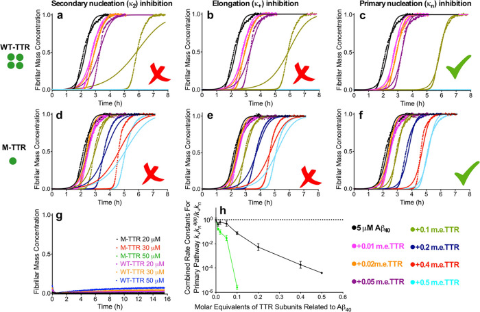
Figure 2.
WT-TTR and M-TTR display strong inhibition of primary nucleation in Aβ40 aggregation. (a–f) Time courses of fibril formation by 5.0 μM Aβ40 in the presence of WT-TTR (a–c) and M-TTR (d–f) at the indicated concentrations (m.e.). The solid lines are fits of the kinetic profiles by a model in which secondary nucleation (a, d), elongation (b, e), or primary nucleation (c, f) is inhibited by TTRs. (g) Incubation of 20, 30, and 50 μM WT-TTR and M-TTR under the same conditions in the absence of Aβ40. (h) Change in the effective rate constants of primary nucleation in Aβ40 aggregation, as derived from (e) and (f), shown with increasing concentrations of WT-TTR (green) and M-TTR (black). Error bars are s.d. of the rates obtained from individual fitting of the repeats. The traces in (a)–(f) show the average traces of those repeats.

