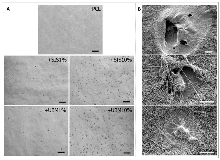
Figure 3.
(A) Gross images of as-spun electrospun fibres for poly(ε-caprolactone) (PCL) and with the addition of 1% or 10% decellularised tissue powder (dECM—small intestinal submucosa (SIS) and urinary bladder matrix (UBM)) (scale = 5 mm). Images converted to greyscale for observation of ‘wet’ droplets on dECM containing fibre scaffolds. (B) Scanning electron microscopy images highlighting the ‘wet’ droplets present within the PCL + SIS10% fibres and the holes these create. Scale bar = 40 μm, magnification ×1000 (top image) and ×2000 (middle and bottom images).

