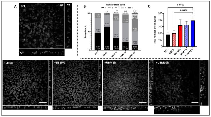
Figure 8.
(A) Representative confocal images of DAPI-stained human conjunctival epithelial cell nuclei cultured on electrospun scaffolds fabricated from poly(ε-caprolactone) (PCL) and PCL with addition of 1% or 10% decellularised tissue powder (small intestinal submucosa (SIS) and urinary bladder matrix (UBM)). Images shown; XY z-stack (scale = 50 μm), XZ side view and YZ side view. (B) Cell layers observed from 6 set regions of interest presented as percentages within each group (n = 3). (C) Total number of cell nuclei counted within 6 regions of interest for each group (n = 3). One-way ANOVA with Tukey’s multiple comparisons, significance for p < 0.05. Statistical differences shown by p values.

