Figure 1.
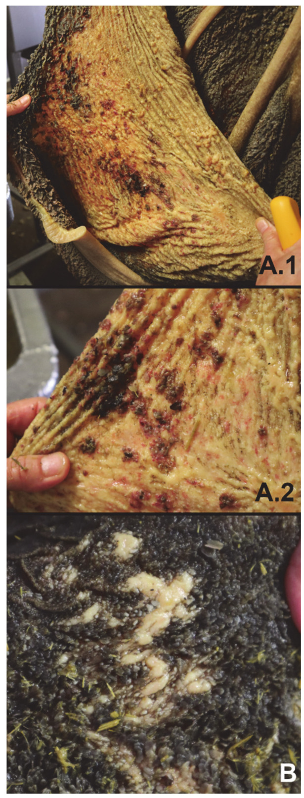
(A.1,A.2) Overview of rumen wall (A.1) and close-up (A.2) of cow L1 indicating the absence of rumen papillae on large parts of the rumen wall and presence of hemorrhages and ulcers. (B) Close-up of rumen wall of cow L6 with local absence of rumen papillae and thickening of the rumen wall (yellowish white spots).

