Figure 6.
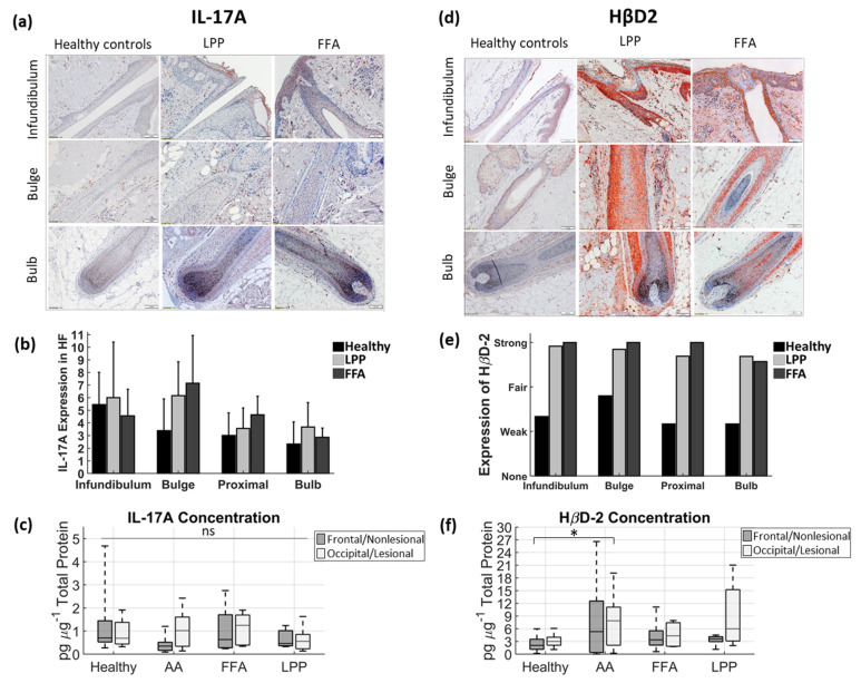
The expression of IL-17A and HβD2 in scalp lesions of patients with cicatricial alopecia and healthy subjects: (a,b) IL-17A; and (d,e) HβD-2 expression in paraffin-embedded sections of healthy scalp and cutaneous lesions (scalp) of cicatricial alopecia patients assessed by immunohistochemistry, followed by a semi-quantitative analysis. The figure panel pair images taken at different hair follicle depths of interest: infundibulum, central part (including bulge region) and bulb. Magnification × 100, scale bars = 100 μm. For (b) IL-17A, the y-axis shows the mean number of positive cells per follicular compartment. Marked presence of (c) IL-17A and (f) HβD2 in infra-infundibular compartments was further confirmed by ELISA analyses of plucked hair follicles from frontal and occipital scalps in healthy and lesional and non-lesional scalp areas in patients with AA (n = 7), LPP (n = 6) and FFA (n = 6). In each box of the box plot, the central line indicates the median, and the bottom and top edges of the box indicate the 25th and 75th percentiles, respectively. The whiskers extend to the maximum and minimum data values. LPP; lichen planopilaris, AA; alopecia areata circumscripta, FFA; frontal fibrosing alopecia; ns, not significant; * p < 0.05 AA (lesional site) compared to healthy controls (frontal site).

