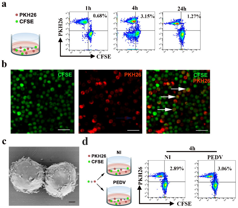
Figure 5.
Cell-to-cell contact in CD3+ T cells of PBMC is not affected in PEDV infection. (a,d) For flow cytometry analysis, CD3+ T cells or CD3+ T cells infected PEDV were stained with the fluorescent probe PKH26 (red) and co-cultivated with target CFSE+ CD3+ T cells at a 1:1 ratio. The percentages of double-fluorescent cells among all cells, which correspond to cell-cell clustering or fusion, are depicted at the indicated time points. Data are representative of three independent experiments. NI, non-infected cells. (b) For confocal microscopy analysis, donors and targets were labeled with distinct membrane-associated fluorescent probes, which emittied in the red (PKH-26) or green (CFSE) wavelength. Formation of double-fluorescent cells in co-culture at a 1:1 ratio was detected by IF. The scale bar represents 20 μm. (c) The structures of lymphocyte-to-lymphocyte contacts were observed using a scanning electron microscope. The scale bar represents 1 μm. PKH26: Red Fluorescent Cell Linker Kits; CFSE: Carboxyfluorescein succinimidyl amino ester.

