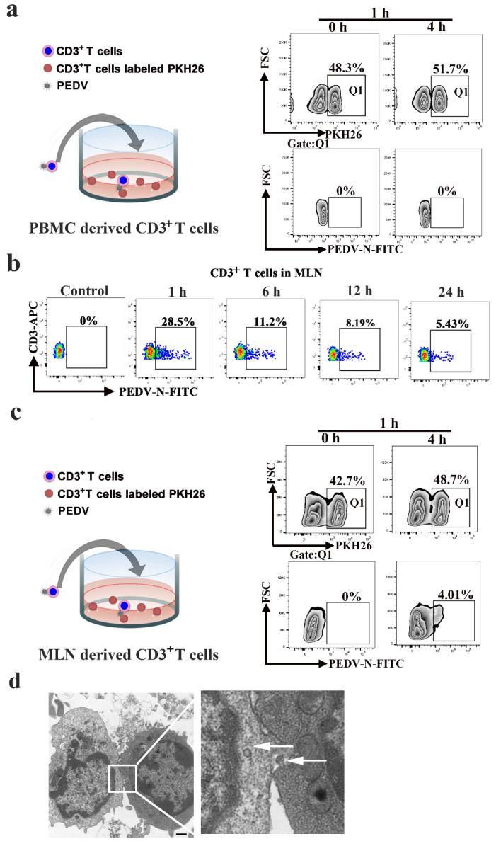
Figure 6.
PEDV cell-to-cell transfer in CD3+ T cells of the lymph node. CD3+ T cells from PBMC (a) or the MLN (c) were infected with PEDV. These cells were then used as donors in the flow cytometry-based assay of viral transfer. Infected cells were co-cultivated with the indicated target PKH26 CD3+ T cells from PBMC or the MLN at a 1:1 ratio at the indicated time points. (b), CD3+ T cells from MLN were infected with PEDV at different times. The viral load values were detected using FACS. (d) The structures of lymphocyte-to-lymphocyte contacts were observed using transmission electron microscopy. In addition, we found complete virions at the site of cell-cell contact (arrowhead). The scale bar represents 1 μm. MLN: mesenteric lymph node. Q1:PKH26+ cells

