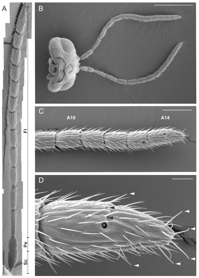
Figure 1.
Scanning electron microscopy (SEM) micrographs showing the filiform antenna of Dryocosmus kuriphilus female. (A) General view of the antenna showing the scape (Sc), the pedicel (Pe) and the flagellum (Fl). The length (mean ± SD) of each antennomere was: Sc = 90.9 ± 5.1; Pe = 125.9 ± 3.3; Fl1 = 182.3 ± 5.4; Fl2 = 164.8 ± 5.9; Fl3 = 139.6 ± 3.6; Fl4 = 135.7 ± 2.9; Fl5 = 122.2 ± 3; Fl6 = 115.7 ± 2.8; Fl7 = 96.6 ± 2.1; Fl8 = 100.4 ± 2.3; Fl9 = 92.1 ± 3; Fl10 = 88.1 ± 3.9; Fl11 = 83.9 ± 4; Fl12 = 143.3 ± 8.8. (B) Ventral view of the head capsule with antennae. (C) Close-up view of the apical part of the flagellum (A10–A14). (D) Detail of the apical antennomere (A14) characterized by a transverse furrow (black arrowheads) positioned in the medial region of the antennomere. White arrowheads indicate the position of sensilla chaetica. Bar scale: (A,B) 500 µm; (C), 100 µm; (D), 20 µm.

