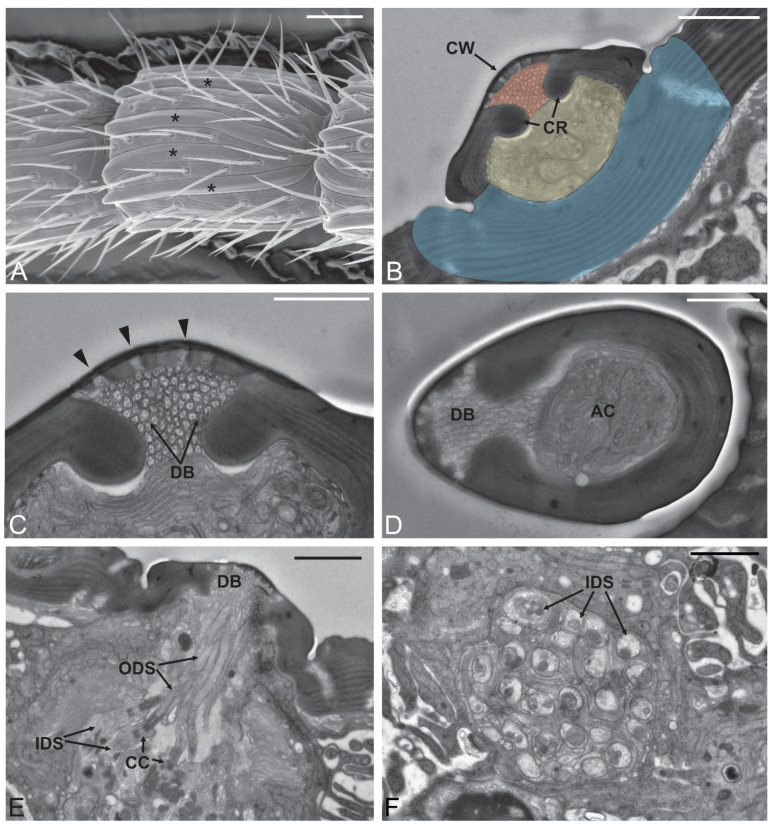
Figure 2.
Sensilla placoidea. (A) SEM micrograph showing A9 with the presence of numerous sensilla placoidea (*): they can be as long as the antennomere itself. (B–E) Transmission electron microscopy (TEM) micrographs showing internal features of sensilla placoidea though sections taken at different levels. In (B), the sensillum is proximal to its distal end. Different regions were colored to better highlight the different areas. In red is the area occupied by the dendritic branches projected by the outer dendritic segments innervating the sensillum. They completely fill the lumen that is externally outlined by the sensillum cuticular wall (CW), while internally there are two cuticular ridges (CR). Just below these two elements, there is a second area (colored in yellow) that is occupied by the sensillum accessory cells, and at this level is separated from the antennal lumen by a thick cuticular wall (colored in blue). More distally (D) the sensillum is separated from the antennal wall but still maintains the separation into two regions. In (C), the cuticular pores (black arrowheads) that open on the sensillum wall are presented. (E) TEM proximal cross-section showing the sensory neurons innervating the sensillum placoideum. Each sensory neuron is clearly divided into a proximal inner dendritic segment (IDS) and an outer dendritic segment (ODS). Between them, typical ciliary constrictions (CC) are visible. ODS enter the sensillum lumen where they organize in numerous dendritic branches (DB). Each sensillum placoideum is innervated by about 25 sensory neurons, as shown in (F). Bar scale: (A), 20 µm; (B,E,F), 2 µm; (C,D), 1 µm.

