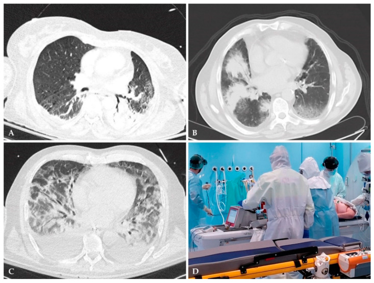
Figure 3.
Role of chest CT in the diagnosis of nosocomial pneumonia. (A) CT scans can accurately differentiate between atelectasis versus pneumonia compared to CXR, especially among critically ill patients. The left lower lobe retrocardiac consolidation, with air bronchogram, consistent with nosocomial pneumonia, was not visualized on portable CXR, but manifested on CT. (B) CT scan may reveal mild infiltrates that are usually missed with conventional CXR. While right lung consolidation shown on this image was visible on CXR, CT allowed for better characterization and revealed a mild infiltrate on the left lower lobe. (C) A wide range of lung pathologies may have similar appearances on CT scan. This image illustrates the difficulty in establishing a differential diagnosis in a patient with acute respiratory distress syndrome (ARDS), with suspected VAP. (D) In-hospital transfer of critically ill patients represents a logistical challenge with potential risks. This image depicts the transfer of a patient with COVID-19 on extracorporeal membrane oxygenation (ECMO) support to a CT scanner, to rule out VAP.

