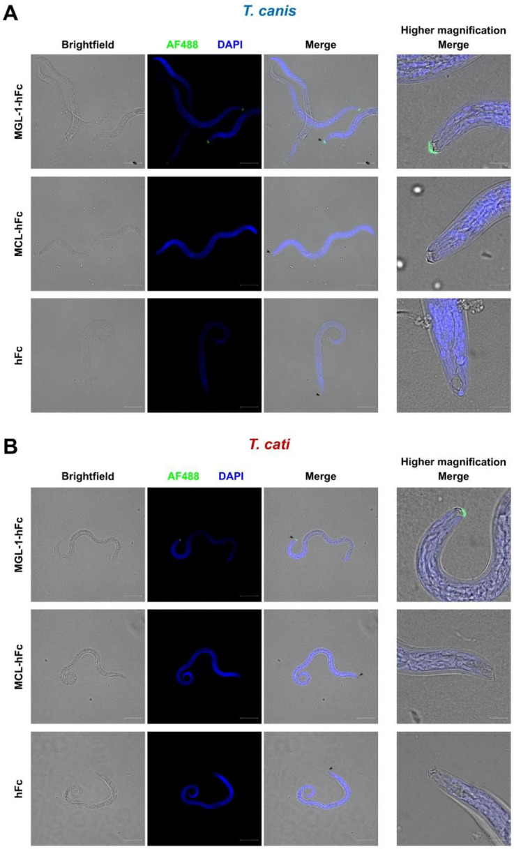
Figure 2.
Fluorescence microscopy reveals binding of macrophage galactose-type lectin-1 (MGL-1) but not macrophage C-type lectin (MCL) to the oral aperture of T. canis (A) and T. cati (B) L3. Scale bar represents 50 µm for lower magnification (left columns) and 10 µm for higher magnification (right column). Green fluorescence: CLR-hFc fusion protein, blue fluorescence: DAPI-stained DNA, hFc: negative control.

