Abstract
Purpose: The purpose of this study was to discover the aqueous humor proteomic changes associated with visual field indices in glaucoma patients. Methods: Aqueous humor samples were analyzed using Liquid Chromatography Tandem Mass Spectrometry (LC-MS/MS). The visual fields were analyzed with the Humphrey Visual Field analyzer. Statistical analyses were performed to discover the relationship between the aqueous humor proteins and visual field parameters including Pattern Standard Deviation (PSD), Visual Field Index (VFI), Mean Deviation (MD) and Glaucoma Hemifield Test (GHT). Results: In total, 222 proteins were identified in 49 aqueous humor samples. A total of 11, 9, 7, and 6 proteins were significantly correlated with PSD, VFI, MD, and GHT respectively. These proteins include apolipoprotein D, members of complement pathway (C1S, C4A, C4B, C8B, and CD14), and immunoglobulin family (IKHV3-9, IGKV2-28). Conclusion: Several proteins involved in immune responses (immunoglobulins and complement factors) and neurodegeneration (apolipoprotein D) were identified to be associated with abnormal visual field parameters. These findings provide targets for future studies investigating precise molecular mechanisms and new therapies for glaucomatous optic neuropathy.
Keywords: aqueous humor, glaucoma, mass spectrometry, proteomics, visual field
1. Introduction
Glaucoma is a progressive optic neuropathy characterized by retinal ganglion cell death resulting in optic nerve atrophy and corresponding visual field loss [1]. The global prevalence of glaucoma is expected to increase to 118 million people in 2040, making it a major health crisis [2]. Functional glaucomatous loss can be assessed via the global indices of Standard Automated Perimetry (SAP) testing such as the Mean Deviation (MD), Pattern Standard Deviation (PSD), Visual Field Index (VFI), and Glaucoma Hemifield Test (GHT) [3]. The MD represents diffuse losses in the visual field compared to age-matched controls. Conversely, PSD represents focal defects that may underlie disease-specific alterations in the visual field [4]. VFI is a global indicator of vision deterioration reflecting retinal ganglion cell loss with values ranging from 0–100% indicating a perimetrically blind eye to perfect vision. GHT measures the asymmetry between the superior and inferior hemifields, categorized as Outside Normal Limits (ONL) or Within Normal Limits (WNL) [3,4].
Aqueous Humor (AH) is a clear fluid that circulates within the anterior segment of the eye and plays an integral role in many ocular health functions, including nutrient and oxygen supply, removal of metabolic waste, ocular immunity, ocular shape, and refraction [5]. Proteins in AH are vital for the maintenance of anterior segment homeostasis and signaling molecules within the AH are implicated in synthesis, degradation, and modification of the Trabecular Meshwork (TM) Extracellular Matrix (ECM) [6]. The elevated levels of TGFβ2 in the AH of glaucoma patients are associated with ECM stiffening within the trabecular meshwork [7,8]. Moreover, AH facilitates the migration of cytokines that stimulate TM cells [9].
Previous studies have demonstrated alterations in several AH proteins in glaucoma patients [10,11,12,13,14]. However, the association between AH protein levels and functional attributes that estimate disease progression is yet to be explored. This study aimed to evaluate the relationship between the AH proteome and visual field indices in glaucoma patients. The study of this relationship may provide us with a better understanding of how molecular alterations (proteins) are linked to changes in functional attributes (visual field metrics).
2. Materials and Methods
2.1. Subjects
The aqueous humor was collected from 49 patients diagnosed with Primary Open Angle Glaucoma (POAG) undergoing phacoemulsification or glaucoma incisional surgery at the Medical College of Georgia, Augusta University. Patients do not have other eye conditions that may influence study outcome. During the surgery, a clear corneal incision is made through which the aqueous humor is normally evacuated from the anterior chamber and discarded. However, instead of discarding the aqueous humor, samples were collected in Eppendorf tubes. This method of sample collection at the beginning of the surgery is efficient and poses no significant risk to the patient.
The Institutional Review Board at Augusta University approved the study (IRB #611480) and written informed consents were obtained from all study participants. Chart reviews were conducted to record age, race, gender, smoking history, presence of systemic diseases, and intraocular pressure (IOP) levels for all subjects (Table 1).
Table 1.
Patient demographic characteristics.
| Patient Characteristics | Count |
|---|---|
| POAG patients | 49 |
| Female/Male | 33/16 |
| AA/Caucasian | 29/20 |
| Smoking, N/Y | 37/12 |
| Hypertension, N/Y | 16/33 |
| Cardiovascular disease, N/Y | 46/3 |
| Cerebrovascular disease, N/Y | 48/1 |
| Collagen vascular disease, N/Y | 43/6 |
| Age (in years) | 68.67 ± 11.4 |
| IOP | 14.9 ± 9.7 |
AA: African American, POAG: Primary Open Angle Glaucoma.
2.2. AH Sample Preparation
Human aqueous humor samples (60 µL) were lyophilized and then reconstituted into 30 µL 8 M urea (Fisher Chemical, Fair Lawn, NJ, USA) in 50 mM Tris-HCl (pH 8; Amersham Biosciences, Uppsala, Sweden). The reduction of the cysteine residues was performed with 20 mM DTT (Sigma-Aldrich; St-Louis, MO, USA) followed by alkylation with 55 mM of iodoacetamide (Sigma-Aldrich; St-Louis, MO, USA). Urea concentration was reduced to below 1 M by adding 240 µL of 50 mM ammonium bicarbonate buffer (Sigma-Aldrich; St-Louis, MO, USA). Protein concentration was measured using a Bradford assay kit according to the manufacturer’s instructions (Pierce, Rockford, IL, USA) and protein digestion was performed using trypsin (1:20 w/w, 37 °C overnight).
2.3. LC-MS/MS Analysis
Digested AH samples were cleaned using a C18 spin plate (Nest Group, Southborough, MA, USA) followed by column separation using an Ultimate 3000 nano-UPLC system (Thermo Scientific Waltham, MA, USA,) and then analyzed with an Orbitrap Fusion tribrid mass spectrometer (Thermo Scientific, Waltham, MA, USA). Reconstituted peptide mixture (6 μL) was trapped and washed on a Pepmap100 C18 trap (5 μm, 0.3 mm × 5 mm; Thermo Scientific, Waltham, MA, USA) using 2% acetonitrile (Fisher Chemical, Fair Lawn, NJ, USA) in water with 0.1% formic acid (Sigma-Aldrich; St-Louis, MO, USA) for 10 min at 20 μL/min and then separated on a Pepman100 RSLC C18 column (2.0 μm, 75 μm × 150 mm) using a solvent gradient of 2 to 40% acetonitrile with 0.1% formic acid at a flow rate of 300 nL/min over 120 min and a column temperature of 40 °C. Eluted peptides were ionized via nano-electrospray ionization with a source temperature of 275 °C, spray voltage of 2000 V, and then introduced into Orbitrap Fusion mass spectrometer. The following instrument settings were used: Data-dependent acquisition in positive mode; orbitrap MS analyzer for precursor scan at 120,000 FWHM from 300 to 1500 m/z; Ion-trap MS analyzer for MS/MS scan in top speed mode (2-s cycle time); dynamic exclusion settings (repeat count 1, repeat duration 15 s, and exclusion duration 30 s); and fragmentation using Collision-induced Dissociation (CID) with a normalized collision energy of 30%.
2.4. Protein Identification and Quantification
The raw MS data were processed using the Proteome Discoverer (v1.4, Thermo Scientific, Waltham, MA, USA) and then submitted for a database search against the manually annotated Uniprot-SwissProt database (20,385 entries) using the SequestHT algorithm. Peptide Spectral Matches (PSMs) were validated using the Perculator PSM validator within the Proteome Discoverer software (v1.4, Thermo Scientific, Waltham, MA, USA). Database search included the following search parameters: Mass tolerances were set at 10 ppm for precursor and 0.6 Da for product ion tolerances; static carbidomethylation (+57.021 Da) for cysteine; and dynamic oxidation (+15.995 Da) for methionine and dynamic phosphorylation (+79.966 Da) for serine, threonine, and tyrosine. Proteins that contained similar peptides that could not be differentiated based on MS/MS analysis alone were grouped based on the Principles of Parsimony. A report comprising the protein identities and PSMs (spectrum counts) for each protein was then exported. The PSM counts serve as a semi-quantitative measure for relative protein expression levels in all samples. A list of 222 proteins identified in AH samples is included in Table S1.
2.5. Visual Field Measurements
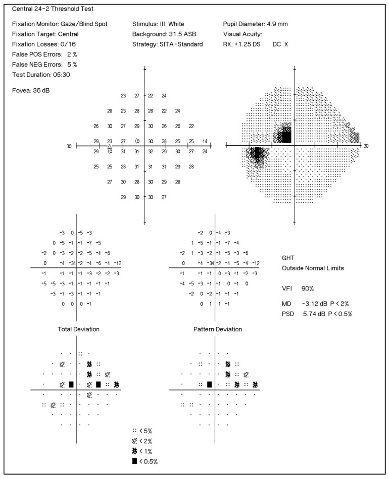
The visual fields were assessed with the Humphrey Field Analyzer (Zeiss Humphrey Systems, Dublin, CA, USA), using the Swedish interactive threshold algorithm (SITA) with the appropriate correction for refractive errors. A trained technician performed all visual field testing. During the procedure, patients respond to a series of white light stimuli of varying brightness. The test assesses retina’s sensitivity to detect stimulus at various points within the visual field. The analyzer prints out results with information about measures indicating the status of visual field for the tested eye. MD is obtained from the total deviation plot and indicates overall mean departure from an age-matched normal. All test values at various points on the visual field are added and divided by the number of test locations. Negative value indicates field loss and might be indicative of glaucoma progression. p value is assigned if MD is significantly outside normal. PSD is obtained from the pattern deviation plot and highlights focal losses. It reflects the degree of departure of the patient’s visual field pattern from the normal hill of vision. A large PSD reflects an irregular hill of vision and is indicative of glaucoma. GHT assesses five corresponding and mirrored areas in the superior and inferior visual fields. If a significant difference is observed between superior and inferior fields, GHT is categorized as outside the normal limit. A representative visual field chart is shown in Figure 1.
Figure 1.
Representative image of the visual field index chart obtained from the Humphrey Visual Field Analyzer II. The test was performed using the standard automated perimetry algorithm with the central 24-2 testing protocol. MD: Mean Deviation, PSD: Pattern Standard Deviation, VFI: Visual Field Index.
2.6. Statistical Analysis
The PSM values obtained from LC-MS/MS analysis were median normalized prior to statistical analysis. Spearman’s correlation analyses were performed between protein levels and PSD, VFI, and MD indices. The Wilcoxon rank sum test was performed to assess proteins differentially expressed between the Outside Normal Limit (ONL) vs. Within Normal Limit (WNL) category of GHT. All statistical analyses were performed using the R Project for Statistical Computing (version 3.6.3, Vienna, Austria).
3. Results
3.1. Aqueous Humor Proteins Associated with PSD
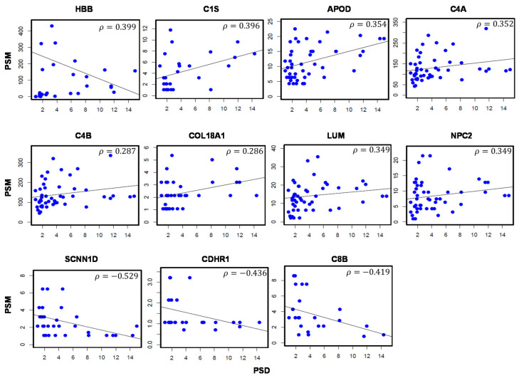
Correlation analysis revealed a total of 11 proteins significantly associated with PSD (Table 2). Plots visualizing the relationship between protein levels and PSD values are shown in Figure 2. Proteins positively correlated with PSD values include the hemoglobin subunit beta (HBB; ρ = 0.399), complement C1s subcomponent (C1S; ρ = 0.396), apolipoprotein D (APOD; ρ = 0.354), complement C4-A (C4A; ρ = 0.352), and complement C4-B (C4B; ρ = 0.349). Proteins with negative correlations include the amiloride-sensitive sodium channel subunit delta (SCNN1D; ρ = −0.529), cadherin-related family member 1 (CDHR1; ρ = −0.436), and complement component C8 beta chain (C8B; ρ = −0.419).
Table 2.
Aqueous humor proteins associated with pattern standard deviation.
| UniProt ID | Gene Symbol | Description | Corr.coeff (ρ) | p-Value |
|---|---|---|---|---|
| P68871 | HBB | Hemoglobin subunit beta | 0.399 | 0.039 |
| P09871 | C1S | Complement C1s subcomponent | 0.396 | 0.041 |
| P05090 | APOD | Apolipoprotein D | 0.354 | 0.013 |
| P0C0L4 | C4A | Complement C4-A | 0.352 | 0.013 |
| P0C0L5 | C4B | Complement C4-B | 0.349 | 0.014 |
| P39060 | COL18A1 | Collagen alpha-1(XVIII) chain | 0.349 | 0.034 |
| P51884 | LUM | Lumican | 0.287 | 0.046 |
| P61916 | NPC2 | NPC intracellular cholesterol transporter 2 | 0.286 | 0.046 |
| P51172 | SCNN1D | Amiloride-sensitive sodium channel subunit delta | −0.529 | 0.001 |
| Q96JP9 | CDHR1 | Cadherin-related family member 1 | −0.436 | 0.026 |
| P07358 | C8B | Complement component C8 beta chain | −0.419 | 0.037 |
Corr.coeff: Correlation coefficient.
Figure 2.
Aqueous humor proteins significantly correlated with Pattern Standard Deviation (PSD) values in glaucoma patients. Scatter plots depict the correlation between protein abundance (PSM; y-axis) and PSD measurements (x-axis). A total of 11 proteins are significantly correlated with PSD values. ρ: Correlation coefficient. p-val < 0.05. Sample size (n) = 49.
3.2. Aqueous Humor Proteins Associated with VFI
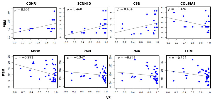
A total of 9 proteins were associated with VFI, which is a measure of overall visual function (Table 3). Four proteins positively correlated to VFI include: CDHR1 (ρ = 0.607), SCNN1D (ρ = 0.460), C8B (ρ = 0.454), Titin (TTN; ρ = 0.399), while 5 proteins with a negative association including Collagen alpha-1(XVIII) chain (COL18A1; ρ = −0.426), APOD (ρ = −0.391), C4B (ρ = −0.347), C4A (ρ = −0.345), and Lumican (LUM; ρ = −0.327). Correlation plots are presented in Figure 3.
Table 3.
Aqueous humor proteins associated with Visual Field Index values.
| UniProt ID | Gene Symbol | Description | Corr.coeff (ρ) | p-Value |
|---|---|---|---|---|
| Q96JP9 | CDHR1 | Cadherin-related family member 1 | 0.607 | 0.002 |
| P51172 | SCNN1D | Amiloride-sensitive sodium channel subunit delta | 0.460 | 0.021 |
| P07358 | C8B | Complement component C8 beta chain | 0.454 | 0.034 |
| Q8WZ42 | TTN | Titin | 0.399 | 0.039 |
| P39060 | COL18A1 | Collagen alpha-1(XVIII) chain | −0.426 | 0.021 |
| P05090 | APOD | Apolipoprotein D | −0.391 | 0.014 |
| P0C0L5 | C4B | Complement C4-B | −0.347 | 0.03 |
| P0C0L4 | C4A | Complement C4-A | −0.345 | 0.032 |
| P51884 | LUM | Lumican | −0.327 | 0.042 |
Corr.coeff: Correlation Coefficient.
Figure 3.
Aqueous humor proteins significantly correlated with Visual Field Index (VFI) values in glaucoma patients. Scatter plots depict the correlation between protein abundance (y-axis) and VFI measurements (x-axis). Nine proteins are significantly correlated with VFI values. ρ: Correlation coefficient. p-val < 0.05. Sample size (n) = 49.
3.3. Aqueous Humor Proteins Associated with MD
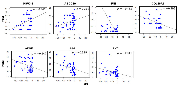
MD represents diffuse vision loss and becomes more negative as glaucoma progresses. Two proteins were positively associated and 5 proteins were negatively associated with MD values (Table 4). Correlation plots visualizing the relationship between protein levels and MD values are presented in Figure 4. These 7 proteins include Immunoglobulin heavy variable 3-9 (IGHV3-9; ρ = 0.342), multidrug resistance-associated protein 7 (ABCC10; ρ = 0.319), fibronectin (FN1; ρ = −0.433), COL18A1 (ρ = −0.395), APOD (ρ = −0.347), LUM (ρ = −0.329), and LYZ (ρ = −0.311).
Table 4.
Aqueous humor proteins associated with Mean Deviation (MD).
| UniProt ID | Gene Symbol | Description | Corr.coeff (ρ) | p-Value |
|---|---|---|---|---|
| P01782 | IGHV3-9 | Immunoglobulin heavy variable 3-9 | 0.342 | 0.048 |
| Q5T3U5 | ABCC10 | Multidrug resistance-associated protein 7 | 0.319 | 0.035 |
| P02751 | FN1 | Fibronectin | −0.433 | 0.015 |
| P39060 | COL18A1 | Collagen alpha-1(XVIII) chain | −0.395 | 0.016 |
| P05090 | APOD | Apolipoprotein D | −0.347 | 0.016 |
| P51884 | LUM | Lumican | −0.329 | 0.023 |
| P61626 | LYZ | Lysozyme C | −0.311 | 0.048 |
Corr.coeff: Correlation coefficient.
Figure 4.
Aqueous humor proteins significantly associated with Mean Deviation (MD) values in glaucoma patients. A total of 7 proteins were significantly correlated with MD values. ρ: Correlation coefficient. p-val < 0.05. Sample size (n) = 49.
3.4. Proteomic Changes Associated with GHT
Analyses were performed to compare the protein levels by dividing the subjects into ONL and WNL groups based on the Glaucoma Hemifield Test (GHT). Four proteins significantly increased with a deviant visual field (ONL) include Monocyte differentiation antigen CD14 (CD14; FC = 2.726), APOD (FC = 1.629), C4B (FC = 1.481), and C4A (FC = 1.476) (Table 5). Two proteins including Immunoglobulin kappa variable 2-28 (IGKV2-28; FC = −2.321) and TTN (FC = −2.061) were decreased in subjects with a deviant visual field. The distribution of protein levels in the two groups is shown in Figure 5.
Table 5.
Aqueous humor proteins associated with abnormal glaucoma hemifield test.
| UniProt ID | Gene Symbol | Description | Fold Change (FC) |
p-Value |
|---|---|---|---|---|
| P08571 | CD14 | Monocyte differentiation antigen CD14 | 2.726 | 0.022 |
| P05090 | APOD | Apolipoprotein D | 1.629 | 0.011 |
| P0C0L5 | C4B | Complement C4-B | 1.481 | 0.046 |
| P0C0L4 | C4A | Complement C4-A | 1.476 | 0.046 |
| A0A075B6P5 | IGKV2-28 | Immunoglobulin kappa variable 2-28 | −2.321 | 0.038 |
| Q8WZ42 | TTN | Titin | −2.016 | 0.028 |
Figure 5.
Six proteins associated with deviant glaucoma hemifield (GHT). The subjects were divided into two groups based on the glaucoma hemifield values, Outside Normal Limits (ONL), and Within Normal Limits (WNL). The boxplots depict the distribution of protein levels. Fold Change (FC) represents the alteration in the protein levels in the ONL group as compared to the WNL group. p-val < 0.05. Sample size (n) = 49.
4. Discussion
Functional assessment of the visual field is employed for the diagnosis and staging of glaucoma. Global visual field indices, including MD and PSD represent diffuse and focal sensitivity losses. In this study, we discovered several AH proteins associated with visual field parameters in glaucoma patients. These proteins are involved in several cellular functions, including neurodegeneration, signaling, metabolism, and immune responses.
Glaucoma is classified as a neurodegenerative disease. The role of apolipoproteins in neurodegenerative disorders, particularly Alzheimer’s disease, is well known [15,16,17]. APOD was positively associated with PSD, while negatively associated with VFI and MD values and elevated in the ONL category of GHT. Overall, the differential association of APOD with each of visual field parameters is indicative of its influence on glaucoma progression. In previous studies, mutations and altered levels of apolipoproteins were observed in glaucoma patients [18,19,20].
Earlier studies have shown that immune-related proteins and several members of the complement components including C1S, C1r, C7-C9, and CFH are activated in glaucoma [21,22,23,24]. In this study, we found 7 proteins to be associated with visual field metrics including C1S, C4A, C4B, C8B, IGHV3-9, CD14, and IGKV2-28. C1S, C4A, and C4B were associated with elevated PSD, lower VFI and ONL category of GHT, suggesting their involvement in glaucoma. On the contrary, C8B was positively correlated with VFI and members of the immunolglobulin family such as IKHV3-9 and IGKV2-28 were positively correlated with MD and presented in lower levels in ONL category patients. The complement family of proteins mediate several immune-related processes, including innate, adaptive, and humoral immunity [25,26].
COL18A1 was associated with elevated PSD, and lower VFI and MD reinforcing the importance of collagens in vision disorders. While the role of COL18A1 in POAG is yet to be established, mutation in this protein is associated with angle closure glaucoma [27]. Lumican is a keratin sulfate proteoglycan involved in the maintenance of collagen fibrils, thus influencing corneal transparency. As a key component of extracellular matrix, this protein is involved in maintaining ocular immunologic status [28]. Similar to COL18A1, lumnican positively correlated with PSD and negatively associated with VFI and MD. Lumican has been implicated in aqueous outflow resistance consistent with its over expression in trabecular meshwork of POAG patients compared to normal eyes [29]. CDHR1 belongs to the cadherin superfamily of calcium-dependent cell adhesion molecules that acts as a photoreceptor. This protein negatively correlated with PSD and positively associated with VFI, indicating lower levels as glaucoma progresses. While the relation between the levels of this protein and glaucoma is yet to be explored, mutations in CDHR1 have been associated with retinal dystrophies [30,31].
5. Conclusions
In conclusion, this study was a novel attempt to correlate the proteomic changes in aqueous humor with the visual field indices. The proteins discovered in this study may be used as potential targets in future studies to discover novel mechanisms of glaucoma progression.
Supplementary Materials
The following are available online at https://www.mdpi.com/2077-0383/10/6/1180/s1, Table S1: List of 222 proteins identified in 49 aqueous humor samples.
Author Contributions
Conceptualization, S.K.K., S.S. and A.S.; Data curation, S.K.K.; Formal analysis, S.K.K. and T.J.L.; Funding acquisition, A.S.; Methodology, S.K.K. and W.Z.; Project administration, A.S.; Resources, K.B., L.U., D.B., A.E., S.S. and A.S.; Software, T.J.L. and A.S.; Supervision, A.S.; Visualization, T.J.L.; Writing—original draft, S.K.K.; Writing—review & editing, L.U., S.S. and A.S. All authors have read and agreed to the published version of the manuscript.
Funding
This work was funded by an NIH/NEI grant (R01-EY029728) awarded to Ashok Sharma.
Institutional Review Board Statement
The study was conducted according to the guidelines of the Declaration of Helsinki, and approved by the Institutional Review Board of Augusta University (IRB # 611480, 17 December 2018).
Informed Consent Statement
Informed consent was obtained from all subjects involved in the study.
Conflicts of Interest
The authors declare no conflict of interest.
Footnotes
Publisher’s Note: MDPI stays neutral with regard to jurisdictional claims in published maps and institutional affiliations.
References
- 1.Weinreb R.N., Aung T., Medeiros F.A. The pathophysiology and treatment of glaucoma: A review. JAMA. 2014;311:1901–1911. doi: 10.1001/jama.2014.3192. [DOI] [PMC free article] [PubMed] [Google Scholar]
- 2.Tham Y.C., Li X., Wong T.Y., Quigley H.A., Aung T., Cheng C.Y. Global prevalence of glaucoma and projections of glaucoma burden through 2040: A systematic review and meta-analysis. Ophthalmology. 2014;121:2081–2090. doi: 10.1016/j.ophtha.2014.05.013. [DOI] [PubMed] [Google Scholar]
- 3.Alencar L.M., Medeiros F.A. The role of standard automated perimetry and newer functional methods for glaucoma diagnosis and follow-up. Indian J. Ophthalmol. 2011;59:S53–S58. doi: 10.4103/0301-4738.73694. [DOI] [PMC free article] [PubMed] [Google Scholar]
- 4.Yaqub M. Visual fields interpretation in glaucoma: A focus on static automated perimetry. Community Eye Health. 2012;25:1–8. [PMC free article] [PubMed] [Google Scholar]
- 5.Grus F.H., Joachim S.C., Pfeiffer N. Proteomics in ocular fluids. Proteom. Clin. Appl. 2007;1:876–888. doi: 10.1002/prca.200700105. [DOI] [PubMed] [Google Scholar]
- 6.Izzotti A., Longobardi M., Cartiglia C., Sacca S.C. Proteome alterations in primary open angle glaucoma aqueous humor. J. Proteome Res. 2010;9:4831–4838. doi: 10.1021/pr1005372. [DOI] [PubMed] [Google Scholar]
- 7.Braunger B.M., Fuchshofer R., Tamm E.R. The aqueous humor outflow pathways in glaucoma: A unifying concept of disease mechanisms and causative treatment. Eur. J. Pharm. Biopharm. 2015;95:173–181. doi: 10.1016/j.ejpb.2015.04.029. [DOI] [PubMed] [Google Scholar]
- 8.Vranka J.A., Kelley M.J., Acott T.S., Keller K.E. Extracellular matrix in the trabecular meshwork: Intraocular pressure regulation and dysregulation in glaucoma. Exp. Eye Res. 2015;133:112–125. doi: 10.1016/j.exer.2014.07.014. [DOI] [PMC free article] [PubMed] [Google Scholar]
- 9.Alvarado J.A., Yeh R.F., Franse-Carman L., Marcellino G., Brownstein M.J. Interactions between endothelia of the trabecular meshwork and of Schlemm’s canal: A new insight into the regulation of aqueous outflow in the eye. Trans. Am. Ophthalmol. Soc. 2005;103:148–162. [PMC free article] [PubMed] [Google Scholar]
- 10.Sharma S., Bollinger K.E., Kodeboyina S.K., Zhi W., Patton J., Bai S., Edwards B., Ulrich L., Bogorad D., Sharma A. Proteomic Alterations in Aqueous Humor From Patients With Primary Open Angle Glaucoma. Investig. Ophthalmol. Vis. Sci. 2018;59:2635–2643. doi: 10.1167/iovs.17-23434. [DOI] [PMC free article] [PubMed] [Google Scholar]
- 11.Chowdhury U.R., Madden B.J., Charlesworth M.C., Fautsch M.P. Proteome analysis of human aqueous humor. Investig. Ophthalmol. Vis. Sci. 2010;51:4921–4931. doi: 10.1167/iovs.10-5531. [DOI] [PMC free article] [PubMed] [Google Scholar]
- 12.Kliuchnikova A.A., Samokhina N.I., Ilina I.Y., Karpov D.S., Pyatnitskiy M.A., Kuznetsova K.G., Toropygin I.Y., Kochergin S.A., Alekseev I.B., Zgoda V.G., et al. Human aqueous humor proteome in cataract, glaucoma, and pseudoexfoliation syndrome. Proteomics. 2016;16:1938–1946. doi: 10.1002/pmic.201500423. [DOI] [PubMed] [Google Scholar]
- 13.Funke S., Perumal N., Bell K., Pfeiffer N., Grus F.H. The potential impact of recent insights into proteomic changes associated with glaucoma. Expert Rev. Proteom. 2017;14:311–334. doi: 10.1080/14789450.2017.1298448. [DOI] [PubMed] [Google Scholar]
- 14.Kaeslin M.A., Killer H.E., Fuhrer C.A., Zeleny N., Huber A.R., Neutzner A. Changes to the Aqueous Humor Proteome during Glaucoma. PLoS ONE. 2016;11:e0165314. doi: 10.1371/journal.pone.0165314. [DOI] [PMC free article] [PubMed] [Google Scholar]
- 15.Zalocusky K.A., Nelson M.R., Huang Y. An Alzheimer’s-disease-protective APOE mutation. Nat. Med. 2019;25:1648–1649. doi: 10.1038/s41591-019-0634-9. [DOI] [PubMed] [Google Scholar]
- 16.Reily C., Stewart T.J., Renfrow M.B., Novak J. Glycosylation in health and disease. Nat. Rev. Nephrol. 2019;15:346–366. doi: 10.1038/s41581-019-0129-4. [DOI] [PMC free article] [PubMed] [Google Scholar]
- 17.Nikolac Perkovic M., Pivac N. Genetic Markers of Alzheimer’s Disease. Adv. Exp. Med. Biol. 2019;1192:27–52. doi: 10.1007/978-981-32-9721-0_3. [DOI] [PubMed] [Google Scholar]
- 18.Nowak A., Rozpedek W., Cuchra M., Wojtczak R., Siwak M., Szymanek K., Szaflik M., Szaflik J., Szaflik J., Majsterek I. Association of the expression level of the neurodegeneration-related proteins with the risk of development and progression of primary open-angle glaucoma. Acta Ophthalmol. 2018;96:e97–e98. doi: 10.1111/aos.13479. [DOI] [PubMed] [Google Scholar]
- 19.Yaylacioglu Tuncay F., Aktas Z., Ergun M.A., Ergun S.G., Hasanreisoglu M., Hasanreisoglu B. Association of polymorphisms in APOE and LOXL1 with pseudoexfoliation syndrome and pseudoexfoliation glaucoma in a Turkish population. Ophthalmic Genet. 2017;38:95–97. doi: 10.3109/13816810.2015.1126617. [DOI] [PubMed] [Google Scholar]
- 20.Carnes M.U., Allingham R.R., Ashley-Koch A., Hauser M.A. Transcriptome analysis of adult and fetal trabecular meshwork, cornea, and ciliary body tissues by RNA sequencing. Exp. Eye Res. 2018;167:91–99. doi: 10.1016/j.exer.2016.11.021. [DOI] [PubMed] [Google Scholar]
- 21.Tezel G., Yang X., Luo C., Kain A.D., Powell D.W., Kuehn M.H., Kaplan H.J. Oxidative stress and the regulation of complement activation in human glaucoma. Investig. Ophthalmol. Vis. Sci. 2010;51:5071–5082. doi: 10.1167/iovs.10-5289. [DOI] [PMC free article] [PubMed] [Google Scholar]
- 22.Gajda-Derylo B., Stahnke T., Struckmann S., Warsow G., Birke K., Birke M.T., Hohberger B., Rejdak R., Fuellen G., Junemann A.G. Comparison of cytokine/chemokine levels in aqueous humor of primary open-angle glaucoma patients with positive or negative outcome following trabeculectomy. Biosci. Rep. 2019;39 doi: 10.1042/BSR20181894. [DOI] [PMC free article] [PubMed] [Google Scholar]
- 23.Anshu A., Price M.O., Richardson M.R., Segu Z.M., Lai X., Yoder M.C., Price F.W., Jr. Alterations in the aqueous humor proteome in patients with a glaucoma shunt device. Mol. Vis. 2011;17:1891–1900. [PMC free article] [PubMed] [Google Scholar]
- 24.Mirzaei M., Gupta V.B., Chick J.M., Greco T.M., Wu Y., Chitranshi N., Wall R.V., Hone E., Deng L., Dheer Y., et al. Age-related neurodegenerative disease associated pathways identified in retinal and vitreous proteome from human glaucoma eyes. Sci. Rep. 2017;7:12685. doi: 10.1038/s41598-017-12858-7. [DOI] [PMC free article] [PubMed] [Google Scholar]
- 25.Hadders M.A., Beringer D.X., Gros P. Structure of C8alpha-MACPF reveals mechanism of membrane attack in complement immune defense. Science. 2007;317:1552–1554. doi: 10.1126/science.1147103. [DOI] [PubMed] [Google Scholar]
- 26.Schroeder H.W., Jr., Cavacini L. Structure and function of immunoglobulins. J. Allergy Clin. Immunol. 2010;125:S41–S52. doi: 10.1016/j.jaci.2009.09.046. [DOI] [PMC free article] [PubMed] [Google Scholar]
- 27.Suri F., Yazdani S., Chapi M., Safari I., Rasooli P., Daftarian N., Jafarinasab M.R., Ghasemi Firouzabadi S., Alehabib E., Darvish H., et al. COL18A1 is a candidate eye iridocorneal angle-closure gene in humans. Hum. Mol. Genet. 2018;27:3772–3786. doi: 10.1093/hmg/ddy256. [DOI] [PubMed] [Google Scholar]
- 28.Amjadi S., Mai K., McCluskey P., Wakefield D. The role of lumican in ocular disease. ISRN Ophthalmol. 2013;2013:632302. doi: 10.1155/2013/632302. [DOI] [PMC free article] [PubMed] [Google Scholar]
- 29.Diskin S., Kumar J., Cao Z., Schuman J.S., Gilmartin T., Head S.R., Panjwani N. Detection of differentially expressed glycogenes in trabecular meshwork of eyes with primary open-angle glaucoma. Investig. Ophthalmol. Vis. Sci. 2006;47:1491–1499. doi: 10.1167/iovs.05-0736. [DOI] [PMC free article] [PubMed] [Google Scholar]
- 30.Stingl K., Mayer A.K., Llavona P., Mulahasanovic L., Rudolph G., Jacobson S.G., Zrenner E., Kohl S., Wissinger B., Weisschuh N. CDHR1 mutations in retinal dystrophies. Sci. Rep. 2017;7:6992. doi: 10.1038/s41598-017-07117-8. [DOI] [PMC free article] [PubMed] [Google Scholar]
- 31.Nikopoulos K., Avila-Fernandez A., Corton M., Lopez-Molina M.I., Perez-Carro R., Bontadelli L., Di Gioia S.A., Zurita O., Garcia-Sandoval B., Rivolta C., et al. Identification of two novel mutations in CDHR1 in consanguineous Spanish families with autosomal recessive retinal dystrophy. Sci. Rep. 2015;5:13902. doi: 10.1038/srep13902. [DOI] [PMC free article] [PubMed] [Google Scholar]
Associated Data
This section collects any data citations, data availability statements, or supplementary materials included in this article.


