Abstract
A series of novel coumarin-3-carboxamide derivatives were designed and synthesized to evaluate their biological activities. The compounds showed little to no activity against gram-positive and gram-negative bacteria but specifically showed potential to inhibit the growth of cancer cells. In particular, among the tested compounds, 4-fluoro and 2,5-difluoro benzamide derivatives (14b and 14e, respectively) were found to be the most potent derivatives against HepG2 cancer cell lines (IC50 = 2.62–4.85 μM) and HeLa cancer cell lines (IC50 = 0.39–0.75 μM). The activities of these two compounds were comparable to that of the positive control doxorubicin; especially, 4-flurobenzamide derivative (14b) exhibited low cytotoxic activity against LLC-MK2 normal cell lines, with IC50 more than 100 μM. The molecular docking study of the synthesized compounds revealed the binding to the active site of the CK2 enzyme, indicating that the presence of the benzamide functionality is an important feature for anticancer activity.
Keywords: coumarin3-carboxamides, coumarins, pyranocoumarins, anticancer activity, antibacterial activity
1. Introduction
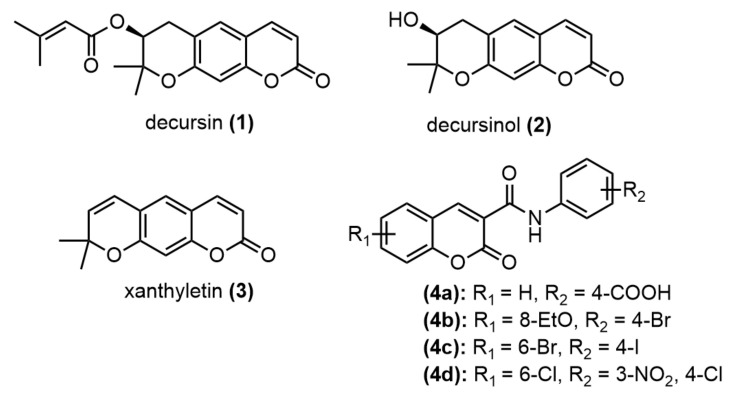
Coumarin is one of the potent secondary metabolites in plants [1,2] and fungi [3], and it is characterized by several pharmacological properties [4]. Like decursin 1 and decursinol 2, these coumarins have pyranocoumarin moiety (Figure 1), having been isolated from the medicinal plant Angelica genus and shown potential for treating inflammatory diseases such as cancer, hepatic fibrosis, diabetic retinopathy, and neurological disorders [5]. The dehydrated derivative of decursinol, xanthyletin 3, has also been shown to possess several biological properties, such as anti-tumor and antibacterial activities [6]. With a benzopyrone skeleton, coumarin is versatile and can be easily transformed into a large variety of functionalized skeletons. As a result, many coumarin derivatives have been designed, synthesized, and evaluated to address broad biological activities [7] such as antibacterial [8], antifungal [9], antioxidant [10], anti-inflammatory [11], anticancer [12], anticoagulant [13], and antiviral activities [14]. The synthetic N-phenyl coumarin carboxamide 4a has been designed and shown to possess high antibacterial activity against Helicobacter pylori (H. pylori), with the minimum inhibitory concentration (MIC) = 1 µg/mL [15], while the benzyl substitution of coumarin carboxamides 4b–d has been shown to arrest breast cancer cell (BT474 and SKBR-3) growth by inhibiting ErbB-2 and ERK1 MAP kinase. Moreover, compounds 4b–d are specific to cancer cell lines, with no cytotoxicity against normal human fibroblasts [16]. In our ongoing search for novel compounds to overcome drug resistance, the diverse biological activities of coumarins have been interesting. In the current study, we designed novel pyranocoumarin-3-carboxamide derivatives with the expectation that the carboxamide part could possess active pharmacological properties 4a–d and that the pyran ring moiety could also show specific biological proteins, as in the case of xanthyletin 3. The synthesized compounds were examined to evaluate their antibacterial activity and cytotoxicity against HepG2 and HeLa cell lines. As the coumarins were attractive casein kinase 2 (CK2) inhibitors [17], molecular docking was used to study the possible interactions of novel coumarin-3-carboxamides with the CK2 enzymes.
Figure 1.
Synthetic and natural-occurring coumarins with biological activities.
2. Results and Discussion
2.1. Chemistry
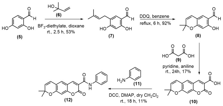
The preparation of pyranocoumarin-3-carboxamide was applied from the previous synthetic strategies reported by Faulgues and colleagues [18] and was described in Scheme 1. Commercially available 2,4-dihydroxy benzyldehyde 5 and 3-hydroxy-3-methyl-1-butene 6 were used as the starting materials and were subjected to Lewis acid–promoted Friedel-Crafts alkylation reaction in dioxane with BF3-diethylate to obtain the aldehyde 7 in 53% yield as a major product together with many unidentified products [19]. The aldehyde 7 was cyclized to form a pyrano ring using 2,3-dichloro-5,6-dicyano-l,4-benzoquinone (DDQ), and the benzopyran 8 was obtained in a very good yield. The cyclization of benzopyran 8 with the malonic anhydride 9 in pyridine and aniline at room temperature for 24 h according to a previous report [20] gave a poor yield of the pyranocoumarin-3-carboxylic acid 10, due to the difficulty of purification. The amidation of the acid 10 with aniline using N,N’-dicyclohexyl carbodiimide (DCC) and 4-dimethylaminopyridine (DMAP) gave the amide 12 in 11% yield after recrystallization.
Scheme 1.
Synthesis of pyranocoumarin-3-carboxamide 12.
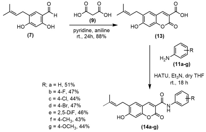
To improve the yield of pyranocoumarin-3-carboxamide 12, the coumarin-3-carboxylic acid 13 was prepared in good yield prior to amidation with appropriate anilines using hexafluorophosphate azabenzotriazole tetramethyl Uronium (HATU) and Et3N to obtain amide 14a–g in 43%–51% yields (Scheme 2). Then, the cyclization of 14a by refluxing with DDQ in benzene gave pyranocoumarin-3-carboxamide 12 in 66% yield (Scheme 3). To study the effect of the substituent at C3 of coumarin ring, the carboxyl group was decarboxylated using Cu powder to give coumarin 15 in 53% yield (Scheme 4). Then, the synthesized coumarins were further evaluated for their antibacterial and anticancer activities.
Scheme 2.
Synthesis of coumarin-3-carboxamides 14a–g.
Scheme 3.
Cyclization of pyranocoumarin-3-carboxamide 12.
Scheme 4.
Decarboxylation of coumarin-3-carboxylic acid.
2.2. Antibacterial Activity
Coumarin derivatives 10, 12, 13, 14a–g, and 15 were evaluated for their antibacterial activity against Bacillus cereus, Bacillus subtilis, Staphylococcus aureus, Escherichia coli, Salmonella typhimuriumthrough using the microbroth dilution method. Penicillin G and solvent were used as positive and negative controls, respectively, and the MIC (µg/mL) values were obtained (Table 1). The results show that only compounds 10 and 13 exhibited moderate antibacterial activities against gram-positive bacteria, while the other tested compounds displayed MIC values of more than 128 µg/mL. This may be because the carboxylic acid at the C3 position played an essential role in the antibacterial activity. Compound 15, without the carboxyl group, showed no antibacterial activity, and the carboxamides 14a–g, displayed no activity. Meanwhile, coumarin-3-carboxylic acid 13 was the most active among the tested compounds, with an MIC value of 32 μg/mL against B. cereus; however, it was less active than the reference drug penicillin G. Moreover, all the tested compounds showed no activity against any gram-negative bacteria.
Table 1.
MIC of coumarin derivatives 10, 12a, 13, 14a–g, 15 and penicillin G against B. cereus, B. subtilis, S. aureus, E. coli, S. typhimurium.
| Compounds | R | MIC (µg/mL) | ||||
|---|---|---|---|---|---|---|
| Gram-Positive | Gram-Negative | |||||
| B. cereus | B. subtilis | S. aureus | E. coli | S. typhimurium | ||
| 10 | - | 128 | 128 | 64 | >128 | > 128 |
| 12 | 4-H | > 128 | > 128 | > 128 | >128 | > 128 |
| 13 | - | 32 | 128 | 64 | >128 | > 128 |
| 14a | 4-H | > 128 | > 128 | > 128 | >128 | > 128 |
| 14b | 4-F | > 128 | > 128 | > 128 | >128 | > 128 |
| 14c | 4-Cl | > 128 | > 128 | > 128 | >128 | > 128 |
| 14d | 4-Br | > 128 | > 128 | > 128 | >128 | > 128 |
| 14e | 2,5-diF | > 128 | > 128 | > 128 | >128 | > 128 |
| 14f | 4-CH3 | > 128 | > 128 | > 128 | >128 | > 128 |
| 14g | 4-OCH3 | > 128 | > 128 | > 128 | >128 | > 128 |
| 15 | - | > 128 | > 128 | > 128 | >128 | > 128 |
| penicillin G | - | 16 | 2 | 16 | 32 | 64 |
2.3. Anticancer Activity
All synthesized compounds were evaluated for in vitro cytotoxic activity against two cancer cell lines (HepG2 and HeLa cell lines) and normal cell lines (LLC-MK2) through an MTT assay, and the results are presented in Table 2. Most of the tested compounds displayed potent anticancer activity. The N-phenyl coumarin-3-carboxamides 12 and 14a showed significantly more potency than the parent acids 10 and 13, respectively, against HepG2 cell lines. Moreover, compound 15, with no substituent at C3, showed better activity than the parent acid 13. The effect of substituents on the phenyl ring was compared with the effect of substituents on the carboxamide 14a and it was found that the phenyl bearing fluorine atoms 14b and 14e possessed similar effects on the potency, while, the phenyl bearing 4-chlorine and 4-bromine atoms showed less activity against both cancer cell lines. Moreover, the phenyl bearing 4-methyl and 4-methoxyl groups displayed weak activity against the test cancer cell lines. From these results, the size and electron-donating group of the para-substituted benzene ring may affect anticancer properties. From these tested compounds, the amide 14e displayed the most potent anticancer activity; however, it exhibited high cytotoxic activity against the normal cell line, with IC50 = 1.33 μM. Interestingly, amide 14b displayed slightly lower activity than 14e, but it showed low cytotoxicity against the normal cell line. This compound possessed anticancer activity comparable to those of the tested anticancer drugs doxorubicin and acridine orange.
Table 2.
In vitro anticancer activities of synthesized coumarins compared with doxorubicin and acridine orange.
| Compounds | R | IC50 (µM) | ||
|---|---|---|---|---|
| Cancer Cell Line | Normal Cell Line | |||
| HepG2 | HeLa | LLC-MK2 | ||
| 10 | - | > 100 | 80.38 | > 100 |
| 12 | 4-H | 2.35 | > 100 | > 100 |
| 13 | - | 81.73 | 26.42 | 37.95 |
| 14a | 4-H | 4.33 | 10.40 | 0.48 |
| 14b | 4-F | 4.85 | 0.75 | >100 |
| 14c | 4-Cl | 30.28 | 14.04 | 66.80 |
| 14d | 4-Br | 45.76 | 37.36 | > 100 |
| 14e | 2,5-diF | 2.62 | 0.39 | 1.33 |
| 14f | 4-CH3 | > 100 | > 100 | 85.31 |
| 14g | 4-OCH3 | > 100 | > 100 | > 100 |
| 15 | - | 9.13 | 13.47 | 6.87 |
| Doxorubicin | - | 1.91 | 1.18 | 63.47 |
| Acridine orange | - | 5.72 | 6.59 | 82.30 |
2.4. Molecular Docking
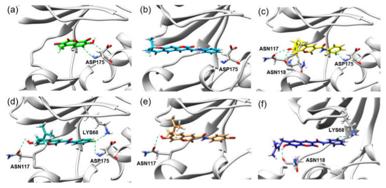
Casein Kinase 2 enzyme is a key player in the pathophysiology of cancer [21,22]. Using the iGEMDOCK v2.1 software [23], molecular docking was performed to investigate binding positions and intermolecular interactions between coumarin 10, 12, 13, 14a–f, and 15 and the binding site of CK2. Coumarins 10, 12, 13, 14a–f, and 15 were docked to the active site of CK2 co-crystallized with G12 (PDB ID: 2QC6). Moreover, G12 was also redocked to CK2, and its total energy and hydrogen bond length were compared with those of coumarins 10, 12, 13, 14a–f, and 15. The molecular docking results show that the binding position of redocked G12 was roughly the same as that of co-crystallized G12 in CK2 (Figure 2a). Moreover, all synthesized compounds were bound to the active site of CK2, and the binding positions were similar to that of G12 (Figure 2b,c), and their binding energies (−118.99 to −89.19 kcal/mol) were lower than that of G12 (−79.10 kcal/mol) (Table 3). Figure 3 also illustrates the hydrogen bond interactions in the binding of coumarin 12, 14a, 14b, 14d, and 14e in the cavity of CK-2 compared with that of G12. Coumarins 14a–f had much lower binding energy (−118.99 to −105.53 kcal/mol) than G12, and their N-phenyl ring was also bound to similar positions of the G12 phenolic ring (Figure 2c). Key amino acid residues, including LYS68, ASN118, ASN117, and ASP175, formed a hydrogen bond with 14a–f, where ASN117 and ASN118 interacted with the hydroxy group of 14a–f, while N-phenyl ring interacted with LYS68 and ASP175. The substitution group on the N-phenyl ring of 14a–f also influenced the number of hydrogen bonds in the CK2 active site. Moreover, a comparison of the halogen substitution groups on the N-phenyl ring showed that the Cl and Br substitution groups on the N-phenyl ring formed no hydrogen bond, while the F substitution group 14b and 14e could form hydrogen bonds with LYS68 and ASP175 (Figure 3d,f), which may be because Cl and Br are larger than F in size. Additionally, the experimental results show that the coumarins 14b and 14e had good anticancer activities.
Figure 2.
(a) Redocked G12 (brown) and G12 (green) in the cavity of CK-2 (PDB ID: 2QC6). (b) Comparison of the bindings of 10 (pink), 12 (sky blue), 13 (orange), 15 (violet), and G12 (green) in the CK-2 cavity. (c) Comparison of the bindings of 14a (yellow), 14e (blue), 14f (purple), 14g (red), and G12 (green) in the CK-2 cavity (PDB ID: 2QC6).
Table 3.
Summary of binding energy, amino acid interactions, and hydrogen bond length of coumarin derivatives in CK2 binding site.
| Compounds | Total Energy (kcal/mol) | Amino Acid Residue | Hydrogen Bond Length (Å) |
|---|---|---|---|
| 10 | −89.19 | LYS68, ASP175 | 2.44, 2.34 |
| 12 | −100.88 | ASP175 | 2.91 |
| 13 | −91.13 | LYS68 | 1.92 |
| 14a | −108.21 | ASN118, ASN117, ASP175 | 2.19, 2.99,2.51 |
| 14b | −107.08 | ASN117, LYS68, ASP175 | 2.93, 2.42, 2.74 |
| 14c | −106.07 | ASN117 | 2.94 |
| 14d | −105.91 | ASN117 | 2.95 |
| 14e | −111.19 | ASN118, LYS68 | 2.27, 2.44 |
| 14f | −105.53 | ASN117 | 2.93 |
| 14g | −118.99 | ASN117, LYS68, GLU81 | 2.74, 1.90, 2.72 |
| 15 | −86.27 | LYS68, GLU114, ASP175, | 2.08, 1.99, 2.42 |
| G12 | −79.10 | ASP175 | 2.43 |
Figure 3.
Hydrogen bond interactions in the bindings of (a) G12,(b) 12,(c) 14a, (d) 14b,(e) 14d, and (f) 14e in the CK-2 cavity.
3. Materials and Methods
3.1. Chemistry
General information: Solvents and reagents were purchased from commercial suppliers TCI Chemicals (Tokyo, Japan), Sigma-Aldrich (Bangalore, India), and Fluka (Dorset, UK). Structure determination was conducted by analyzing the 1H, 13C, and 19F NMR spectra (Bruker 300 apparatus) and the infrared (IR) spectrum was determined using PerkinElmer Frontier Fourier-transform infrared spectrometer. Melting point was conducted using Stuart SMP2 melting point apparatus and high-resolution mass spectroscopy was analyzed by Thermo scientific, Orbitrap Q Exactive Focus.
3.1.1. Synthesis of 2,4-Dihydroxy-5-(3-methylbut-2-en-1-yl)-benzaldehyde 7
First, 2,4-dihydroxy benzaldehyde 5 (1.5 g, 10.8 mmol) in dioxane (5 mL) was added to a stirred solution of 3-hydroxy-3-methyl-1-butene 6 (1.5 mL, 14.3 mmol) and boron trifluoride diethyl etherate (BF3-OEt2, 1.5 mL) in dioxane (3 mL), and stirring was continued for 2.5 h at room temperature. Dichloromethane (50 mL) was added, and the resulting solution was extracted with water (3 × 50 mL). The combined organic layer was dried over Na2SO4 before evaporation to dryness and then purified via column chromatography (silica gel, 4:1 hexane:EtOAc) to obtain a white solid of 2,4-dihydroxy-5-(3-methylbut-2-en-1-yl) benzaldehyde 7 (0.48 g, 53% yield): m.p. 124–125 °C (lit. [19] 121–123 °C), 1H-NMR (300 MHz, CDCl3): δ 11.27 (s, 1H), 9.69 (s, 1H, OH), 7.26 (s, 1H), 6.37 (s, 1H), 6.10 (s, 1H, OH), 5.30 (tt, J = 4.53, 1.35 Hz, 1H), 3.30 (d, J = 7.2 Hz, 2H), 1.77 (s, 3H), 1.61 (s, 3H) ppm.
3.1.2. Synthesis of 7-Hydroxy-2,2-dimethyl-2H-chromene-6-carbaldehyde 8
A mixture of compound 7 (1.0 g, 4.85 mmol) and DDQ (1.2 g, 5.28 mmol) in benzene (10 mL) was refluxed for 6 h, and the precipitate was filtered off. The filtrate was evaporated to dryness to afford the crude product, which was purified via column chromatography (silica gel, 10:1 hexane:EtOAc) to obtain a white solid of 7-hydroxy-2,2-dimethyl-2H-chromene-6-carbaldehyde 8 (0.91 g, 92% yield): m.p. 82–83 °C (lit. [20] 95–96 °C), 1H NMR (300 MHz, CDCl3): δ 11.41 (br, OH), 9.68 (s, 1H), 7.16 (s, 1H), 6.35 (s, 1H), 6.30 (d, J = 7.86 Hz, 1H), 5.59 (d, J = 9.93 Hz, 1H), 1.59 (s, 3H) 1.48 (s, 3H) ppm.
3.1.3. Synthesis of 8,8-Dimethyl-2-oxo-2H,8H-pyrano[3,2-g]chromene-3-carboxylic acid 10
Compound 8 (1.0 g, 4.90 mmol) and malonic acid 9 (1.0 g, 9.60 mmol) were dissolved in pyridine (5.5 mL) containing aniline (0.5 mL) and stirred for 24 h at room temperature. Afterward, the rection mixture was poured into ice-cold 10% HCl (80 mL). The yellow precipitate was washed with cold water to remove mineral acid and then air-dried to yield a yellow solid (recrystallization by 2:1:2; EtOAc:EtOH:hexane) of 8,8-dimethyl-2-oxo-2H,8H-pyrano[3,2-g]chromene-3-carboxylic acid 10 (0.23 g, 17% yield): m.p. 187–188 °C, 1H NMR (300 MHz, CDCl3): δ 8.80 (s, 1H), 7.29 (s, 1H), 6.85 (s, 1H), 6.40 (d, J = 10.02 Hz, 1H), 5.81 (d, J = 10.02 Hz, 1H), 1.52 (s, 6H) ppm., 13C NMR (75 MHz, CDCl3): δ 164.54 (C), 163.28 (C), 160.87 (C), 156.60 (C), 150.99 (CH), 132.34 (C), 127.13 (CH), 120.17 (CH), 120.06 (CH), 112.50 (C), 110.59 (C), 104.38 (CH), 79.33 (C), 28.75 (2CH3) ppm. IR: 3051.36 (C-H, aromatic), 2922.46 (C-H, aliphatic), 1743.12 (C=O, acid) cm−1; HREI-MS (m/z) calculated for C15H13O5 (M)+ 273.0758, found 273.0757.
3.1.4. Synthesis of 8,8-Dimethyl-2-oxo-N-phenyl-2H,8H-pyrano[3,2-g]chromene-3-carboxamide 12
A mixture of compound 10 (0.10 g, 0.36 mmol), aniline 11 (0.040 mL, 0.43 mmol), DCC (0.10 g, 0.44 mmol), and DMAP (8 mg, 0.065 mmol) in dry CH2Cl2 (5 mL) was stirred at room temperature for 18 h. Afterward, the reaction mixture was filtered, and the filtrate was evaporated under vacuum. The residue was recrystallized using EtOH to obtain a yellow solid of 8,8-dimethyl-2-oxo-N-phenyl-2H,8H-pyrano[3,2-g]chromene-3-carboxamide 12 (14 mg, 11% yield): m.p. 187–188 °C, 1H NMR (300 MHz, CDCl3): δ 10.81(s, 1H), 8.89 (s, 1H), 7.75 (d, J = 1.14 Hz, 2H), 7.39 (t, J = 6.54 Hz, 2H), 7.30 (s, 1H), 7.18 (t, J = 1.14 Hz, 1H), 6.80 (s, 1H), 6.40 (d, J = 9.90 Hz, 1H), 5.78 (d, J = 9.96 Hz, 1H), 1.55 (s, 6H) ppm., 13C NMR (75 MHz, CDCl3): δ 162.20 (C), 160.03 (C), 159.55 (C), 156.35 (C), 148.67 (CH), 137.30 (C), 131.82 (CH), 129.02 (2CH), 126.72 (CH), 124.57 (CH), 120.53 (2CH), 120.43 (CH), 119.58 (C), 114.71 (C), 112.73 (C), 75.72 (C), 28.64 (2CH3) ppm. IR: 3198.94 (N-H), 3059.35 (C-H, aromatic), 2969.10 (C-H, aliphatic), 1699.85 (C=O, amide) cm−1; HREI-MS (m/z) calculated for C21H18O4N (M)+ 348.1230, found 348.1230.
3.1.5. Synthesis of 3-Carboxy-6-(3-methyl-2-butenyl)-7-hydroxy-coumarin 13
Compound 7 (1.0 g, 4.85 mmol) and malonic acid 9 (1.0 g, 9.60 mmol) were dissolved in pyridine (5.5 mL) containing aniline (0.5 mL), and stirred for 24 h at room temperature. Afterward, the reaction mixture was poured into ice-cold 10% HCl (80 mL), and the yellow precipitate obtained was washed with cold water to remove mineral acid and then air-dried to yield a yellow solid of 3-carboxy-6-(3-methyl-2-butenyl)-7-hydroxy-coumarin 13 (1.10g, 88% yield): m.p. 237–238 °C (lit. [20] 218–224 °C), 1H NMR (300 MHz, CDCl3+methanol-d4) δ 8.79 (s. 1H), 7.40 (s, 1H), 6.85 (s, 1H), 5.32 (tt, J = 7.4, 1.3 Hz, 1H), 3.34 (d, J = 7.29 Hz, 2H), 1.79 (s, 3H), 1.70 (s, 3H) ppm., 13C-NMR (75 MHz, CDCl3+methanol-d4): δ 163.58 (C), 162.79 (C), 162.37 (C), 154.78 (C), 150.43 (CH), 133.49 (C), 129.32 (CH), 127.92 (C), 119.16 (CH), 110.24 (C), 107.85 (C), 100.76 (CH), 26.34 (CH2), 24.42 (CH3), 16.50 (CH3) ppm., IR: 3303.61 (O-H), 3049.81 (C-H, aromatic), 2911.92 (C-H, aliphatic), 1733.10 (C=O, acid), 1718.83 (C=O, lactone) cm−1.
3.1.6. General Procedure of Coumarin-3-carboxamides Preparation (14a–g)
Triethanolamine (TEA) (0.1 mL, 1.36 mmol) was added to a solution of compound 13 (70 mg, 0.26 mmol) and HATU (0.12 g, 0.36 mmol) in dry THF (5 mL), and the mixture was stirred at room temperature for 30 min. The obtained dark clear mixture was treated with aniline derivatives (11a–g) (1.2 eq.). The resulting mixture was stirred at room temperature for 18 h. Dichloromethane (50 mL) was added, and the resulting solution was extracted with sat. NaCl (3 × 30 mL). The remaining organic layer was dried over Na2SO4 before evaporation to dryness. After evaporation of the solvent in vacuo, the crude product was purified via preparative thin-layer chromatography (silica gel, 4:1 hexane:EtOAc) to give yellow solids of coumarin-3-carboxamide (14a–g)
7-Hydroxy-6-(3-methylbut-2-en-1-yl)-2-oxo-N-phenyl-2H-chromene-3-carboxamide 14a (47 mg, 51% yield): m.p. 258–259 °C, 1H NMR (300 MHz, CDCl3+pyridine-d5): δ 10.86 (s, NH), 8.90 (s, 1H), 7.73 (d, J = 7.62 Hz, 2H), 7.42 (s, 1H), 7.35 (t, J = 7.62 Hz, 2H), 7.12 (t, J = 7.38 Hz, 1H), 6.84 (s, 1H), 5.40 (tt, J = 7.26, 1.35 Hz, 1H), 3.43 (d, J = 7.26 Hz, 2H), 1.81 (s, 3H), 1.74 (s, 1H) ppm., 13C NMR (75 MHz, CDCl3+pyridine-d5): δ 163.38 (C), 162.81 (C), 160.56 (C), 155.49 (C), 149.36 (CH), 138.08 (CH), 134.13(C), 130.04 (CH), 128.97 (2CH), 128.68 (C), 124.35 (CH), 121.14 (CH), 120.49 (2CH), 112.94 (C), 111.33 (C), 101.68 (CH), 27.85 (CH2), 25.84 (CH3), 17.85 (CH3) ppm., IR: 3195.27 (O-H), 2917.31 (C-H, aliphatic), 1695.54 (C=O, amide) cm−1; HREI-MS (m/z) calculated for C21H20O4N (M)+ 350.1387, found 350.1386.
N-(4-Fluorophenyl)-7-hydroxy-6-(3-methylbut-2-en-1-yl)-2-oxo-2H-chromene-3-carboxamide 14b (45 mg, 47% yield): m.p. 259–261 °C, 1H NMR (300 MHz, CDCl3): δ 10.56 (br, NH), 8.89 (s, 1H), 7.68 (dd, J = 8.97, 4.92 Hz, 2H), 7.42 (s, 1H), 7.07 (dd, J = 9.12, 8.79 Hz 2H), 6.83 (s, 1H), 5.34 (t, J = 7.23 Hz, 1H), 3.35 (d, J = 7.29 Hz, 2H), 1.80 (s, 3H), 1.72 (s, 1H) ppm., 13C NMR (75 MHz, CDCl3): δ 163.04 (C), 162.57 (C), 160.72 (C), 159.64 (d, J = 242.5 Hz, C), 155.46 (C), 149.71 (CH), 134.43 (C), 133.84 (d, J = 3.0 Hz, C), 130.23 (CH), 128.80 (C), 120.91 (CH), 115.72 (d, J = 22.5 Hz, 2CH), 112.86 (C), 112.31 (d, J = 7.5 Hz, 2CH), 111.68 (C), 101.70 (CH), 27.74 (CH2), 25.87 (CH3), 17.82 (CH3) ppm., 19F NMR (282 MHz, CDCl3, std. TFA): −118.08 (s, 1F) ppm., IR: 3208.66 (O-H), 3155.21 (N-H), 3048.54 (C-H, aromatic), 2913.86 (C-H, aliphatic), 1698.12 (C=O, amide) cm−1; HREI-MS (m/z) calculated for C21H19O4NF (M)+ 368.1293, found 368.1293.
N-(4-Chlorophenyl)-7-hydroxy-6-(3-methylbut-2-en-1-yl)-2-oxo-2H-chromene-3-carboxamide 14c (44 mg, 44% yield): m.p. 282–283 °C, 1H NMR (300 MHz, CDCl3+pyridine-d5): δ 10.92 (s, NH), 8.89 (s, 1H), 7.69 (d, J = 8.88 Hz, 2H), 7.37 (s, 1H), 7.30 (dd, J = 8.89, 2.01 Hz, 2H), 6.83 (s, 1H), 5.41 (t, J = 7.23 Hz, 1H), 3.43 (d, J = 7.23 Hz, 2H), 1.81 (s, 3H), 1.74 (s, 1H) ppm., 13C NMR (75 MHz, CDCl3+pyridine-d5): δ 163.65 (C), 162.81 (C), 160.64 (C), 155.57 (C), 149.50 (CH), 136.76 (C), 134.06 (C), 130.09 (CH), 129.15 (C), 128.95 (2CH), 128.83 (C), 121.64 (2CH), 121.17 (CH), 112.56 (C), 111.27 (C), 101.68 (CH), 27.85 (CH2), 25.83 (CH3), 17.84 (CH3) ppm. IR: 3196.03 (O-H), 3122.34 (N-H), 3073.18 (C-H, aromatic), 2911.85 (C-H, aliphatic), 1698.47 (C=O, amide) cm−1; HREI-MS (m/z) calculated for C21H19O4N35Cl (M)+ 384.0997, found 384.0996.
N-(4-Bromophenyl)-7-hydroxy-6-(3-methylbut-2-en-1-yl)-2-oxo-2H-chromene-3-carboxamide 14d (52 mg, 47% yield)): m.p. 276–277 °C, 1H NMR (300 MHz, CDCl3+pyridine-d5): δ 10.92 (s, NH), 8.89 (s, 1H), 7.69 (d, J = 8.88 Hz, 2H), 7.44 (d, J = 9.66 Hz, 2H), 7.39 (s, 1H), 6.83 (s, 1H), 5.40 (t, J = 7.14 Hz, 1H), 3.43 (d, J = 7.26 Hz, 2H), 1.81 (s, 3H), 1.73 (s, 1H) ppm., 13C NMR (75 MHz, CDCl3+pyridine-d5): δ 163.67 (C), 162.80 (C), 160.65 (C), 155.57 (C), 149.51 (CH), 137.25 (C), 134.04 (C), 131.89 (2CH), 130.09 (CH), 128.83 (C), 121.96 (2CH), 121.16 (CH), 116.80 (C), 112.52 (C), 111.25 (C), 101.67 (CH), 27.85 (CH2), 25.83 (CH3), 17.84 (CH3) ppm. IR: 3192.94 (O-H), 3070.45 (C-H, aromatic), 2915.14 (C-H, aliphatic), 1697.23 (C=O, amide) cm−1; HREI-MS (m/z) calculated for C21H19O4N79Br (M)+ 428.0492, found 428.0492.
N-(2,5-Difluorophenyl)-7-hydroxy-6-(3-methylbut-2-en-1-yl)-2-oxo-2H-chromene-3-carboxamide 14e (46 mg, 46% yield): m.p. 248–250 °C, 1H NMR (300 MHz, CDCl3+pyridine-d5): δ 12.27 (s, NH), 8.90 (s, 1H), 8.40 (ddd, J = 10.46, 6.66, 3.15 Hz, 1H), 7.43 (s, 1H), 7.04 (ddd, J = 9.57, 9.18, 4.89 Hz, 1H), 6.86 (s, 1H), 6.65–6.75 (m,1H), 5.41 (t, J = 7.35 Hz, 1H), 3.43 (d, J = 7.23 Hz, 2H), 1.81 (s, 3H), 1.74 (s, 1H) ppm., 13C NMR (75 MHz, CDCl3+pyridine-d5): δ 163.90 (C), 162.63 (C), 160.90 (C), 158.57, (d, J = 238.5 Hz, C), 158.55, (d, J = 239.3 Hz, C), 155.75 (C), 149.73 (C), 134.03 (C), 130.18 (CH), 128.88 (C), 127.65 (d, J = 11.3 Hz, CH), 121.19 (CH), 115.20 (dd, J = 27.4, 9 Hz, CH), 112.28 (C), 111.18 (C), 110.03 (dd, J = 24.0, 7.5 Hz, CH), 109.09 (d, J = 30 Hz, CH), 101.74 (CH), 27.83 (CH2), 25.82 (CH3), 17.83 (CH3) ppm. 19F NMR (282 MHz, CDCl3+pyridine-d5, std. TFA): −117.72 (d, J = 14.10 Hz, 1F), -136.03 (d, J = 14.10 Hz, 1F) ppm., IR: 3252.42 (O-H), 3130.54 (N-H), 2073.1 (C-H, aromatic), 2915.45 (C-H, aliphatic), 1701.64 (C=O, amide) cm−1; HREI-MS (m/z) calculated for C21H17O4NF2 (M+Na)+ 408.1018, found 408.1013.
7-Hydroxy-6-(3-methylbut-2-en-1-yl)-2-oxo-N-(p-tolyl)-2H-chromene-3-carboxamide 14f (41 mg, 43% yield): m.p. 276–277 °C, 1H NMR (300 MHz, CDCl3+pyridine-d5): δ 10.80 (s, NH), 8.90 (s, 1H), 7.65 (d, J = 7.44 Hz, 2H), 7.41 (s, 1H), 7.15 (d, J = 8.31 Hz, 2H), 6.85 (s, 1H), 5.41 (tt, J = 7.32, 1.35 Hz, 1H), 3.43 (d, J = 7.20 Hz, 2H), 2.31 (s, 3H), 1.81 (s, 3H), 1.74 (s, 1H) ppm., 13C NMR (75 MHz, CDCl3+pyridine-d5): δ 163.33 (C), 162.79 (C), 160.40 (C), 155.45 (C), 149.19 (CH), 135.56 (C), 134.01 (C), 133.91 (C), 130.00 (CH), 129.47 (2CH), 121.22 (CH), 120.45 (2CH), 113.03 (C), 111.31 (C), 101.66 (CH), 27.86 (CH2), 25.83 (CH3), 20.91 (CH3), 17.84 (CH3) ppm. IR: 3187.07 (O-H), 3130.54 (N-H), 3073.13 (C-H, aromatic), 2913.70 (C-H, aliphatic), 1698.29 (C=O, amide) cm−1; HREI-MS (m/z) calculated for C22H22O4N (M)+ 364.1543, found 364.1540.
7-Hydroxy-N-(4-methoxyphenyl)-6-(3-methylbut-2-en-1-yl)-2-oxo-2H-chromene-3-carboxamide 14g (43 mg, 44% yield): m.p. 259–261 °C, 1H NMR (300 MHz, CDCl3+pyridine-d5): δ 10.74 (s, NH), 8.88 (s, 1H), 7.63 (d, J = 9.03 Hz, 2H), 7.41 (s, 1H), 6.91 (d, J = 9.06 Hz, 2H), 6.82 (s, 1H), 5.34 (tt, J = 7.35, 1.47 Hz, 1H), 3.82 (s, 3H), 3.36 (d, J = 7.20 Hz, 2H), 1.80 (s, 3H), 1.72 (s, 1H) ppm., 13C NMR (75 MHz, CDCl3+pyridine-d5): δ 163.00 (C), 162.28 (C), 160.44 (C), 156.65 (C), 156.65 (C), 155.34 (C), 149.37 (CH), 134.40 (C), 131.01 (C), 130.16 (CH), 128.33 (C), 122.29 (2CH), 120.94 (CH), 113.20 (C), 111.74 (C), 101.71 (CH), 55.56 (CH3), 27.76 (CH2), 25.81 (CH3), 17.82 (CH3) ppm. IR: 3182.53 (O-H), 3111.40 (N-H), 3073.14 (C-H, aromatic), 2911.91 (C-H, aliphatic), 1695.83 (C=O, amide) cm−1; HREI-MS (m/z) calculated for C22H22O5N (M)+ 380.1493, found 380.1490.
3.1.7. Synthesis of 8,8-Dimethyl-2-oxo-N-phenyl-2H,8H-pyrano[3,2-g]chromene-3-carboxamide 12
A mixture of compound 14a (1.7 g, 4.85 mmol) and DDQ (1.2 g, 5.28 mmol) in benzene (10 mL) was refluxed for 6 h, and the precipitate was filtered off. The filtrate was evaporated to dryness to afford the crude product, which was purified via column chromatography (silica gel, 10:1 hexane:EtOAc) to obtain a white solid of 8,8-dimethyl-2-oxo-N-phenyl-2H,8H-pyrano[3,2-g]chromene-3-carboxamide 12 (1.11 g, 66% yield): m.p. 187–188 °C, 1H NMR (300 MHz, CDCl3): δ 10.81(s, 1H), 8.89 (s, 1H), 7.75 (d, J = 1.14 Hz, 2H), 7.39 (t, J = 6.54 Hz, 2H), 7.30 (s, 1H), 7.18 (t, J = 1.14 Hz, 1H), 6.80 (s, 1H), 6.40 (d, J = 9.90 Hz, 1H), 5.78 (d, J = 9.96 Hz, 1H), 1.55 (s, 6H) ppm.
3.1.8. Synthesis of 6-(3-Methyl-2-buteny1)-7-hydroxycoumarin 15
Compound 13 (0.20 g, 0.73 mmol) in 2 mL quinoline containing 0.3 g Cu powder was heated for 3 min at 215–220 °C in an oil bath. The mixture was cooled to room temperature and diluted with CH2Cl2 (30 mL) prior to extraction with 10% HCI (2 × 30 mL) and then with water (30 mL). The solvent was evaporated, leaving a tacky orange solid, which was purified via column chromatography (silica gel, 1:1 hexane:EtOAc) to yield a cream solid of 6-(3-Methyl-2-buteny1)-7-hydroxycoumarin 15 (0.09 g, 53% yield), m.p. 134–136 °C (lit. 133 °C [24]), 1H NMR (300 MHz, CDCl3): δ 7.67 (d, J = 9.42 Hz, 1H), 7.20 (s, 1H), 7.06 (s, 1H), 6.24 (d, J = 9.42 Hz, 1H), 5.33 (tt, J = 8.73, 1.41 Hz, 1H), 3.38 (d, J = 7.23 Hz, 2H), 1.78 (s, 3H), 1.75 (s, 3H).
3.2. Determination of Antibacterial Activity
The antibacterial activities of coumarin derivatives 10, 12, 13, 14a–g, and 15 were evaluated against five reference standard bacteria, both gram-positive and gram-negative: B. cereus TISTR 2372, B. subtilis TISTR 001, S. aureus TISTR 2392, E. coli TISTR 073, and S. typhimurium TISTR 2519, using a standard microbroth dilution method [25].
The MIC values of coumarin derivatives 10, 12, 13, 14a–g, and 15 were determined through the microbroth dilution method in 96-well microtitre plates. The bacterial cultures were prepared from overnight cultures on nutrient broth (NB) at 37 °C for 24 h by diluting in NB compared with 0.5 McFarland. Coumarin derivatives 10, 12, 13, 14a–g, and 15 (5000 µg/mL) were prepared in EtOH, and 128 µg/mL of these were added to the first wells. Two-fold serial dilutions were prepared, and final concentrations of 128 to 1 µg/mL were achieved. The positive controls for penicillin G were determined, with the final concentrations from 128 to 1 µg/mL. In addition, an extra row of EtOH was used as a vehicle control to determine its possible inhibitory activity. Finally, 10 µL of bacterial suspension was added to each well. After the bacteria were incubated at 37 °C for 24 h, the microtitre plates were visually examined for bacterial growth; the growth rate was monitored at the optical density at 600 nm with a microplate reader. In each row, the well containing the lowest concentration that showed no visible growth was considered the MIC.
3.3. Cell Viability Assay
The cell viability assays of coumarin derivatives 10, 12, 13, 14a–g, and 15 were conducted against three cancer cell lines (HepG2, HeLa, and MDA-MB-231) and one normal cell line (LLC-MK2) using an MTT assay [26].
Stock solutions of coumarin derivatives 10, 12a, 13, 14a–g, and 15 were prepared in EtOH at a concentration of 5000 µg/mL. Prior to use, the stock solutions were further diluted to 128 µg/mL and added to the first wells. Two-fold serial dilutions were prepared, and final concentrations of 128 to 1 µg/mL in culture medium were achieved. Cells were seeded at a density of 5 × 10⁴ cells/well in a 96-well plate and incubated for 16 h, followed by treatment with the test compounds. The control culture contained the carrier solvent of 2.5% dimethyl sulfoxide (DMSO). After 24 h, HepG2, HeLa, MDA-MB-231, and LLC-MK2 cells were then incubated with MTT (500 μg/mL) for 4 h. Then, DMSO was added to dissolve the blue formazan crystals formed, which were formed as a result of the action of cellular oxidoreductase enzymes on the MTT dye. Finally, the optical density at 450 nm was determined using a microplate reader.
4. Conclusions
We designed and synthesized a series of coumarin-3-carboxamides and evaluated their antibacterial and anticancer activities. The carboxylic acid at the C3 position of coumarins was necessary for the antibacterial activity, as seen for compounds 10 and 13, which showed moderate antibacterial activities against the tested gram-positive bacteria. Meanwhile, most of the tested compounds showed potent anticancer activity, and the 4-fluorophenyl coumarin-3′-carboxazine 4b was by far the most active anticancer, with activity comparable to that of the anticancer drug doxorubicin, and it had low cytotoxicity against a normal cell line. The molecular docking study revealed the binding to the active site of the CK2 enzyme, indicating that the presence of the phenyl carboxamide is important for anticancer activity.
Acknowledgments
We gratefully acknowledge the Department of Chemistry and the Department of Microbiology, Faculty of Science, Silpakorn University, for the financial support and antibacterial and anticancer assay. We also thank the Chulabhorn Research Institute for the measurements of the HR-ESI mass spectroscopy investigation and the Rice Department.
Author Contributions
Conceptualization, project administration, supervision, W.P.; Methodology, investigation, validation, writing—original draft, A.C.; conceptualization, methodology, microbiology testing supervision, T.T.; molecular modeling supervision, J.S.; methodology, conceptualization, project administration, writing—review and editing, supervision, W.S.P. All authors have read and agreed to the published version of the manuscript.
Funding
This research received no external funding.
Institutional Review Board Statement
Not applicable.
Informed Consent Statement
Not applicable.
Data Availability Statement
The data presented in this study are available on request from the corresponding author.
Conflicts of Interest
The authors declare no conflict of interest.
Footnotes
Publisher’s Note: MDPI stays neutral with regard to jurisdictional claims in published maps and institutional affiliations.
References
- 1.Murray R.D.H. Coumarins. Nat. Prod. Rep. 1995;12:477–505. doi: 10.1039/np9951200477. [DOI] [Google Scholar]
- 2.Xu J., Kjer J., Sendker J., Wray V., Guan H., Edrada R., Muller W.E., Bayer M., Lin W., Wu J., et al. Cytosporones, coumarins, and an alkaloid from the endophytic fungus Pestalotiopsis sp. isolated from the Chinese mangrove plant Rhizophoramucronata. Bioorg. Med. Chem. 2009;17:7362–7367. doi: 10.1016/j.bmc.2009.08.031. [DOI] [PubMed] [Google Scholar]
- 3.Do Nascimento J.S., Conceição J.C.S., de Oliveira Silva E. Biotransformation of Coumarins by Filamentous Fungi: An Alternative Way for Achievement of Bioactive Analogs. Mini. Rev. Org. Chem. 2019;16:568–577. doi: 10.2174/1570193X15666180803094216. [DOI] [Google Scholar]
- 4.Penta S. Advances in Structure and Activity Relationship of Coumarin Derivatives. Academic Press; Cambridge, MA, USA: 2016. Introduction to Coumarin and SAR; pp. 1–8. [DOI] [Google Scholar]
- 5.Shehzad A., Parveen S., Qureshi M., Subhan F., Lee Y.S. Decursin and decursinol angelate: Molecular mechanism and therapeutic potential in inflammatory diseases. Inflamm. Res. 2018;67:209–218. doi: 10.1007/s00011-017-1114-7. [DOI] [PubMed] [Google Scholar]
- 6.Chun-Ching C., Ming-Jen C., Peng C.F., Huang H.Y., Chen I.S. A Novel Dimeric Coumarin Analog and Antimycobacterial Constituents from Fatoua pilosa. Chem. Biodivers. 2010;7:1728–1736. doi: 10.1002/cbdv.200900326. [DOI] [PubMed] [Google Scholar]
- 7.Stefanachi A., Leonetti F., Pisani L., Catto M., Carotti A. Coumarin: A Natural, Privileged and Versatile Scaffold for Bioactive Compounds. Molecules. 2018;23:250. doi: 10.3390/molecules23020250. [DOI] [PMC free article] [PubMed] [Google Scholar]
- 8.Al-Majedy Y.K., Kadhum A.A.H., Al-Amiery A.A., Mohamad A.B. Coumarins: The Antimicrobial agents. Sys. Rev. Pharm. 2017;8:62–70. doi: 10.5530/srp.2017.1.11. [DOI] [Google Scholar]
- 9.Montagner C., de Souza S.M., Groposoa C., Delle Monache F., Smania E.F., Smania A., Jr. Antifungal activity of coumarins. Z. Naturforsch. C J. Biosci. 2008;63:21–28. doi: 10.1515/znc-2008-1-205. [DOI] [PubMed] [Google Scholar]
- 10.Kadhum A.A., Al-Amiery A.A., Musa A.Y., Mohamad A.B. The antioxidant activity of new coumarin derivatives. Int. J. Mol. Sci. 2011;12:5747–5761. doi: 10.3390/ijms12095747. [DOI] [PMC free article] [PubMed] [Google Scholar]
- 11.Kirsch G., Abdelwahab A.B., Chaimbault P. Natural and Synthetic Coumarins with Effects on Inflammation. Molecules. 2016;21:1322. doi: 10.3390/molecules21101322. [DOI] [PMC free article] [PubMed] [Google Scholar]
- 12.Song X.F., Fan J., Liu L., Liu X.F., Gao F. Coumarin derivatives with anticancer activities: An update. Arch. Pharm. (Weinheim) 2020;353:e2000025. doi: 10.1002/ardp.202000025. [DOI] [PubMed] [Google Scholar]
- 13.Golfakhrabadi F., Abdollahi M., Ardakani M.R., Saeidnia S., Akbarzadeh T., Ahmadabadi A.N., Ebrahimi A., Yousefbeyk F., Hassanzadeh A., Khanavi M. Anticoagulant activity of isolated coumarins (suberosin and suberenol) and toxicity evaluation of Ferulago carduchorum in rats. Pharm. Biol. 2014;52:1335–1340. doi: 10.3109/13880209.2014.892140. [DOI] [PubMed] [Google Scholar]
- 14.Neyts J., Cleucq E., Singha R., Chang Y.H., Das A.R., Chakraborty S.K., Hong S.C., Tsay S.C., Hsu M.H., Hwu J.R. Structure-Activity Relationship of New Anti-Hepatitis C Virus Agents: Heterobicycle-Coumarin Conjugates. J. Med. Chem. 2009;52:1486–1490. doi: 10.1021/jm801240d. [DOI] [PubMed] [Google Scholar]
- 15.Chimenti F., Bizzarri B., Bolasco A., Secci D., Chimenti P., Carradori S., Granese A., Rivanera D., Lilli D., Scaltrito M.M., et al. Synthesis and in vitro selective anti-Helicobacter pylori activity of N-substituted-2-oxo-2H-1-benzopyran-3-carboxamides. Eur. J. Med. Chem. 2006;41:208–212. doi: 10.1016/j.ejmech.2005.11.001. [DOI] [PubMed] [Google Scholar]
- 16.Reddy N.S., Gumireddy K., Mallireddigari M.R., Cosenza S.C., Venkatapuram P., Bell S.C., Reddy E.P., Reddy M.V. Novel coumarin-3-(N-aryl)carboxamides arrest breast cancer cell growth by inhibiting ErbB-2 and ERK1. Bioorg. Med. Chem. 2005;13:3141–3147. doi: 10.1016/j.bmc.2005.02.051. [DOI] [PubMed] [Google Scholar]
- 17.Xiao Y.H., Zhi J.S., Hong L.Z., Li S., Xiao Y.Z. Study on the Anticancer Activity of Coumarin Derivatives by Molecular Modeling. Chem. Biol. Drug Des. 2011;78:651–658. doi: 10.1111/j.1747-0285.2011.01195.x. [DOI] [PubMed] [Google Scholar]
- 18.Faulques M., Rene L., Royer R., Averbeck D., Moradi M. Synthesis and photo induced biological properties of demethyl derivatives of natural pyranocoumarins xanthyletin or seselin. Eur. J. Med. Chem. 1983;18:9–14. doi: 10.1002/chin.198325350. [DOI] [Google Scholar]
- 19.Wang Z., Cao Y., Paudel S., Yoon G., Cheon S.H. Concise synthesis of licochalcone C and its regioisomer, licochalcone H. Arch. Pharm. Res. 2013;36:1432–1436. doi: 10.1007/s12272-013-0222-3. [DOI] [PubMed] [Google Scholar]
- 20.Steck W. New Syntheses of Demethylsuberosin, Xanthyletin, ( ± )-Decursinol, (+)-Marmesin, ( - )-Nodakenetin, ( L- )-Decursin, and ( + )-Prantschimginl. Can. J. Chem. 1971;49:2297–2301. doi: 10.1139/v71-371. [DOI] [Google Scholar]
- 21.Trembley J.H., Wang G., Unger G., Slaton J., Ahmed K. Protein kinase CK2 in health and disease: CK2: A key player in cancer biology. Cell Mol. Life Sci. 2009;66:1858–1867. doi: 10.1007/s00018-009-9154-y. [DOI] [PMC free article] [PubMed] [Google Scholar]
- 22.Chilin A., Battistutta R., Bortolato A., Cozza G., Zanatta S., Poletto G., Mazzorana M., Zagotto G., Uriarte E., Guiotto A., et al. Coumarin as Attractive Casein Kinase 2 (CK2) Inhibitor Scaffold: An Integrate Approach To Elucidate the Putative Binding Motif and Explain Structure–Activity Relationships. J. Med. Chem. 2008;51:752–759. doi: 10.1021/jm070909t. [DOI] [PubMed] [Google Scholar]
- 23.Hsu K.-C., Chen Y.-F., Lin S.-R., Yang J.-M. iGEMDOCK: A graphical environment of enh ancing GEMDOCK using pharmacological interactions and post-screening analysis. BMC Bioinform. 2011;12:S33. doi: 10.1186/1471-2105-12-S1-S33. [DOI] [PMC free article] [PubMed] [Google Scholar]
- 24.Trumble J.T., Millar J.G. Biological Activity of Marmesin and Demethylsuberosin against a Generalist Herbivore, Spodoptera exigua (Lepidoptera: Noctuidae) J. Agric. Food Chem. 1996;44:2859–2864. doi: 10.1021/jf960156b. [DOI] [Google Scholar]
- 25.Mbaveng A.T., Ngameni B., Kuete V., Simo I.K., Ambassa P., Roy R., Bezabih M., Etoa F.X., Ngadjui B.T., Abegaz B.M., et al. Antimicrobial activity of the crude extracts and five flavonoids from the twigs of Dorstenia barteri (Moraceae) J. Ethnopharmacol. 2008;116:483–489. doi: 10.1016/j.jep.2007.12.017. [DOI] [PubMed] [Google Scholar]
- 26.Tim M. Rapid Colorimetric Assay for Cellular Growth and Survival: Application to Proliferation and Cytotoxicity Assays. J. lmmunol. Methods. 1983;65:55–63. doi: 10.1016/0022-1759(83)90303-4. [DOI] [PubMed] [Google Scholar]
Associated Data
This section collects any data citations, data availability statements, or supplementary materials included in this article.
Data Availability Statement
The data presented in this study are available on request from the corresponding author.


