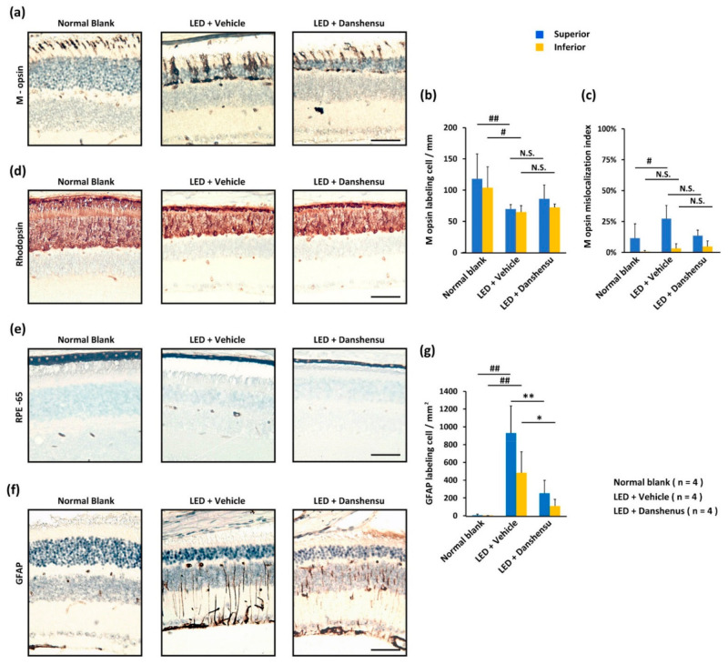
Figure 4.
Effect of danshensu on cellular protection after light-evoked retinal damage. (a) IHC staining showing the alterations of the photoreceptor-specific function of M opsin protein; (d) and Rhodopsin in the retinas; (b) Representation of M opsin-labeled cell density; (c) and the percentage of M opsin mislocalization; (e) Alterations in the pigment cell layer of outer blood-retinal-barrier (outer BRB) when labeled with RPE65 protein; (f) The pathologic Müller cells of inner blood-retinal-barrier (inner BRB) when labeled with GFAP protein; (g) Representation of the density of GFAP-labeled cells. Data are mean ± SD. Mann–Whitney U test, N.S., non-significant. ## p < 0.01, # p < 0.05, compared with the blank group. ** p < 0.01, and * p < 0.05, compared with the vehicle group. Scale bar: 35 μm.

