Abstract
Reverse transcriptase polymerase chain reaction (RT-PCR) negative results in the upper respiratory tract represent a major concern for the clinical management of coronavirus disease 2019 (COVID-19) patients. Herein, we report the case of a 43-years-old man with a strong clinical suspicion of COVID-19, who resulted in being negative to multiple severe acute respiratory syndrome coronavirus 2 (SARS-CoV-2) RT-PCR tests performed on different oropharyngeal and nasopharyngeal swabs, despite serology having confirmed the presence of SARS-CoV-2 IgM. The patient underwent a chest computed tomography (CT) that showed typical imaging findings of COVID-19 pneumonia. The presence of viral SARS-CoV-2 was confirmed only by performing a SARS-CoV-2 RT-PCR test on stool. Performing of SARS-CoV-2 RT-PCR test on fecal samples can be a rapid and useful approach to confirm COVID-19 diagnosis in cases where there is an apparent discrepancy between COVID-19 clinical symptoms coupled with chest CT and SARS-CoV-2 RT-PCR tests’ results on samples from the upper respiratory tract.
Keywords: COVID-19, SARS-CoV-2, RT-PCR, feces, oropharyngeal swab, nasopharyngeal swab, fecal swab, viral pneumonia, chest, computed tomography
1. Introduction
Coronavirus disease 2019 (COVID-19), caused by the severe acute respiratory syndrome coronavirus 2 (SARS-CoV-2), is a pandemic disease that can manifest with fever, pneumonia and, in severe cases, with acute respiratory distress symptoms (ARDS) [1,2]. Gastrointestinal symptoms with vomiting and diarrhea are often reported as other manifestations of the disease [3,4,5,6]. It is widely accepted that SARS-CoV-2 is mostly transmitted by respiratory droplets and fomite, and there is also evidence of fecal-oral transmission [1,3,4,5,6,7]. Asymptomatic and pauci-symptomatic individuals represent a major concern for the virus spread. Real-time reverse transcriptase polymerase chain reaction (RT-PCR) on oropharyngeal and nasopharyngeal (OP/NP) swab samples is considered the gold standard routine method for detection of SARS-CoV-2. However, SARS-CoV-2 RT-PCR tests giving negative results represent a diagnostic challenge for clinicians in the management of patients, especially when these negative results do not confirm clinical manifestations. In this context, computed tomography (CT) represents the most effective tool to diagnose pneumonia, and it can aid in supporting the diagnosis of COVID-19 in symptomatic cases, in the presence of multiple negative RT-PCR results, when conducted together with the SARS-CoV-2 serological tests [8,9,10]. However, recent studies reported that the virus can persist for a long time in feces, and it has been proposed to perform SARS-CoV-2 RT-PCR testing on fecal specimen as part of routine analyses for the detection of SARS-CoV-2, especially before the release of COVID-19 hospitalized patients [7,11,12,13,14,15,16,17,18,19].
2. Case Presentation
A 43-years-old patient with a contact history with COVID-19 patients came to the emergency room of our hospital because of the worsening of dyspnea, chills, and fever (38.5 °C). Upon clinical examination, he showed a normal pressure value PA 120/75 mmHg, cardiac palpitations, and diffuse reduced vesicular breathing. Arterial blood gas revealed respiratory failure with O2 saturation (SaO2) not higher than 90%. Blood tests revealed: mild leukopenia (3000/mm3), thrombocytopenia (96,000/mm3,), mild increase of the D- Dimer (0.7 mg/L), mild high levels of transaminases; all other blood tests were within their normal ranges and are listed in Table 1.
Table 1.
Main laboratory analyses result with the reporting systemic unit (SU) of measurements at the hospital admission and the normal value range.
| Laboratory Parameters | SU | Patient’s Value at Hospital Admission | Normal Value Range |
|---|---|---|---|
| Hemoglobin | mg/dL | 16.0 | 13.0–17.5 |
| Mean cell volume | fL | 89.1 | 80.0–98.0 |
| Platelets count | X1000/μL | 96.0 | 140.00–450.00 |
| White blood count | X1000/μL | 3.00 | 4.00–11.0 |
| Neutrophils | % | 57.6 | 40.0–75.0 |
| Lymphocytes | % | 33.7 | 20.0–50.0 |
| Monocytes | % | 6.1 | 0.0–11.0 |
| Eosinophils | % | 0.4 | 0.0–0.7 |
| Basophiles | 0.6 | 0.0–0.2 | |
| Aspartate transaminases | U/L | 40 | <37 |
| Alanine transaminases | U/L | 44 | <41 |
| Glycemia | mg/dL | 90 | 60–110 |
| Creatinine | mg/dL | 1.01 | 0.7–1.3 |
| Lactate dehydrogenase | U/L | 196 | 135–225 |
| C Reactive Protein | mg/dL | 0.47 | <0.5 |
| D-Dimer | mg/dL | 0.7 | <0.3 |
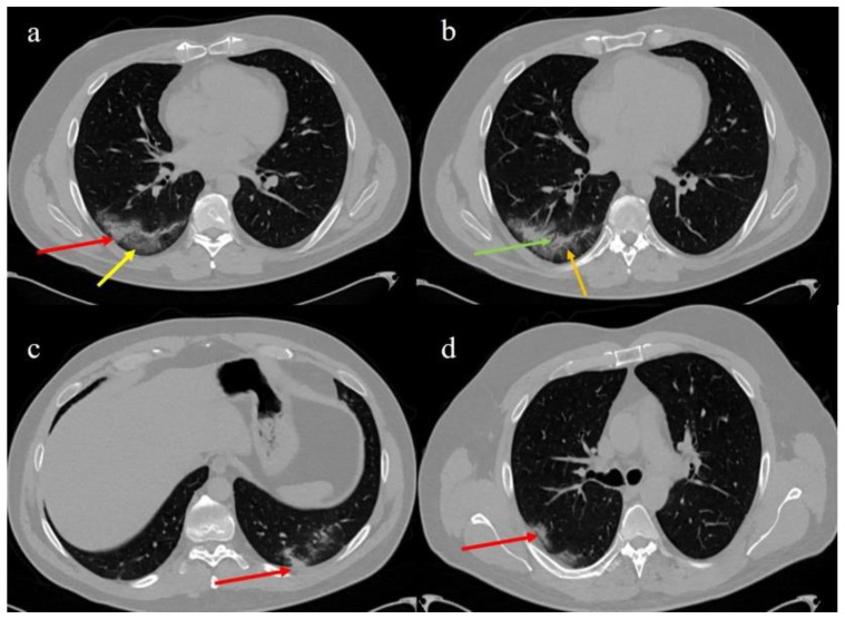
The patient reported that fever had started 12 days before and in the first days was associated to dyspnea, diarrhea, and myalgia. On the second day of fever, a RT-PCR testing on OP/NP wab sample (Allplex™ SARS-CoV-2/FluA/FluB/RSV Assay, Seegene, Italian distributor Arrow Diagnostics S.r.l, Genova (ITA)) was conducted by the local health authority and the result was negative. Due to the initial presence of diarrhea and the emerging studies that revealed the persistence of SARS-CoV-2 in the stool, the family doctor prescribed a SARS-CoV-2 RT-PCR test on the stool sample, which was carried out in an authorized testing laboratory. The RT- PCR on stool (adapted Allplex™ SARS-CoV-2/FluA/FluB/RSV Assay) resulted to be positive. The family doctor administered metronidazole (200 mg, 3 times/die) for one day. At the hospital, two days after an additional negative SARS-CoV-2 RT-PCR testing on an OP/NP swab (Easy® SARS-CoV-2), it was agreed to perform a chest CT scan, due also to the worsening of the dyspnea, coupled with a SARS-CoV-2 serological test. The chest CT scan showed typical COVID-19 findings with areas of ground glass in a crazy paving pattern with consolidation in the right inferior lobe (Figure 1a,b) in a peripheral posterior distribution and with small areas of consolidation in the left inferior lobe (Figure 1c) and a small consolidation in the right superior lobe (Figure 1d).
Figure 1.
Chest computed tomography (CT) images of the patient with the typical distribution of COVID-19 pneumonia: (a) consolidation (red arrow) and crazy paving area (yellow arrow) with a posterior and peripheral distribution in the right inferior lobe; (b) observed air bronchogram (green arrow) in the consolidation together with small vascular vessel enlargement (orange arrow); (c) small consolidations in the left inferior lobe (red arrow); (d) small consolidations in the posterior segment of the right superior lobe (red arrow).
The serological test, done using a chemiluminescent assay (Abbott Alinity i SARS-CoV-2 IgG) detected the presence of SARS-CoV-2 IgM (34.07 AU/mL) and the absence of SARS-CoV-2 IgG (cut-off 1.4 Index S/C as suggested by manufacturer).
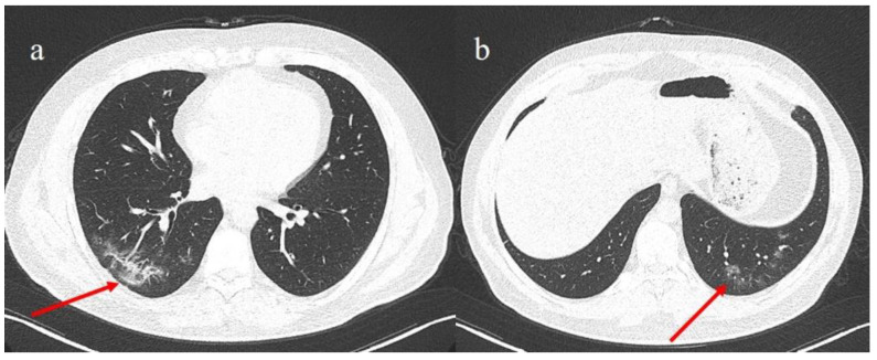
As the patient’s symptoms worsened, it was decided to perform an additional SARS-CoV-2 RT-PCR testing on an OP/NP swab sample (Easy® SARS-CoV-2), but the result was again negative. Due to the previous positive RT-PCR results for SARS-CoV-2 in the stool sample, the observation of pneumonia by chest CT scan, and the concomitant serological SARS-CoV-2 IgM positivity, at the hospital it was decided to initiate a therapeutical treatment with azithromycin (Azithromycin 500 mg/day, intravenous administration), corticosteroid (Methylprednisolone 400 mg/dL, intravenous administration), and low molecular weight heparin (4000 UI), in addition to oxygen therapy. After three days, the clinical conditions of the patient improved. At the end of this period, a SARS-CoV-2 RT-PCR test on OP/NP and faecal swab samples (Allplex™ SARS-CoV-2/FluA/FluB/RSV Assay) were conducted, which resulted in being negative and positive, respectively. A chest CT was repeated a week later which showed a reduction of the consolidations previously reported (Figure 2). Therefore, a week later another fecal swab sample (Allplex™ SARS-CoV-2/FluA/FluB/RSV Assay) was also repeated that finally resulted negative.
Figure 2.
On the left panel (a), reduction of the consolidation the right inferior lobe (red arrow) after therapy; on the right panel (b), the reduction of areas previously observed as small zones of consolidations with some ground glass (GGO) area (red arrow) in the absorptive phase after therapy.
3. Discussion
Several researchers pointed out the importance of chest CT in presence of negative SARS-CoV-2 RT-PCR tests results, especially in the pandemic context, where the risk of underestimating the disease can increase the risk of virus transmission among individuals [9,20,21,22]. The Fleishner Society guidelines [8] suggests that chest imaging can be helpful in the patients triage in the presence of high pre-test probability. High sensitivity (67–100%) and relatively low specificity (25–80%) are reported for the CT scans [23], and CT specificity is higher only in the presence of high prevalence of the disease [23]. In the context of COVID-19, the Fleishner Society suggests the use of chest imaging in the presence of moderate and severe COVID-19 symptoms, in the follow-up of patients at high risk of pneumonia progression, in worsening patients, and in symptomatic patients when SARS-CoV-2 RT-PCR test results are negative, or not available, and in the presence of a high probability pre-test [8]. COVID-19-associated pneumonia has specific radiological image patterns consisting of ground glass areas (GGO), consolidation, and crazy paving motifs with a multifocal and posterior distribution or central and peripheral distribution. Vascular enlargement and bronchial distortion are also other reported radiological characteristics [24]. These radiological patterns vary according to the phase of the disease [21,25,26]. However, cases with normal CT scans in patients who resulted positive for SARS-CoV-2 RT-PCR in OP/NP swabs [27] have also been reported. For this reason, the current guidelines [8,28,29] suggest that chest CT cannot be used as a screening tool to diagnose COVID-19. Although RT-PCR on OP/NP swabs is considered the standard method to diagnose SARS-CoV-2, negative false results ranging from 1% to 30% [30,31]. The bronchoalveolar lavage fluid (BALF) specimen test might be the most accurate method, but the risk of exposure for healthcare personnel is relatively high [32]. It has been reported that in more than 30% of SARS-CoV-2 RT-PCR tests, confirmed COVID-19 patients have detectable SARS-CoV-2 RNA in stool, and recent studies revealed that viral RNA can be found in fecal specimens [19,33,34]. In addition, there is indication that SARS-CoV-2 RNA can persist more in stool than in the upper respiratory tract, and that patients with both severe and mild disease showed viral SARS-CoV-2 RNA in the fecal sample for more than four weeks after symptom onset [12,13,14,15,16,17,18,19]. Tang et al. [17] have also described the case of a child diagnosed as a COVID-19 patient thanks to a positive SARS-CoV-2 RT-PCR test on his fecal sample, in contrast to a negative SARS-CoV-2 RT-PCR tests result on the child’s OP/NP samples. Performing a SARS-CoV-2 RT-PCR test on stool of symptomatic patients with repetitive negative SARS-CoV-2 RT-PCR tests results can be used as an additional rapid test to support diagnosis by chest CT coupled with a serological test, or to support diagnosis by serological tests only, when chest CT is not possible. Our case report is in line with the collection of findings made by Neu et al. [35], who reviewed evidence of interactions between viruses and gut bacteria, and how these interactions can have a fundamental role in the pathogenic phase against the eukaryotic cells. It is also coherent with what was described by G. Petruk et al. [36] about the identification of interaction between SARS-CoV-2 spike protein and E. Coli lipopolysaccharides and with the identification of the persistence of SARS-CoV-2 nucleic acids and immunoreactivity in the intestinal biopsies (small bowels) obtained from asymptomatic individuals four months after the onset of COVID-19 [37].
4. Conclusions
The here reported case provides additional evidence that the use of SARS-CoV-2 RT-PCR tests on stool will help to confirm diagnosis of COVID-19 in highly suspicious cases, especially when SARS-CoV-2 RT-PCR tests on OP/NP swabs end with negative results. These tests can be used to support, or in combination with, CTs and serological tests to speed-up COVID-19 diagnosis.
Acknowledgments
Authors would like to thank Giuliano Marino, Director of Marsan Consulting Srl, clinical governance, Italy, marino@marsanconsulting.it for the funding acquisition
Author Contributions
Conceptualization B.B., C.B., and M.P. (Mauro Petrillo); methodology, A.M.C.; software, B.B., M.P. (Mauro Petrillo), G.B.; validation, C.B., M.P. (Mauro Petrillo), B.B., L.M., M.P. (Marina Piscopo); formal analysis, B.B., M.P. (Mauro Petrillo) original draft preparation, B.B.; writing—review and editing, M.P. (Mauro Petrillo), C.B.; visualization, A.M.C., M.P. (Mauro Petrillo), C.B.; project administration, G.B.; funding acquisition. All authors have read and agreed to the published version of the manuscript.
Funding
This research was funded by Marsan Consulting srl, Pineta Grande Hospital.
Institutional Review Board Statement
The authors comply with international and national ethical standards.
Informed Consent Statement
Written informed consent has been obtained from the patient to publish this paper.
Data Availability Statement
Data available on request due to restrictions eg privacy or ethical.
Conflicts of Interest
The authors declare no conflict of interest.
Footnotes
Publisher’s Note: MDPI stays neutral with regard to jurisdictional claims in published maps and institutional affiliations.
References
- 1.Wu D., Wu T., Liu Q., Yang Z. The SARS-CoV-2 outbreak: What we know. Int. J. Infect. Dis. 2020;94:44–48. doi: 10.1016/j.ijid.2020.03.004. [DOI] [PMC free article] [PubMed] [Google Scholar]
- 2.Stawicki S.P., Jeanmonod R., Miller A.C., Paladino L., Gaieski D.F., Yaffee A.Q., De Wulf A., Grover J., Papadimos T.J., Yafer Y., et al. The 2019–2020 novel coronavirus (severe acute respiratory syndrome coronavirus 2) pandemic: A joint american college of academic international medicine-world academic council of emergency medicine multidisciplinary COVID-19 working group consensus paper. J. Glob. Infect. Dis. 2020;12:47–93. doi: 10.4103/jgid.jgid_86_20. [DOI] [PMC free article] [PubMed] [Google Scholar]
- 3.Yang L., Tu L. Implications of gastrointestinal manifestations of COVID-19. Lancet Gastroenterol. Hepatol. 2020;5:629–630. doi: 10.1016/S2468-1253(20)30132-1. [DOI] [PMC free article] [PubMed] [Google Scholar]
- 4.Kopel J., Perisetti A., Gajendran M., Boregowda U., Goyal H. Clinical insights into the gastrointestinal manifestations of COVID-19. Dig. Dis. Sci. 2020;65:1932–1939. doi: 10.1007/s10620-020-06362-8. [DOI] [PMC free article] [PubMed] [Google Scholar]
- 5.Gu J., Han B., Wang J. COVID-19: Gastrointestinal manifestations and potential fecal-oral transmission. Gastroenterology. 2020;158:1518–1519. doi: 10.1053/j.gastro.2020.02.054. [DOI] [PMC free article] [PubMed] [Google Scholar]
- 6.Dhar D., Mohanty A. Gut microbiota and Covid-19-possible link and implications. Virus Res. 2020;285:198018. doi: 10.1016/j.virusres.2020.198018. [DOI] [PMC free article] [PubMed] [Google Scholar]
- 7.Petrillo M., Brogna C., Cristoni S., Querci M., Piazza O., Van den Eede G. Increase of Sars-Cov2 RNA load in fecal samples prompts for rethinking of Sars-Cov-2 biology and epidemiology. Lancet. 2020;5:434–435. doi: 10.5281/zenodo.4088208.svg. [DOI] [PMC free article] [PubMed] [Google Scholar]
- 8.Rubin G.D., Ryerson C.J., Haramati L.B., Sverzellati N., Kanne J.P., Raoof S., Schluger S.W., Volpi A., Yim J.J., Martin I.B.K., et al. The role of chest imaging in patient management during the COVID-19 pandemic: A multinational consensus statement from the Fleischner Society. Chest. 2020;158:106–116. doi: 10.1016/j.chest.2020.04.003. [DOI] [PMC free article] [PubMed] [Google Scholar]
- 9.Brogna B., Bignardi E., Brogna C., Alberigo M., Grappone M., Musto L., Megliola A., Salvatore P., Fontanella G. Typical CT findings of COVID-19 pneumonia in patients presenting with repetitive negative RT-PCR. Radiography. 2020 doi: 10.1016/j.radi.2020.09.012. [DOI] [PMC free article] [PubMed] [Google Scholar]
- 10.Zhang W., Du R.H., Li B., Zheng X.S., Yang X.L., Zhou P., Ben H., Wang Y.Y., Xiao G.F., Yan B., et al. Molecular and serological investigation of 2019-nCoV infected patients: Implication of multiple shedding routes. Emerg. Microbes Infect. 2020;9:386–389. doi: 10.1080/22221751.2020.1729071. [DOI] [PMC free article] [PubMed] [Google Scholar]
- 11.Chen Y., Chen L., Deng Q., Zhang G., Wu K., Yang J., Liu B., Wang W., Wei C. The presence of SARS-CoV-2 RNA in the feces of COVID-19 patients. J. Med. Virol. 2020;92:833–840. doi: 10.1002/jmv.25825. [DOI] [PubMed] [Google Scholar]
- 12.Wu Y., Guo C., Tang L., Hong Z., Zhou J., Kuang L., Fang X., Mishra N., Shan H. Prolonged presence of SARS-CoV-2 viral RNA in faecal samples. Lancet Gastroenterol. Hepatol. 2020;5:434–435. doi: 10.1016/S2468-1253(20)30083-2. [DOI] [PMC free article] [PubMed] [Google Scholar]
- 13.Kipkorir V., Cheruiyot I., Ngure B., Misiani M., Munguti J. Prolonged SARS-Cov-2 RNA detection in anal/rectal swabs and stool specimens in COVID-19 patients after negative conversion in nasopharyngeal RT-PCR test. J. Med. Virol. 2020;92:2328–2331. doi: 10.1002/jmv.26007. [DOI] [PMC free article] [PubMed] [Google Scholar]
- 14.Xie J., Long X., Ren C., He R., Yan X., Liu E., Xu H., Li Q., Li W. Follow-up study of long-time positive RT-PCR in stool specimens from asymptomatic children infected with SARS-CoV-2. Pediatr. Infect. Dis. J. 2020;39:e315–e317. doi: 10.1097/INF.0000000000002837. [DOI] [PubMed] [Google Scholar]
- 15.Zhang J., Wang S., Xue Y. Fecal specimen diagnosis 2019 novel coronavirus-infected pneumonia. J. Med. Virol. 2020;92:680–682. doi: 10.1002/jmv.25742. [DOI] [PMC free article] [PubMed] [Google Scholar]
- 16.Jiang X., Luo M., Zou Z., Wang X., Chen C., Qiu J. Asymptomatic SARS-CoV-2 infected case with viral detection positive in stool but negative in nasopharyngeal samples lasts for 42 days. J. Med. Virol. 2020;92:1807–1809. doi: 10.1002/jmv.25941. [DOI] [PMC free article] [PubMed] [Google Scholar]
- 17.Tang A., Tong Z.D., Wang H.L., Dai Y.X., Li K.F., Liu J.N., Wu W.J., Yuan C., Yu M.Y., Li P., et al. Detection of novel Coronavirus by RT-PCR in stool specimen from asymptomatic child, China. Emerg. Infect. Dis. 2020;26:1337–1339. doi: 10.3201/eid2606.200301. [DOI] [PMC free article] [PubMed] [Google Scholar]
- 18.Mim M.A., Rakhi N.N., Saha O., Rahaman M.M. Recommendation of fecal specimen for routine molecular detection of SARS-CoV-2 and for COVID-19 discharge criteria. Pathog. Glob. Health. 2020;114:168–169. doi: 10.1080/20477724.2020.1765651. [DOI] [PMC free article] [PubMed] [Google Scholar]
- 19.Szymczak W.A., Goldstein D.Y., Orner E.P., Fecher R.A., Yokoda R.T., Skalina K.A., Fox A.S., Gendlina S. Utility of stool PCR for the diagnosis of COVID-19: Comparison of two commercial platforms. J. Clin. Microbiol. 2020;58:e01369–e01420. doi: 10.1128/JCM.01369-20. [DOI] [PMC free article] [PubMed] [Google Scholar]
- 20.Chen L.D., Li H., Ye Y.M., Wu Z., Huang Y.P., Zhang W.L., Li L. A COVID-19 patient with multiple negative results for PCR assays outside Wuhan, China: A case report. BMC Infect. Dis. 2020;20:517. doi: 10.1186/s12879-020-05245-7. [DOI] [PMC free article] [PubMed] [Google Scholar]
- 21.Long C., Xu H., Shen Q., Zhang X., Fan B., Li H., Wang C., Zeng B., Li Z. Diagnosis of the coronavirus disease (COVID-19): RRT-PCR or CT? Eur. J. Radiol. 2020;126:108961. doi: 10.1016/j.ejrad.2020.108961. [DOI] [PMC free article] [PubMed] [Google Scholar]
- 22.Xie X., Zhong Z., Zhao W., Zheng C., Wang F., Liu J. Chest CT for typical 2019-nCoV pneumonia: Relationship to negative RTPCR testing. Radiology. 2020;296:E41–E45. doi: 10.1148/radiol.2020200343. [DOI] [PMC free article] [PubMed] [Google Scholar]
- 23.Kim H., Hyunsook H., Soon H.Y. Diagnostic performance of CT and reverse transcriptase-polymerase chain reaction for coronavirus disease 2019: A meta-analysis. Radiology. 2020;296:E145–E155. doi: 10.1148/radiol.2020201343. [DOI] [PMC free article] [PubMed] [Google Scholar]
- 24.Ojha V., Mani A., Pandey N.N., Sharma S., Kumar S. CT in coronavirus disease 2019 (COVID-19): A systematic review of chest CT findings in 4410 adult patients. Eur. Radiol. 2020;30:6129–6138. doi: 10.1007/s00330-020-06975-7. [DOI] [PMC free article] [PubMed] [Google Scholar]
- 25.Pan F., Ye T., Sun P., Gui S., Liang B., Zheng C., Li L., Zheng D., Wang J., Hesketh R.L., et al. Time course of lung changes on chest CT during recovery from 2019 novel coronavirus (COVID-19) pneumonia. Radiology. 2020;295:715–721. doi: 10.1148/radiol.2020200370. [DOI] [PMC free article] [PubMed] [Google Scholar]
- 26.Bernheim A., Mei X., Huang M., Yang Y., Fayad Z.A., Zhang N., Diao K., Lin B., Zhu X., Li K., et al. Chest CT findings in coronavirus disease-19 (COVID-19): Relationship to duration of infection. Radiology. 2020;295:200463. doi: 10.1148/radiol.2020200463. [DOI] [PMC free article] [PubMed] [Google Scholar]
- 27.Chung M., Bernheim A., Mei X., Yang Y., Fayad Z.A., Zhang N., Huang M., Zeng X., Cui J., Hu H., et al. CT imaging features of 2019 novel coronavirus (2019-nCoV) Radiology. 2020;295:202–207. doi: 10.1148/radiol.2020200230. [DOI] [PMC free article] [PubMed] [Google Scholar]
- 28.Centers for Disease Control and Prevention Interim Clinical Guidance for Management of Patients with Confirmed Coronavirus Disease (COVID-19) [(accessed on 16 April 2020)]; Available online: https://www.cdc.gov/coronavirus/2019-ncov/hcp/clinical-guidance-managementpatients.html.
- 29.ACR Recommendations for the Use of Chest Radiography and Computed Tomography (CT) for Suspected COVID-19 Infection. [(accessed on 16 April 2020)]; Available online: https://www.acr.org/Advocacy-and-Economics/ACR-PositionStatements/Recommendations-for-Chest-Radiography-and-CT-for-Suspected-COVID19-Infection.
- 30.Long D.R., Gombar S., Hogan C.A., Greninger A.L., O’Reilly-Shah V., BrysonCahn C., Stevens B., Rustagi A., Jerrome K., Kong K.S., et al. Occurrence and timing of subsequent SARS-CoV-2 RT-PCR positivity among initially negative patients. Clin. Infect. Dis. 2020 doi: 10.1093/cid/ciaa722. [DOI] [PMC free article] [PubMed] [Google Scholar]
- 31.Arevalo-Rodriguez I., Buitrago-Garcia D., Simancas-Racines D., Zambrano-Achig P., Del Campo R., Zamora J., Molina J.A.P., Low N., Bossuyt P.M. False-negative results of initial RT-PCR assays for COVID-19: A systematic review. PLoS ONE. 2020;15:e0242958. doi: 10.1371/journal.pone.0242958. [DOI] [PMC free article] [PubMed] [Google Scholar]
- 32.Chen D., Xu W., Lei Z., Huang Z., Liu J., Gao Z., Peng L. Recurrence of positive SARS-CoV-2 RNA in COVID-19: A case report. Int. J. Infect. Dis. 2020;93:297–931. doi: 10.1016/j.ijid.2020.03.003. [DOI] [PMC free article] [PubMed] [Google Scholar]
- 33.Rokkas T. Gastrointestinal involvement in COVID-19: A systematic review and meta-analysis. Ann. Gaestrenterol. 2020;33:355. doi: 10.20524/aog.2020.0506. [DOI] [PMC free article] [PubMed] [Google Scholar]
- 34.Cheung K.S., Hung I.F., Chan P.P., Lung K.C., Tso E., Liu L.W.K., Ng Y.Y., Chu M.Y., Chung T., Tam R., et al. Gastrointestinal manifestations of SARS-CoV-2 infection and virus load in fecal samples from a Hong Kong cohort: Systematic review and meta-analysis. Gastroenterology. 2020;159:81–95. doi: 10.1053/j.gastro.2020.03.065. [DOI] [PMC free article] [PubMed] [Google Scholar]
- 35.Neu U., Mainou B.A. Virus interactions with bacteria: Partners in the infectious dance. PLoS Pathog. 2020;16:e1008234. doi: 10.1371/journal.ppat.1008234. [DOI] [PMC free article] [PubMed] [Google Scholar]
- 36.Petruk G., Puthia M., Petrlova J., Strömdahl A.C., Kjellström S., Schmidtchen A. SARS-CoV-2 spike protein binds to bacterial lipopolysaccharide and boosts proinflammatory activity. J. Mol. Cell. Biol. 2020;10:mjaa067. doi: 10.1093/jmcb/mjaa067. [DOI] [PMC free article] [PubMed] [Google Scholar]
- 37.Gaebler C., Wang Z., Lorenzi J.C.C., Muecksch F., Finkin S., Tokuyama M., Cho A., Jankovik M., Oliveira T., Cipolla M., et al. Evolution of antibody immunity to SARS-CoV-2. Nature. 2021 doi: 10.1038/s41586-021-03207-w. [DOI] [PMC free article] [PubMed] [Google Scholar]
Associated Data
This section collects any data citations, data availability statements, or supplementary materials included in this article.
Data Availability Statement
Data available on request due to restrictions eg privacy or ethical.


