Abstract
Aedes mosquitoes are vectors for many pathogenic viruses. Cell culture systems facilitate the investigation of virus growth in the mosquito vector. We found Zika virus (ZIKV) growth to be consistent in A. albopictus cells but hypervariable in A. aegypti cell lines. As a potential explanation of this variability, we tested the hypothesis that our cells harbored opportunistic viruses. We screened Aedes cell lines for the presence of insect specific viruses (ISVs), Cell-fusing agent virus (CFAV) and Phasi charoen-like virus (PCLV). PCLV was present in the ZIKV-growth-variable A. aegypti cell lines but absent in A. albopictus lines, suggesting that these ISVs may interfere with ZIKV growth. In support of this hypothesis, PCLV infection of CFAV-positive A. albopictus cells inhibited the growth of ZIKV, dengue virus and La Crosse virus. These data suggest ISV infection of cell lines can impact arbovirus growth leading to significant changes in cell permissivity to arbovirus infection.
Keywords: Arbovirus, Aedes, coinfection, superinfection, mosquito cell lines, Zika virus, Phasi Charoen like virus, Cell fusing agent virus, insect specific virus
Introduction
Diseases caused by arboviruses are a global threat perpetuated by mosquito transmission. Arboviruses are defined by their arthropod vector and capacity to cause disease in animals. Over ninety percent of arboviruses which cause human disease are specifically vectored by mosquitoes (McGraw and O’Neill, 2013). Arboviruses that cause disease in humans include both negative-sense and positive-sense RNA viruses spanning diverse families such as Flaviviridae and Bunyaviridae (Cleton et al., 2012). Annual outbreaks of dengue virus (DENV) (Flaviviridae) have been consistent in the Americas for over 20 years (Cafferata et al., 2013; Donalisio et al., 2017). Meanwhile, Zika virus (ZIKV) has rapidly emerged as a threat to human health in the Americas (Fauci and Morens, 2016). Negative-sense arboviruses, such as La Crosse virus (LACV), which is a leading cause of pediatric encephalitis in the United States (Bewick et al., 2016), have been circulating in the Americas for a long time. The emergence and re-emergence of these arboviruses causes a global disease burden and it is estimated that greater than half of the world’s population at risk of infection (Fauci and Morens, 2016; Messina et al., 2014) demonstrating an urgent need for novel arbovirus control.
DENV, ZIKV, and LACV are all transmitted by Aedes sp. mosquitoes and there is a continuing effort to reduce infection through targeting the mosquito host. One approach is the identification and application of ways to reduce arbovirus replication within the Aedes mosquito vector. Initial efforts in this vein have encouraging results. Studies have identified genetic modification of mosquito immunity (Jupatanakul et al., 2017) or modification of mosquito symbionts (Jupatanakul et al., 2014) to efficiently reduce virus transmission. Further, modification of mosquitoes (transgenesis) or their associated microorganisms (paratransgenesis) have identified genetic modification as viable approaches to reduce the insect capacity to transmit disease (Coutinho-Abreu et al., 2010; Ren et al., 2008). Modification of the mosquito microbiome, including gut commensal bacteria or vertically transmitted endosymbionts (e.g. Wolbachia) can reduce arbovirus replication within mosquitoes (Bourtzis et al., 2014; Cirimotich et al.; Schultz et al., 2018). Furthermore, it has been recently suggested that modulation of the insect virome may have important implications for arbovirus control (Bolling et al., 2015a).
There are a limited number of cell lines commonly used to study the virus/vector interactions. Many studies utilize C6/36 cells, an Aedes albopictus cell line. This cell line is limited in application because C6/36 cells are defective in antiviral immunity (Brackney et al., 2010). Innate immune competent cells such as the A. aegypti cell lines (Aag2 and CCL-125s) represent additional cell lines but have shown variability in virus susceptibility (Singh, 1967; Singh and Paul, 1968; Wikan et al., 2009). The issue of variability of virus growth in A. aegypti cell permissibility has not been extensively studied but one hypothesis is that opportunistic viruses in some cell culture lines may interfere with consistent virus growth.
Insect-specific viruses (ISV) are viruses specific to insects which are unable to infect mammalian cells. ISV infection of cell culture has been recognized for over 40 years. Cell fusing agent virus (CFAV) was first identified in 1975 in A. aegypti cells, and was shown to cause a cell fusing phenotype in A. albopictus cells (Stollar and Thomas, 1975). CFAV is a positive-sense RNA virus of the family Flaviviridae and genus Flavivirus. Several ISVs have been shown to interfere with arbovirus growth, reviewed by (Bolling et al., 2015a). Sequencing has identified additional opportunistic viruses including Phasi Charoen like virus (PCLV) in cell lines and in mosquitoes (Bolling et al., 2015a; Bolling et al., 2015b; Maringer et al., 2017). PCLV is a negative-sense segmented RNA virus of the family Bunyaviridae and genus Phasivirus. RNA-sequencing has identified coinfection of CFAV and PCLV infections in an Aag2 cell line stocks (Schnettler et al., 2016). This raises the possibility that these two ISVs could interfere with the growth of arboviruses in the same culture system (Bolling et al., 2015b), as this cell line demonstrates variable growth of ZIKV.
We assessed the kinetics of ZIKV growth in two Aedes cells lines persistently infected with CFAV alone or CFAV and PCLV. We screened A. aegypti and A. albopictus cells to assess the prevalence of CFAV and PCLV infection in other mosquito cell lines. We then determined the ability of various arboviruses to grow in the presence or absence of CFAV-PCLV coinfection and if this ISV coinfection could interfere with arbovirus studies. We hypothesize that the presence of coinfection of CFAV and PCLV in Aag2 cells is responsible for high variability in arbovirus growth. Supporting our hypothesis, we demonstrate that the introduction of PCLV into A. albopictus cells to generate a CFAV-PCLV coinfection interferes with the growth of two flaviviruses, ZIKV and DENV, and the bunyavirus, LACV.
Materials and methods
Insect cell culture
All insect cells were grown at 28°C with 5% CO2. A. albopictus derived C710 cells from Ann Fallon were cultured in E-5 media (Schultz et al., 2017) and subcultured 1:10 weekly. A. albopictus derived C6/36 cells were cultured in minimal essential media with 10% fetal bovine serum, 1X nonessential amino acids, and 2mM glutamine. C6/36 cells from the lab of Sharon Isern and Scott Michael were subcultured weekly at a 1:10 dilution. A. albopictus derived Aa23 cells were grown in Schneider’s media with 10% fetal bovine serum and 50ug/mL penicillin and 50ug/mL streptomycin.
A. aegypti Aag2 cells were cultured in Schneider’s media with 10% fetal bovine serum and 50ug/mL penicillin and 50ug/mL streptomycin at 28°C with 5% CO2. Cells were subcultured 1:10 weekly. A. aegypti CCL-125 cells were grown in minimal essential media with 20% fetal bovine serum as recommended by ATCC. CCL-125 cells were subcultured at a 1:5 dilution weekly. A. aegypti Aag2 cells were provided by Zhiyong Xi and Tonya Colpitts. CCL-125 cell lines were obtained from Marshall Bloom and the ATCC.
Virus stocks
ZIKV strains MR766 and PRVABC59 were obtained from BEI Resources (Biological and Emerging Infections Resources Program, NIAID). Virus was grown in C6/36 cells infected at an MOI 0.01 and harvested after 7 days. Virus supernatant was spun at 4,000 RCF, filtered and aliquoted. To concentrate virus for high MOI experiment, 8% polyethylene glycol 8000 was incubated with virus overnight at 4 °C. Virus was pelleted at 30,000 RCF and resuspended in NTE (NaCl-Tris-EDTA) buffer. DENV serotype 2 strain NGC was obtained from Scott Michael and Sharon Isern and grown in LLC-MK2 cells as previously described (Nicholson et al., 2011). LACV H78 was grown at an MOI of 0.01 in Vero E6 cells for 24 hours. Supernatant was spun at 4,000 RCF, filtered, aliquoted, and stored at −80°C.
Mammalian cell culture
Macaca mulatta kidney LLC-MK2 cells were cultured in Dulbecco’s Modified Eagle Medium with 10% fetal bovine serum and 2mM glutamine. LLC-MK2 cells were subcultured weekly at a 1:10 dilution.
ZIKV Growth kinetics in Aag2 and Aa23 cells
To determine growth kinetics, viral RNA from the supernatant of Aag2 and Aa23 cells was assessed over one week after a low MOI infection. Aag2 and Aa23 cells were plated in a 24 well plate at 5×10⁴ cells/well. One day post plating, cells were infected with ZIKV MR766 or PRVABC59 at an MOI of 0.1 for 1 hour at 28°C in serum-free medium (Schneider’s Drosophila media). Virus inoculum was removed and complete media was added to each well. Cells were incubated at 28°C with 5% CO2 and supernatant time points were taken from a fresh well every day for 6 days. Supernatant RNA was extracted by Qiagen Viral RNA extraction kit per manufacturer recommendations. Quantitative RT-PCR was carried out with Roche One-Step SYBR green kit. ZIKV primers (ZIKV_For AARTACACATACCARAACAAAGTGGT and ZIKV_Rev TCCRCTCCCYCTYTGGTCTTG) were previously published (Faye et al., 2008).
To determine the production of infectious virions from Aag2 and Aa23 cells, viral supernatant was assayed for infectious virus at day 6 post a low MOI infection. Aag2 and Aa23 cells were plated in a 12 well plate at 1×10⁵ cells/well. One day post plating, cells were infected with ZIKV MR766 or PRVABC59 at an MOI of 0.1 for 1 hour at 28°C in serum-free medium (Schneider’s Drosophila media). Virus inoculum was removed and complete media was added to each well. Cells were incubated at 28°C with 5% CO2 for 6 days. The supernatant was harvested and analyzed by focus forming assay on LLC-MK2 cells (described below).
Focus forming Assay (ZIKV and DENV-2)
Focus forming assay protocol was adapted from previously described (Paul et al., 2016). Briefly, LLC-MK2 cells were incubated with dilutions of virus for 1 hour. Virus was removed and cells were rinsed one time before adding 1% agar in Temin’s modified minimal essential media with 10% fetal bovine serum. Plates were incubated for 72 hours, fixed with 10% formalin for 1 hour at room temperature, and permeabilized with 70% ethanol for 30 minutes. Cell were stained with cross-reactive ZIKV and DENV primary human anti-E protein antibody D11C in PBS with 0.01% tween 20 and 5% NFDM followed by goat anti-human HRP. Foci were developed with 0.5mg/mL diaminobenzidine in 25mM Tris-HCl, pH 7.2. Foci were counted and graphed in GraphPad Prism.
Generation of PCLV persistently infected Aa23 cells
PCLV containing supernatant from Aag2 cells was confirmed by sequencing of the full genome from the supernatant of these cells. Sequencing primers are shown in Table 2. This supernatant from confluent Aag2 cells was collected and centrifuged at maximum speed, filtered through a 0.2uM filter, and aliquoted. A. albopictus Aa23 cells were infected with the PCLV stock at a 1:10 dilution. As a control for nutrient deprivation, PCLV-free Aa23 cells were incubated with 1:10 dilution of Aa23 spent media. Media was replaced at 24 hours post infection. Six days post infection, cells were observed for cytopathic effect and plated in 12 well plates at 1×10⁵cells per well. One day post plating, PCLV and CFAV infection was determined by RT PCR (below) in cells and supernatant.
Table 2. Primers used to sequence the full genome of PCLV from the supernatant of Aag2 cells.
Primers were designed against PCLV Bristol Aag2 sequences (Accession numbers: KU936057.1, KU936056.1, and KU936055.1).
| forward | reverse | |
|---|---|---|
| PCLV_A | GTTGGACATGAAGCAGTGGC | TTTCGCCAGTTCAGACTGCT |
| PCLV_B | AATTAAGATGTCCACTTTGCTCAAT | TTCAGTTGCATCTTCTGGCA |
| PCLV_C | AGCAGTCTGAACTGGCGAAA | AGTGTGGATGCGTTTCTGGT |
| PCLV_D | AACAAGCTGTTTGGAGGAGAGA | ACTGAGCGCAAAAACATGAAGT |
| PCLV_E | ACTTCATGTTTTTGCGCTCAGT | TCCCTGCATCATTCCTGTGG |
| PCLV_F | GAGCAGGGAGCCCTTACATT | CGCCATTCTTGGTCAACATTCA |
| PCLV_G | ACCCATAACCCTGACAGATGC | TCACTTTCTGATGCTGTGTGA |
| PCLV_H | ACACTGATATCATGACTCCCCG | AACCCAGCCCATGTTCTCAAT |
| PCLV_I | ATTGAGAACATGGGCTGGGTT | GTCTCCCCCAGCTAATTCACA |
| PCLV_J | GTCGATTTCGAAGAAGTAGGTGC | TGCTAAACAATACCCTTCTACGTT |
| PCLV_K | TGATATATGCAGCGGCGAGT | AAGCCGTAATGAGAGTGAGC |
| PCLV_L | AAGAAACCATAGACCCGGAGTT | TGTTTTCCCCTGGATGGAGC |
| PCLV_M | CAACGGTGTTGACTGTGACAA | TTCCCCTTACCATCGCTTCTG |
| PCLV_N | ACTGCAGGCTGATAGACGAAC | GTGCAACCATGAAACCCTGC |
| PCLV_O | GCCTGTCCCATCTGCGAATA | GCACAGCCTCTCAGTTTCCT |
| PCLV_P | GGAAACTGAGAGGCTGTGCT | AGCCCCACCATTGGAAAACA |
| PCLV_Q | CAGAATTACTGCGCTCAGAAACATA | TCACAAAGACCAGCCCCAAA |
Reverse transcription PCR
Cells (1.0 ×10⁵) were collected for RNA extraction. RNA was extracted by Qiagen RNeasy kit per manufacturer recommendations. RT-PCR was carried out with Superscript III One-step Reverse transcription PCR kit per manufacturer’s recommendations. PCLV primers were previously published: PCLV-N-FW: CAGTTAAAGCATTTAATCGTATGATAA, PCLVN-RV: CACTAAGTGTTACAGCCCTTGGT (Schnettler et al., 2016). Rps6 primers used target a conserved region of A. aegypti and A. albopictus Rps6 gene: mRpS6_F AGTTGAACGTAT CGTTTCCCGCTAC and mRpS6_R GAAGTGACGCAGCT TGTGGTCGTCC (Lee et al., 2012). Products were visualized by SYBR green nucleic acid dye after running on a 1% agarose gel in Tris-acetate EDTA (TAE).
PCLV Growth curve Quantitative Reverse transcription PCR
To determine PCLV growth kinetics in Aa23 cells, cells were infected with a 1:10, 1:1000, or UV inactivated 1:10 dilution of PCLV from Aag2 supernatant. The 1:10 dilution of PCLV stock was UV inactivated by UV-treatment at 100μJ/cm2 for 10 minutes rocking every 3.3 minutes to inactivate infectious virus. Virus was infected for 24 hours and then removed and replaced with fresh media. From here, time points were collected every 24 hours up until 144 hours. Supernatant RNA was extracted by Qiagen Viral RNA extraction kit per manufacturer recommendations. Quantitative RT-PCR was carried out with BioRad SYBR green kit. PCLV primers were previously published: [PCLV-N-qRT-FW: ATAGTGTGGGACGAGGAGGG, PCLV-N-qRT-RV: AGGTGCCAACAGGAAACACT] (Schnettler et al., 2016). All reactions were annealed at 60°C.
Arbovirus infection of Aa23 with dual ISV infection
Post infection with PCLV or nutrient deprivation (above), Aa23 cells were infected at a low (MOI 0.1) or high (MOI 10) of ZIKV PRVABC59, LACV H78, or DENV-2 NGC for 1 hour at 28°C with 5% CO2. After incubation, cells were rinsed 1 time with PBS and 1mL of complete media was added. Cells were incubated for 6 days at 28 °C with 5% CO2. Supernatant was collected for focus forming or plaque assay to determine infectious virus.
LACV Plaque assays
Vero E6 cells were incubated with dilutions of virus for 1 hour. Virus was removed and cells were rinsed one time before adding 1.2% avicel in Temin’s modified minimal essential media with 10% fetal bovine serum. Plates were incubated for 72 hours. Overlay was removed and cells were fixed with 4% formaldehyde for 1 hour at room temperature. Cells were then stained with 1% crystal violet for 10 minutes, rinsed, and plaques were counted.
Results
Kinetics of Zika virus growth in Aedes albopictus Aa23 and Aedes aegypti Aag2 cells
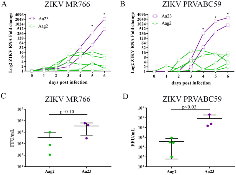
We have previously shown that A. albopictus Aa23 cells produce high titers of infectious ZIKV (Schultz et al., 2017). However, the kinetics of ZIKV growth in Aa23 cells has not yet been shown. Since A. albopictus and A. aegypti are established vectors for ZIKV, we were interested to determine the kinetics of viral infection in our Aa23 (A. albopictus) and our Aag2 (A. aegypti) cell lines. Cells were infected with the African ZIKV MR766 and Puerto Rican ZIKV PRVABC59 strains at a low MOI (MOI 0.1) and incubated for six days. Supernatant was collected for six days and ZIKV RNA was assessed by quantitative reverse transcription polymerase chain reaction (qRT-PCR). In Aa23 cells, ZIKV MR766 grew along logistic curve peaking in viral RNA titer at six days post infection with greater than a 500 fold increase in viral RNA (Figure 1A). Likewise, in Aa23 ZIKV PRVABC59 replicated to greater than a 500 fold increase along a similar growth trend (Figure 1B). In contrast ZIKV MR766 (Figure 1A) and ZIKV PRVABC59 (Figure 1B) viral RNA titer increased by only 16 fold in Aag2 cells suggesting that virus production was unsuccessful in Aag2 cells.
Figure 1. Growth of ZIKV MR766 and PRVABC59 in Aag2 and Aa23 cells.
Aag2 and Aa23 cells infected with (A) ZIKV MR766 and (B) PRVABC59 (MOI 0.1). Supernatant was assayed for viral RNA for up to 6 days post infection. Each line is an independent biological replicate. Aa23 cells are depicted in purple. Aag2 cells are depicted in green. Statistical difference of the mean of Aa23 extracellular ZIKV RNA and Aag2 extracellular ZIKV RNA was compared at each day by a Student’s T test. P values are as follows for (A) MR766: day 1 p<0.23, day 2 p<0.38, day 3 p<0.23, day 4 p<0.08, day 5 p<0.03, day 6 p<0.02 and (B) PRVABC59: day 1 p<0.37, day 2 p<0.30, day 3 p<0.37, day 4 p<0.04, day 5 p<0.02, day 6 p<0.01. * indicates p value is less than 0.05 (C-D) Infectious virus production in the supernatant was assayed after six days by focus forming assay. Each individual dot is an independent experiment. Statistical significance was determined by an unpaired, one-tailed Mann-Whitney test. C: p=0.10, D: p<0.03 Experiments in A/B were conducted independent of experiments shown in C/D and thus cannot be correlated to determine particle: PFU ratio.
To determine viral RNA was representative of ZIKV infectious virus at the peak viral RNA titer, viral growth in the supernatant was assessed by focus forming assay at six days post infection in independent experiments. There was high variability of viral growth spanning two logs variance for ZIKV MR766 and PRVABC59 in Aag2 cells (Figure 2C and 2D). In two independent experiments, ZIKV MR766 growth in Aag2 cells was below 1.0 × 10⁴ FFU/mL (the initial inoculum of virus) indicating little to no virus production (Figure 2C). Viral growth was consistently observed in Aa23 cells. ZIKV PRVABC 59 grew to higher titers than ZIKV MR766 in Aa23 cells suggesting increased fitness in this cell type.
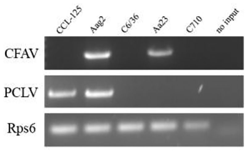
Figure 2. Aedes cell lines are infected with an insect specific flavivirus and phasivirus.
A. aegypti (lanes 1–2) and A. albopictus (lanes 3–5) cell lines were screened for PCLV and CFAV viral RNA by RT-PCR and visualized on a 1% agarose gel in TAE. PCLV and CFAV bands were confirmed by Sanger sequencing of gel extracted amplicons.
Phasi Charoen-like virus and Cell fusing agent virus broadly infect of Aedes cell lines
Following previous studies that have suggested ISV coinfection contributes to decreased arbovirus production in mosquito (Hall-Mendelin et al., 2016; Hall et al., 2016) and the recent identification of CFAV and PCLV as circulating insect viruses found in some cultured insect cells (Schnettler et al., 2016), we screened A. aegypti (Aag2 and CCL-125) and A. albopictus (C710, Aa23, C6/36) cell lines for CFAV and PCLV RNA. DNase treated RNA was isolated from cell lysates. The presence of mosquito Rps6 transcripts confirmed successful RNA extraction and reverse transcription polymerase chain reaction (RT PCR). CFAV RNA was found in Aa23 and Aag2 cells but not in CCL-125, C6/36, or C710 cells (Figure 2). Primers designed to amplify the small (S) segment were used to probe for the presence of PCLV RNA by RT PCR. A. aegypti cell lines Aag2 and CCL-125 each independently obtained from two sources (Table 1) were positive for PCLV genome (Figure 2). Aag2 cells were screened at varying passages depending on source. The two sources of CCL125 cells were screened at passage one after receipt. PCLV RNA was detected in CCL-125 and Aag2 cells over multiple cell passages. PCLV was not detected in A. albopictus cell lines (C710, Aa23, and C6/36) (Figure 2). These results suggest that multiple A. aegypti but not A. albopictus cell line cultures contain PCLV.
Table 1. Screening of CFAV and PCLV in different Aedes cell populations.
Cells were screened for virus by RT PCR followed by agarose gel for visualization of amplicons. Numerical distinction after each cell line indicates a different source of cells.
| Cell line | Species | PCLV detected | CFAV detected |
|---|---|---|---|
| CCL-125 1 | A. aegypti | Yes | No |
| CCL-125 2 | A. aegypti | Yes | No |
| Aag2 1 | A. aegypti | Yes | Yes |
| Aag2 2 | A. aegypti | Yes | Yes |
| Aa23 | A. albopictus | No | Yes |
| C6/36 | A. albopictus | No | No |
| C710 | A. albopictus | No | No |
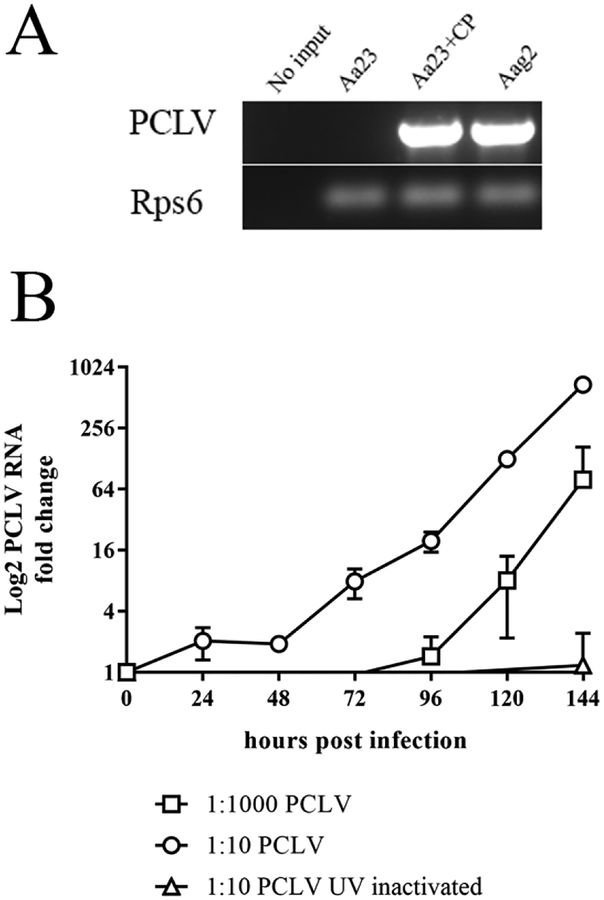
PCLV has previously been reported in A. aegypti mosquitoes in Thailand (Chandler et al., 2014), Australia, and Brazil (Hall et al., 2016) but has not been reported in A. albopictus mosquitoes, leaving open the question of whether A. albopictus cells are permissive to PCLV infection. To test this, filtered cell culture media from PCLV RNA positive Aag2 cells was diluted 1:10 and added to A. albopictus Aa23 cells. Six days post inoculation, PCLV infection was determined by RT PCR in cell lysates and in cell culture media. PCLV was detected in Aa23 cells treated with Aag2 supernatant and in Aag2 cells (Figure 3A). PCLV was not detected in uninfected Aa23 cells which had been incubated with Aa23 spent media as a control for nutrient deprivation (Figure 3A). RT PCR cannot discern RNA presence from a productive RNA infection. Thus, we determined that PCLV grows in Aa23 cells by infecting Aa23 cells with diluted 1:10, 1:1000, or UV inactivated PCLV from Aag2 supernatant (Figure 3B). The 1:10 dilution of the PCLV stock from Aag2 cell supernatant grew to greater than a 500 fold increase in viral RNA in the supernatant six days post infection in three independent biological replicates (Figure 3B). The 1:1000 dilution of PCLV grew to a 64 fold increase in viral RNA six days post infection. UV-inactivated PCLV (1:10 dilution) did not lead to an increase in viral RNA (Figure 3B). No cytopathic effect was observed in A. albopictus cells infected with PCLV. These data suggest that the PCLV in Aag2 cells is infectious and that Aa23 A. albopictus cells are permissive to PCLV.
Figure 3. Aedes albopictus Aa23 cells are permissive to PCLV growth.
(A) Six days post infection with PCLV (1:10 dilution of Aag2 supernatant), Aa23 cells were DNase treated, assayed for PCLV RNA by RT-PCR, and visualized on a 1% agarose gel in TAE. No RNA was provided in the no input control lane. Aa23 are untreated cells serving as a negative control for PCLV infection. Aa23 CP have been infected with CFAV and PCLV from Aag2 supernatant. Aag2 are untreated cells serving as positive control for PCLV infection. (B) Growth curve of PCLV emulating from Aa23 infected cells. Aag2 supernatant containing PCLV was diluted 1:10 or 1:1000 before infecting Aa23 cells. To confirm that PCLV is infectious virus, a 1:10 dilution of supernatant was UV inactivated. Supernatant from infected cells was collected at 24 hour intervals for 144 hours post infection. PCLV growth was determined by viral RNA in the supernatant of cells relative to time 0 hours (pre-infection).
Dual ISV infection impedes the growth of arboviruses
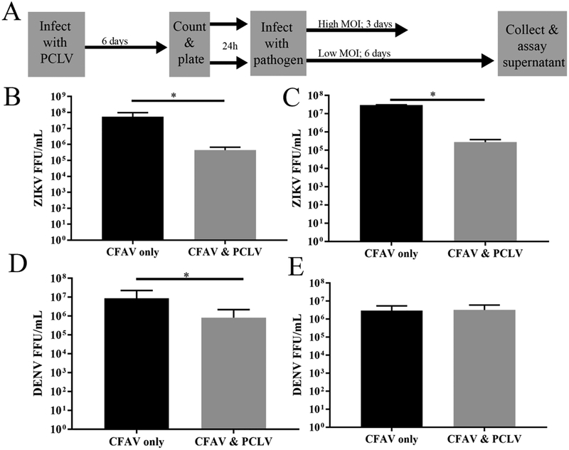
Upon identifying PCLV in some but not all of our cultured cells stocks we became interested in how PCLV and CFAV coinfection might impact the replication of different arboviruses. Since PCLV is consistently present in A. aegypti cell lines that are variable or non-permissive to ZIKV and not in in the A. albopictus cell lines that are established as ZIKV permissive (Aa23 (Schultz et al., 2017), C710 (Schultz et al., 2017), and C6/36 (Offerdahl et al., 2017)), we hypothesized PCLV affects the growth of ZIKV. To test this hypothesis, we established a persistent infection of PCLV in Aa23 cells and challenged cells with arbovirus infection (Figure 4A). Aa23 cells are CFAV positive and PCLV negative and are permissive to ZIKV infection (Schultz et al., 2017). We challenged Aa23 control cells without PCLV (CFAV only) and PCLV-infected Aa23 cells (CFAV and PCLV) cells with a low MOI (MOI 0.1) of ZIKV PRVABC59 to determine if ISV coinfection confers resistance to ZIKV infection and growth. Six days post infection, we observed a significant (p<0.05) 1 log reduction (90%) in ZIKV growth in the dual infected (CFAV and PCLV) infected cells assayed by focus forming assay (Figure 4B). When Aa23 CFAV only and Aa23 CFAV and PCLV positive cells were infected with a high MOI (MOI 10) of ZIKV PRVABC59, we observed a similar 1 log (90%) reduction in ZIKV titer in Aa23 CFAV and PCLV infected cells compared to Aa23 CFAV only positive cells (Figure 4C). These data suggest dual virus infection limits the growth of ZIKV.
Figure 4. Dual ISV infection limits flavivirus growth in A. albopictus cells.
(A) Experimental design to investigate tri-infection of A. albopictus Aa23 cells. Aa23 cells (CFAV positive) were infected with PCLV (1:10 dilution of Aag2 supernatant). PCLV was allowed 6 days to grow in Aa23 cells. Cells were then plated and infected with either ZIKV PRVABC59 or DENV-2 NGC at a low (MOI=0.1) or high MOI (MOI=10). After 6 days (low MOI) or 3 days (high MOI), supernatant was collected and the growth of ZIKV and DENV was assayed by focus forming assay. (B) ZIKV MOI 0.1 (C) ZIKV MOI 10 (D) DENV MOI 0.1 (E) DENV MOI 10. All data shown are the combined means and standard deviation of at least three independent experiments. Statistical significance determined by paired T Test one-tailed, alpha =0.05 on the natural log of FFU/mL accounting for non-normal distribution. * indicates p<0.05. Statistical tests were calculated by GraphPad Prism.
We next wanted to determine if PCLV restriction of viral growth was specific to ZIKV. Aag2 cells are more frequently utilized for DENV virus studies (Walker et al., 2014) and CFAV has previously been shown to promote DENV infection (Zhang et al., 2017) suggesting that PCLV may have a more mild effect on DENV growth in a coinfection setting. We infected Aa23 CFAV only and Aa23 CFAV and PCLV positive cells with DENV-2 at an MOI of 0.1 or 10 and assayed DENV-2 growth six or three days post infection, respectively, by focus forming assay. Consistent with ZIKV data, we observe a one log reduction (90%) of DENV growth in persistently infected Aa23 CFAV and PCLV positive cells infected at a low MOI (Figure 4D). In contrast, a high multiplicity of DENV infection (MOI=10), overcame the effect of PCLV on virus growth (Figure 4E).
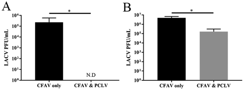
Since viruses of the same family often require the same host resources, we hypothesizeda dual ISV infection which includes a negative-sense segmented RNA virus may have a most pronounced effect on arboviruses with similar genome structure to itself. PCLV is in the Bunyaviridae family so we looked for a mosquito-transmitted Bunyaviridae family virus to co-infect with PCLV in Aa23 cells. LACV is an arbovirus which can be transmitted by Aedes sp. mosquitoes (Bara et al., 2016; Westby et al., 2015) and grown in A. albopictus cell lines (White, 1987). We infected Aa23 CFAV only and Aa23 CFAV and PCLV positive cells lines to determine if LACV virus could grow in the presence of a PCLV co-infection. Cells were infected at a low MOI of 0.1 and a high MOI of 10 with LACV followed by a six or three day incubation, respectively, as depicted in Figure 4A. LACV virus growth was undetectable in Aa23 CFAV and PCLV positive cells compared to Aa23 CFAV only cells (Figure 5A) following a low MOI exposure. LACV infection was reduced by 1–3 log (90–99.9%) following high MOI infection in Aa23 CFAV and PCLV positive cells (Figure 5B). These results show a robust inhibition of LACV replication in mosquito cells with both CFAV and PCLV.
Figure 5: Dual ISV infection limits LACV growth in A. albopictus cells.
(A and B) LACV H78 was grown in Aa23 PCLV-free (CFAV only) or PCLV-infected (CFAV & PCLV) cells at either a LACV MOI 0.1 (A) or LACV MOI 10 (B) by the schematic depicted in Figure 4. All data shown are the combined means and standard deviation of at least three independent experiments. Statistical significance determined by paired T Test one-tailed, alpha =0.05 on the natural log of FFU/mL accounting for non-normal distribution. * indicates p<0.05. N.D. indicates none detected. Statistical tests were calculated by GraphPad Prism.
Discussion
The data shown here has significant implications for arbovirus interactions with ISVs. A. aegypti cell lines, Aag2 and CCL-125, each originating from different mosquito isolation approaches (Singh, 1971) and different sources were positive for PCLV infection. We screened three different A. albopictus cell lines, C6/36, C710, and Aa23, and found PCLV was not present in any of these cell lines, demonstrating that infection was not universal within laboratory cell lines. This is not due to lack of permissivity, as Aa23 cells were readily infected with PCLV present in Aag2 supernatants. We show that PCLV grows in Aa23 cells consistent with previous findings that PCLV RNA produces protein in Aedes cells (Maringer et al., 2017). Unlike other reports of ISV infection including CFAV (Stollar and Thomas, 1975) and Kamiti River virus (Crabtree et al., 2003; Sang et al., 2003), we observed no cytopathic effect in CFAV and PCLV co-infected cells explaining why co-infection has remained undetected in most stocks.
Recreating the conditions of Aag2 dual ISV infection are imperfect yet highlight the potential ISV-arbovirus interference. PCLV alone from the supernatant of CCL-125s (which contained PCLV and not CFAV) did not generate a productive PCLV infection in any A. albopictus line tested. This limited our investigation to dual infection with CFAV and PCLV rather than PCLV infection alone. We infected Aa23 cells which already had a persistent CFAV infection with PCLV from Aag2 supernatants. In this system, CFAV is a long established symbiont of the cells and PCLV, although stable, was a relatively recent introduction. Our findings demonstrate a reduction of arbovirus growth in dual ISV infected Aa23 cells but do not recapitulate the high variability of ZIKV growth observed in Aag2 cells. This variability may be due to PCLV adaptation to Aag2 cells or an alternative variable not yet identified.
Our findings have relevance for ongoing cell culture work with arboviruses. Few A. aegypti cell lines are broadly available. CCL-125 and Aag2 A. aegypti cells can be requested from the ATCC (CCL-125 only) or other researchers (Walker et al., 2014). Recently, there has been contradictory data about cell line support of arbovirus replication in the two A. aegypti cell lines, CCL-125 and Aag2. A. aegypti cell line CCL-125 has been reported to be non-permissive to ZIKV (Offerdahl et al., 2017). Separate reports have suggested A. aegypti CCL-125 cells are (Wikan et al., 2009) and are not (Singh, 1971; Singh and Paul, 1968) susceptible to DENV. ZIKV growth in Aag2 cells has variable growth with reported peak titers ranging from 104 PFU/mL (Weger-Lucarelli et al., 2016) to 106 PFU/mL(Fulton et al., 2017; Weger-Lucarelli et al., 2017). Further, peak DENV growth in Aag2 cells has been published as low as 102 FFU/mL (Geoghegan et al., 2017) to 104 FFU/mL(Terradas et al., 2017). The identification or generation of an A. aegypti mosquito cell line permissive to a broad array of arboviruses with would benefit mechanistic studies of antiviral response in the mosquito.
Our data suggest growth of flaviviruses in A. aegypti Aag2 cell lines is variable and that co-infection of ISVs reduces arbovirus growth. This suggests that the low virus growth noted may be in part due to PCLV co-infection. Thus, the removal of PCLV or identification of a PCLV-free A. aegypti cell line may improve the function of A. aegypti cell culture system for arbovirus studies. It is unclear at this time if PCLV directly or simply the presence of dual ISV infection is the causative agent limiting arbovirus growth. An alternative hypothesis is that any two insect viruses persisting in cell culture exclude the infection of a third infection by an arbovirus. Future studies are needed to delineate the exact role of PCLV in this tripartite microbial infection.
Previous studies have also suggested that ISVs can limit the growth of arboviruses in a co-infection scenario in vitro and in vivo, reviewed by (Blitvich and Firth, 2015; Bolling et al., 2015a; Bolling et al., 2015b). ISVs of the Flavivirus genus have been the primary ISVs shown to limit the vector competence studies of other flaviviruses. Palm Creek virus restricts the growth of West Nile Virus (WNV) and Murray Valley encephalitis virus in vitro by approximately 1–1.5 logs (Hobson-Peters et al., 2013). Nhumirim virus has been shown to limit WNV, St Louis encephalitis virus, and Japanese encephalitis virus (Kenney et al., 2014). Consistent with the repression caused by flavivirus ISVs, we show CFAV and PCLV co-infection causes a repression of flaviviruses, ZIKV and DENV. Because CFAV has previously been reported to be beneficial to DENV replication (Zhang et al., 2017), it is likely that PCLV or a synergistic interaction between PCLV and CFAV causes the observed block in flavivirus replication. Perhaps the most striking observation of these studies is the almost complete inhibition of LACV replication by CFAV and PCLV coinfection. Since PCLV and LACV are both members of the Bunyaviridae family, we hypothesize this strong repression of LACV replication in the presence of PCLV and CFAV coinfection is likely due to similar genetics and lifecycles of LACV and PCLV.
There are many hypotheses about potential mechanisms for arbovirus/ISV competition in mosquito cells. One hypothesis, is that ISV mediated suppression is caused by superinfection exclusion. Sindbis virus infection has been shown to prevent cell line infection with other alphaviruses (Karpf et al., 1997). However, the underlying molecular mechanism of this exclusion is unknown. Further, superinfection exclusion is thought to drive exclusion of viruses within a family. CFAV and PCLV are in the Flaviviridae and Bunyaviridae families, respectively. There is a limited availability of antibodies and other molecular tools to investigate the exact mechanism of ISV suppression. Thus, expansion of ISV tools would aid our understanding of ISV-arbovirus competition.
ISVs have been suggested for the control of arbovirus transmission (Bolling et al., 2015a) based on their inhibition of arboviruses and wide geographic distribution (Cook et al., 2012; Farfan-Ale et al., 2009; Vasilakis et al., 2013; Vasilakis et al., 2014). They are stably maintained in populations by a high incidence of vertical transmission (Bolling et al., 2011; Haddow et al., 2013; Yamanaka et al., 2013). Our study is the first to report a negative-sense segmented RNA ISV that shows the possibility of acting as a broad reaching tool for arbovirus suppression. If the cell-culture inhibition observed is maintained in vivo, PCLV infection could be the basis of bunyavirus exclusion, expanding the tools for vector control and arbovirus disease prevention strategies. PCLV circulation has been reported in Thailand and Brazil but not in the United States where LACV cases are the highest and is a leading cause of pediatric encephalitis (Bewick et al., 2016; Taber et al., 2017; Westby et al., 2015). Further studies investigating the prevalence of PCLV and if this virus is co-circulating with LACV would inform efforts to control LACV.
This study provides insight into in vitro arbovirus growth variability potentially caused by ISV contaminants. Here, we have surveyed the extent of cell line infection with two ISVs, CFAV and PCLV, across A. aegypti and A. albopictus cells showing that both are permissive for PCLV, and A. aegypti cells are regularly infected with PCLV. To prevent confounding data, we recommend screening for CFAV and PCLV infection in future studies. The high prevalence of PCLV in A. aegypti cell lines makes this virus an important consideration for in vivo studies and arbovirus control through direct inhibition or as an agent for paratransgenic approaches.
Insect-specific viruses can be detected in an array of Aedes mosquito cell lines.
Phasi Charoen-like virus is found in A. aegypti but not A. albopictus cell lines.
albopictus cell lines are permissive to Phasi Charoen-like virus.
Zika, dengue, and La crosse virus growth are hindered by insect virus coinfection.
Acknowledgments
We thank Marshall Bloom, Tonya Colpitts, Ann Fallon, Sharon Isern, Scott Michael and
Zhiyong Xi for kindly sharing cell lines.
Funding: The Directors Fund, BU NEIDL
R21 NS101151, National Institute of Allergy and Infectious Diseases
1R56AI097589–01A1, National Institute of Allergy and Infectious Diseases
The funders had no role in study design, data collection and analysis, decision to publish, or preparation of the manuscript.
Footnotes
Publisher's Disclaimer: This is a PDF file of an unedited manuscript that has been accepted for publication. As a service to our customers we are providing this early version of the manuscript. The manuscript will undergo copyediting, typesetting, and review of the resulting proof before it is published in its final citable form. Please note that during the production process errors may be discovered which could affect the content, and all legal disclaimers that apply to the journal pertain.
References
- Bara JJ, Parker AT, Muturi EJ, 2016. Comparative Susceptibility of Ochlerotatus japonicus, Ochlerotatus triseriatus, Aedes albopictus, and Aedes aegypti (Diptera: Culicidae) to La Crosse Virus. Journal of medical entomology 53, 1415–1421. [DOI] [PubMed] [Google Scholar]
- Bewick S, Agusto F, Calabrese JM, Muturi EJ, Fagan WF, 2016. Epidemiology of La Crosse Virus Emergence, Appalachia Region, United States. Emerging Infectious Diseases 22, 1921–1929. [DOI] [PMC free article] [PubMed] [Google Scholar]
- Blitvich BJ, Firth AE, 2015. Insect-specific flaviviruses: a systematic review of their discovery, host range, mode of transmission, superinfection exclusion potential and genomic organization. Viruses 7. [DOI] [PMC free article] [PubMed] [Google Scholar]
- Bolling B, Weaver S, Tesh R, Vasilakis N, 2015a. Insect-Specific Virus Discovery: Significance for the Arbovirus Community. Viruses 7, 2851. [DOI] [PMC free article] [PubMed] [Google Scholar]
- Bolling BG, Eisen L, Moore CG, Blair CD, 2011. Insect-specific flaviviruses from Culex mosquitoes in Colorado, with evidence of vertical transmission. American Journal of Tropical Medicine & Hygeine 85. [DOI] [PMC free article] [PubMed] [Google Scholar]
- Bolling BG, Vasilakis N, Guzman H, Widen SG, Wood TG, Popov VL, Thangamani S, Tesh RB, 2015b. Insect-Specific Viruses Detected in Laboratory Mosquito Colonies and Their Potential Implications for Experiments Evaluating Arbovirus Vector Competence. The American Journal of Tropical Medicine and Hygiene 92, 422–428. [DOI] [PMC free article] [PubMed] [Google Scholar]
- Bourtzis K, Dobson SL, Xi Z, Rasgon JL, Calvitti M, Moreira LA, Bossin HC, Moretti R, Baton LA, Hughes GL, Mavingui P, Gilles JR, 2014. Harnessing mosquito-Wolbachia symbiosis for vector and disease control. Acta Trop 132 Suppl, S150–163. [DOI] [PubMed] [Google Scholar]
- Brackney DE, Scott JC, Sagawa F, Woodward JE, Miller NA, Schilkey FD, Mudge J, Wilusz J, Olson KE, Blair CD, Ebel GD, 2010. C6/36 Aedes albopictus cells have a dysfunctional antiviral RNA interference response. PLoS neglected tropical diseases 4, e856. [DOI] [PMC free article] [PubMed] [Google Scholar]
- Cafferata ML, Bardach A, Rey-Ares L, Alcaraz A, Cormick G, Gibbons L, Romano M, Cesaroni S, Ruvinsky S, 2013. Dengue Epidemiology and Burden of Disease in Latin America and the Caribbean: A Systematic Review of the Literature and Meta-Analysis. Value in Health Regional Issues 2, 347–356. [DOI] [PubMed] [Google Scholar]
- Chandler JA, Thongsripong P, Green A, Kittayapong P, Wilcox BA, Schroth GP, Kapan DD, Bennett SN, 2014. Metagenomic shotgun sequencing of a Bunyavirus in wild-caught Aedes aegypti from Thailand informs the evolutionary and genomic history of the Phleboviruses. Virology 464–465, 312–319. [DOI] [PMC free article] [PubMed] [Google Scholar]
- Cirimotich CM, Ramirez JL, Dimopoulos G, 2011. Native microbiota shape insect vector competence for human pathogens. Cell Host Microbe 10, 307–310. [DOI] [PMC free article] [PubMed] [Google Scholar]
- Cleton N, Koopmans M, Reimerink J, Godeke GJ, Reusken C, 2012. Come fly with me: review of clinically important arboviruses for global travelers. Journal of clinical virology : the official publication of the Pan American Society for Clinical Virology 55, 191–203. [DOI] [PubMed] [Google Scholar]
- Cook S, Moureau G, Kitchen A, Gould EA, de Lamballerie X, Holmes EC, Harbach RE, 2012. Molecular evolution of the insect-specific flaviviruses. Journal of General Virology 93, 223–234. [DOI] [PMC free article] [PubMed] [Google Scholar]
- Coutinho-Abreu IV, Zhu KY, Ramalho-Ortigao M, 2010. Transgenesis and paratransgenesis to control insect-borne diseases: current status and future challenges. Parasitol Int 59, 1–8. [DOI] [PMC free article] [PubMed] [Google Scholar]
- Crabtree MB, Sang RC, Stollar V, Dunster LM, Miller BR, 2003. Genetic and phenotypic characterization of the newly described insect flavivirus. Archives of Virology 148. [DOI] [PubMed] [Google Scholar]
- Donalisio MR, Freitas ARR, Zuben A, 2017. Arboviruses emerging in Brazil: challenges for clinic and implications for public health. Revista de saude publica 51, 30. [DOI] [PMC free article] [PubMed] [Google Scholar]
- Farfan-Ale JA, Loroño-Pino MA, Garcia-Rejon JE, Hovav E, Powers AM, Lin M, Dorman KS, Platt KB, Bartholomay LC, Soto V, Beaty BJ, Lanciotti RS, Blitvich BJ, 2009. Detection of RNA from a Novel West Nile-like Virus and High Prevalence of an Insect-specific Flavivirus in Mosquitoes in the Yucatan Peninsula of Mexico. The American Journal of Tropical Medicine & Hygiene 80, 85–95. [PMC free article] [PubMed] [Google Scholar]
- Fauci AS, Morens DM, 2016. Zika Virus in the Americas — Yet Another Arbovirus Threat. New England Journal of Medicine 374, 601–604. [DOI] [PubMed] [Google Scholar]
- Faye O, Faye O, Dupressoir A, Weidmann M, Ndiaye M, Alpha Sall A, 2008. One-step RT-PCR for detection of Zika virus. Journal of Clinical Virology 43, 96–101. [DOI] [PubMed] [Google Scholar]
- Fulton BO, Sachs D, Schwarz MC, Palese P, Evans MJ, 2017. Transposon Mutagenesis of the Zika Virus Genome Highlights Regions Essential for RNA Replication and Restricted for Immune Evasion. Journal of virology 91. [DOI] [PMC free article] [PubMed] [Google Scholar]
- Geoghegan V, Stainton K, Rainey SM, Ant TH, Dowle AA, Larson T, Hester S, Charles PD, Thomas B, Sinkins SP, 2017. Perturbed cholesterol and vesicular trafficking associated with dengue blocking in Wolbachia-infected Aedes aegypti cells. Nature communications 8, 526. [DOI] [PMC free article] [PubMed] [Google Scholar]
- Haddow AD, Guzman H, Popov VL, Wood TG, Widen SG, Tesh RB, Weaver SC, 2013. First isolation of Aedes flavivirus in the Western Hemisphere and evidence of vertical transmission in the mosquito Aedes (Stegomyia) albopictus (Diptera: Culicidae). Virology 440. [DOI] [PubMed] [Google Scholar]
- Hall-Mendelin S, McLean BJ, Bielefeldt-Ohmann H, Hobson-Peters J, Hall RA, van den Hurk AF, 2016. The insect-specific Palm Creek virus modulates West Nile virus infection in and transmission by Australian mosquitoes. Parasites & Vectors 9, 414. [DOI] [PMC free article] [PubMed] [Google Scholar]
- Hall RA, Bielefeldt-Ohmann H, McLean BJ, O’Brien CA, Colmant AMG, Piyasena TBH, Harrison JJ, Newton ND, Barnard RT, Prow NA, Deerain JM, Mah MGKY, Hobson-Peters J, 2016. Commensal Viruses of Mosquitoes: Host Restriction, Transmission, and Interaction with Arboviral Pathogens. Evolutionary Bioinformatics Online 12, 35–44. [DOI] [PMC free article] [PubMed] [Google Scholar]
- Hobson-Peters J, Yam AW, Lu JW, Setoh YX, May FJ, Kurucz N, Walsh S, Prow NA, Davis SS, Weir R, Melville L, Hunt N, Webb RI, Blitvich BJ, Whelan P, Hall RA, 2013. A new insect-specific flavivirus from northern Australia suppresses replication of West Nile virus and Murray Valley encephalitis virus in co-infected mosquito cells. PLoS One 8. [DOI] [PMC free article] [PubMed] [Google Scholar]
- Jupatanakul N, Sim S, Angleró-Rodríguez YI, Souza-Neto J, Das S, Poti KE, Rossi SL, Bergren N, Vasilakis N, Dimopoulos G, 2017. Engineered Aedes aegypti JAK/STAT Pathway-Mediated Immunity to Dengue Virus. PLoS neglected tropical diseases 11, e0005187. [DOI] [PMC free article] [PubMed] [Google Scholar]
- Jupatanakul N, Sim S, Dimopoulos G, 2014. The insect microbiome modulates vector competence for arboviruses. Viruses 6, 4294–4313. [DOI] [PMC free article] [PubMed] [Google Scholar]
- Karpf AR, Lenches E, Strauss EG, Strauss JH, Brown DT, 1997. Superinfection exclusion of alphaviruses in three mosquito cell lines persistently infected with Sindbis virus. Journal of virology 71, 7119–7123. [DOI] [PMC free article] [PubMed] [Google Scholar]
- Kenney JL, Solberg OD, Langevin SA, Brault AC, 2014. Characterization of a novel insect-specific flavivirus from Brazil: potential for inhibition of infection of arthropod cells with medically important flaviviruses. Journal of General Virology 95, 2796–2808. [DOI] [PMC free article] [PubMed] [Google Scholar]
- Lee SF, White VL, Weeks AR, Hoffmann AA, Endersby NM, 2012. High-throughput PCR assays to monitor Wolbachia infection in the dengue mosquito (Aedes aegypti) and Drosophila simulans. Applied and environmental microbiology 78, 4740–4743. [DOI] [PMC free article] [PubMed] [Google Scholar]
- Maringer K, Yousuf A, Heesom KJ, Fan J, Lee D, Fernandez-Sesma A, Bessant C, Matthews DA, Davidson AD, 2017. Proteomics informed by transcriptomics for characterising active transposable elements and genome annotation in Aedes aegypti. BMC genomics 18, 101. [DOI] [PMC free article] [PubMed] [Google Scholar]
- McGraw EA, O’Neill SL, 2013. Beyond insecticides: new thinking on an ancient problem. Nature Reviews Microbiology 11, 181–193. [DOI] [PubMed] [Google Scholar]
- Messina JP, Brady OJ, Scott TW, Zou C, Pigott DM, Duda KA, Bhatt S, Katzelnick L, Howes RE, Battle KE, Simmons CP, Hay SI, 2014. Global spread of dengue virus types: mapping the 70 year history. Trends in Microbiology 22, 138–146. [DOI] [PMC free article] [PubMed] [Google Scholar]
- Nicholson CO, Costin JM, Rowe DK, Lin L, Jenwitheesuk E, Samudrala R, Isern S, Michael SF, 2011. Viral entry inhibitors block dengue antibody-dependent enhancement in vitro. Antiviral research 89, 71–74. [DOI] [PubMed] [Google Scholar]
- Offerdahl DK, Dorward DW, Hansen BT, Bloom ME, 2017. Cytoarchitecture of Zika virus infection in human neuroblastoma and Aedes albopictus cell lines. Virology 501, 54–62. [DOI] [PMC free article] [PubMed] [Google Scholar]
- Paul LM, Carlin ER, Jenkins MM, Tan AL, Barcellona CM, Nicholson CO, Michael SF, Isern S, 2016. Dengue virus antibodies enhance Zika virus infection. Clinical & Translational Immunology 5, e117. [DOI] [PMC free article] [PubMed] [Google Scholar]
- Ren X, Hoiczyk E, Rasgon JL, 2008. Viral paratransgenesis in the malaria vector Anopheles gambiae. PLoS Pathog 4, e1000135. [DOI] [PMC free article] [PubMed] [Google Scholar]
- Sang RC, Gichogo A, Gachoya J, Dunster MD, Ofula V, Hunt AR, Crabtree MB, Miller BR, Dunster LM, 2003. Isolation of a new flavivirus related to cell fusing agent virus (CFAV) from field-collected flood-water Aedes mosquitoes sampled from a dambo in central Kenya. Archives of Virology 148. [DOI] [PubMed] [Google Scholar]
- Schnettler E, Sreenu VB, Mottram T, McFarlane M, 2016. Wolbachia restricts insect-specific flavivirus infection in Aedes aegypti cells. Journal of General Virology 97, 3024–3029. [DOI] [PMC free article] [PubMed] [Google Scholar]
- Schultz MJ, Connor JH, Frydman HM, 2018. Group B Wolbachia Strain-Dependent Inhibition of Arboviruses. DNA and cell biology 37, 2–6. [DOI] [PMC free article] [PubMed] [Google Scholar]
- Schultz MJ, Isern S, Michael SF, Corley RB, Connor J, Frydman HM, 2017. Variable inhibition of Zika virus replication by different Wolbachia strains in mosquito cell cultures. Journal of virology. [DOI] [PMC free article] [PubMed] [Google Scholar]
- Singh KR, 1971. Propagation of arboviruses in Singh’s Aedes cell lines. I. Growth of arboviruses in Aedes albopictus and A. aegypti cell lines. Current topics in microbiology and immunology 55, 127–133. [DOI] [PubMed] [Google Scholar]
- Singh KRP, 1967. Cell cultures derived from larvae of Aedes albopictus (Skuse) and Aedes aegypti (L.). Current Science 36. [Google Scholar]
- Singh KRP, Paul SD, 1968. Multiplication of arboviruses in cell lines from aedes albopictus and aedes aegypti. Current Science 37, 65–67. [Google Scholar]
- Stollar V, Thomas VL, 1975. An agent in the Aedes aegypti cell line (Peleg) which causes fusion of Aedes albopictus cells. Virology 64. [DOI] [PubMed] [Google Scholar]
- Taber ED, Hutchinson ML, Smithwick EAH, Blanford JI, 2017. A decade of colonization: the spread of the Asian tiger mosquito in Pennsylvania and implications for disease risk. Journal of vector ecology 42, 3–12. [DOI] [PubMed] [Google Scholar]
- Terradas G, Joubert DA, McGraw EA, 2017. The RNAi pathway plays a small part in Wolbachia-mediated blocking of dengue virus in mosquito cells. Scientific reports 7, 43847. [DOI] [PMC free article] [PubMed] [Google Scholar]
- Vasilakis N, Forrester NL, Palacios G, Nasar F, Savji N, Rossi SL, Guzman H, Wood TG, Popov V, Gorchakov R, Gonzalez AV, Haddow AD, Watts DM, da Rosa AP, Weaver SC, Lipkin WI, Tesh RB, 2013. Negevirus: a proposed new taxon of insect-specific viruses with wide geographic distribution. Journal of virology 87, 2475–2488. [DOI] [PMC free article] [PubMed] [Google Scholar]
- Vasilakis N, Guzman H, Firth C, Forrester NL, Widen SG, Wood TG, Rossi SL, Ghedin E, Popov V, Blasdell KR, Walker PJ, Tesh RB, 2014. Mesoniviruses are mosquito-specific viruses with extensive geographic distribution and host range. Virology Journal 11, 97. [DOI] [PMC free article] [PubMed] [Google Scholar]
- Walker T, Jeffries CL, Mansfield KL, Johnson N, 2014. Mosquito cell lines: history, isolation, availability and application to assess the threat of arboviral transmission in the United Kingdom. Parasites & Vectors 7, 382. [DOI] [PMC free article] [PubMed] [Google Scholar]
- Weger-Lucarelli J, Duggal NK, Bullard-Feibelman K, Veselinovic M, Romo H, Nguyen C, Ruckert C, Brault AC, Bowen RA, Stenglein M, Geiss BJ, Ebel GD, 2017. Development and Characterization of Recombinant Virus Generated from a New World Zika Virus Infectious Clone. Journal of virology 91. [DOI] [PMC free article] [PubMed] [Google Scholar]
- Weger-Lucarelli J, Rückert C, Chotiwan N, Nguyen C, Garcia Luna SM, Fauver JR, Foy BD, Perera R, Black WC, Kading RC, Ebel GD, 2016. Vector Competence of American Mosquitoes for Three Strains of Zika Virus. PLoS neglected tropical diseases 10, e0005101. [DOI] [PMC free article] [PubMed] [Google Scholar]
- Westby KM, Fritzen C, Paulsen D, Poindexter S, Moncayo AC, 2015. La Crosse Encephalitis Virus Infection in Field-Collected Aedes albopictus, Aedes japonicus, and Aedes triseriatus in Tennessee. Journal of the American Mosquito Control Association 31, 233–241. [DOI] [PubMed] [Google Scholar]
- White LA, 1987. Susceptibility of Aedes albopictus C6/36 cells to viral infection. Journal of clinical microbiology 25, 1221–1224. [DOI] [PMC free article] [PubMed] [Google Scholar]
- Wikan N, Kuadkitkan A, Smith DR, 2009. The Aedes aegypti cell line CCL-125 is dengue virus permissive. Journal of virological methods 157, 227–230. [DOI] [PubMed] [Google Scholar]
- Yamanaka A, Thongrungkiat S, Ramasoota P, Konishi E, 2013. Genetic and evolutionary analysis of cell-fusing agent virus based on Thai strains isolated in 2008 and 2012. Infection, Genetics and Evolution 19, 188–194. [DOI] [PubMed] [Google Scholar]
- Zhang G, Asad S, Khromykh AA, Asgari S, 2017. Cell fusing agent virus and dengue virus mutually interact in Aedes aegypti cell lines. Scientific reports 7, 6935. [DOI] [PMC free article] [PubMed] [Google Scholar]


